Advertisement

Main menu

  • Home
  • Content
    • Current Issue
    • Accepted Manuscripts
    • Article Preview
    • Past Issue Archive
    • Video Articles
    • AJNR Case Collection
    • Case of the Week Archive
    • Case of the Month Archive
    • Classic Case Archive
  • Special Collections
    • AJNR Awards
    • Low-Field MRI
    • Alzheimer Disease
    • ASNR Foundation Special Collection
    • Photon-Counting CT
    • View All
  • Multimedia
    • AJNR Podcasts
    • AJNR SCANtastic
    • Trainee Corner
    • MRI Safety Corner
    • Imaging Protocols
  • For Authors
    • Submit a Manuscript
    • Submit a Video Article
    • Submit an eLetter to the Editor/Response
    • Manuscript Submission Guidelines
    • Statistical Tips
    • Fast Publishing of Accepted Manuscripts
    • Graphical Abstract Preparation
    • Imaging Protocol Submission
    • Author Policies
  • About Us
    • About AJNR
    • Editorial Board
    • Editorial Board Alumni
  • More
    • Become a Reviewer/Academy of Reviewers
    • Subscribers
    • Permissions
    • Alerts
    • Feedback
    • Advertisers
    • ASNR Home

User menu

  • Alerts
  • Log in

Search

  • Advanced search
American Journal of Neuroradiology

American Journal of Neuroradiology

ASHNR American Society of Functional Neuroradiology ASHNR American Society of Pediatric Neuroradiology ASSR
  • Alerts
  • Log in

Advanced Search

  • Home
  • Content
    • Current Issue
    • Accepted Manuscripts
    • Article Preview
    • Past Issue Archive
    • Video Articles
    • AJNR Case Collection
    • Case of the Week Archive
    • Case of the Month Archive
    • Classic Case Archive
  • Special Collections
    • AJNR Awards
    • Low-Field MRI
    • Alzheimer Disease
    • ASNR Foundation Special Collection
    • Photon-Counting CT
    • View All
  • Multimedia
    • AJNR Podcasts
    • AJNR SCANtastic
    • Trainee Corner
    • MRI Safety Corner
    • Imaging Protocols
  • For Authors
    • Submit a Manuscript
    • Submit a Video Article
    • Submit an eLetter to the Editor/Response
    • Manuscript Submission Guidelines
    • Statistical Tips
    • Fast Publishing of Accepted Manuscripts
    • Graphical Abstract Preparation
    • Imaging Protocol Submission
    • Author Policies
  • About Us
    • About AJNR
    • Editorial Board
    • Editorial Board Alumni
  • More
    • Become a Reviewer/Academy of Reviewers
    • Subscribers
    • Permissions
    • Alerts
    • Feedback
    • Advertisers
    • ASNR Home
  • Follow AJNR on Twitter
  • Visit AJNR on Facebook
  • Follow AJNR on Instagram
  • Join AJNR on LinkedIn
  • RSS Feeds

AJNR Awards, New Junior Editors, and more. Read the latest AJNR updates

Research ArticleNeurointervention
Open Access

Quantifying Intracranial Aneurysm Wall Permeability for Risk Assessment Using Dynamic Contrast-Enhanced MRI: A Pilot Study

P. Vakil, S.A. Ansari, C.G. Cantrell, C.S. Eddleman, F.H. Dehkordi, J. Vranic, M.C. Hurley, H.H. Batjer, B.R. Bendok and T.J. Carroll
American Journal of Neuroradiology May 2015, 36 (5) 953-959; DOI: https://doi.org/10.3174/ajnr.A4225
P. Vakil
aFrom the Departments of Radiology (P.V., S.A.A., J.V., M.C.H., T.J.C.)
cBiomedical Engineering (P.V., C.G.C., T.J.C.), Northwestern University, Chicago, Illinois
  • Find this author on Google Scholar
  • Find this author on PubMed
  • Search for this author on this site
S.A. Ansari
aFrom the Departments of Radiology (P.V., S.A.A., J.V., M.C.H., T.J.C.)
  • Find this author on Google Scholar
  • Find this author on PubMed
  • Search for this author on this site
C.G. Cantrell
cBiomedical Engineering (P.V., C.G.C., T.J.C.), Northwestern University, Chicago, Illinois
  • Find this author on Google Scholar
  • Find this author on PubMed
  • Search for this author on this site
C.S. Eddleman
dDepartment of Neurological Surgery (C.S.E., H.H.B.), University of Texas-Southwestern, Dallas, Texas
  • Find this author on Google Scholar
  • Find this author on PubMed
  • Search for this author on this site
F.H. Dehkordi
eDepartment of Economics and Decision Sciences (F.H.D.), Western Illinois University, Macomb, Illinois.
  • Find this author on Google Scholar
  • Find this author on PubMed
  • Search for this author on this site
J. Vranic
aFrom the Departments of Radiology (P.V., S.A.A., J.V., M.C.H., T.J.C.)
  • Find this author on Google Scholar
  • Find this author on PubMed
  • Search for this author on this site
M.C. Hurley
aFrom the Departments of Radiology (P.V., S.A.A., J.V., M.C.H., T.J.C.)
  • Find this author on Google Scholar
  • Find this author on PubMed
  • Search for this author on this site
H.H. Batjer
dDepartment of Neurological Surgery (C.S.E., H.H.B.), University of Texas-Southwestern, Dallas, Texas
  • Find this author on Google Scholar
  • Find this author on PubMed
  • Search for this author on this site
B.R. Bendok
bNeurosurgery (B.R.B.)
  • Find this author on Google Scholar
  • Find this author on PubMed
  • Search for this author on this site
T.J. Carroll
aFrom the Departments of Radiology (P.V., S.A.A., J.V., M.C.H., T.J.C.)
cBiomedical Engineering (P.V., C.G.C., T.J.C.), Northwestern University, Chicago, Illinois
  • Find this author on Google Scholar
  • Find this author on PubMed
  • Search for this author on this site
  • Article
  • Figures & Data
  • Supplemental
  • Info & Metrics
  • Responses
  • References
  • PDF
Loading

Article Figures & Data

Figures

  • Tables
  • Fig 1.
    • Download figure
    • Open in new tab
    • Download powerpoint
    Fig 1.

    Aneurysm permeability modeling in a 14-mm right ICA aneurysm. In the IA, the signal time course from DCE imaging (A) is used to measure the plasma concentration, Cp(t) (red curve, B). The permeability signal, C(t), is subsequently modeled and fit for every voxel to Equation 2. The signal from a single voxel selected from the white circled region adjacent to the IA (white diamonds, B) and fit (yellow curve, B) are shown. The fitting of all voxels derives permeability parameters (Ktrans, VL, and Vp, C). Note the observed signal next to the aneurysm has nearly 3-fold the signal intensity as background (BG) enhancement (blue curve, B). ROIs were placed adjacent to a healthy artery (left panel, D) and the area of highest permeability, next to the IA (right panel, D).

  • Fig 2.
    • Download figure
    • Open in new tab
    • Download powerpoint
    Fig 2.

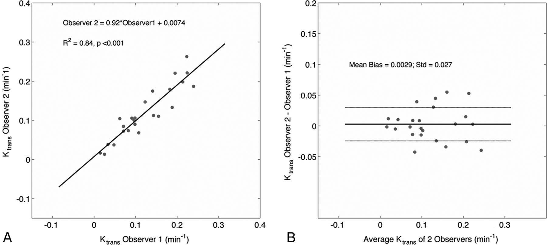
    Correlation and Bland-Altman analysis between measured Ktrans values of both observers reveal strong agreement in derived values. VL is not shown.

  • Fig 3.
    • Download figure
    • Open in new tab
    • Download powerpoint
    Fig 3.

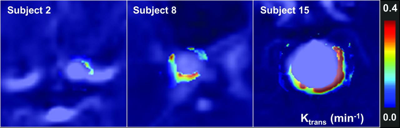
    DSA and Ktrans permeability images demonstrate a broad distribution of wall permeability values. Notice the heterogeneity in Ktrans both among subjects and within a single IA.

  • Fig 4.
    • Download figure
    • Open in new tab
    • Download powerpoint
    Fig 4.

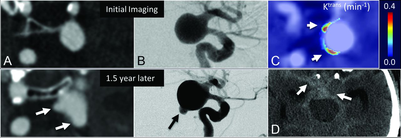
    The evolution of an untreated posterior communicating artery IA in a 61-year-old man demonstrates that elevated Ktrans at baseline correlated with bleb formation. Morphologic changes during 1.5 years are observed on CTA (A) and DSA (B); however, DCE–MR imaging demonstrates 2 regions with high Ktrans (arrows, C) at the time of imaging—apparently correlating with eventual SAH seen on CT (arrows, D).

  • Fig 5.
    • Download figure
    • Open in new tab
    • Download powerpoint
    Fig 5.

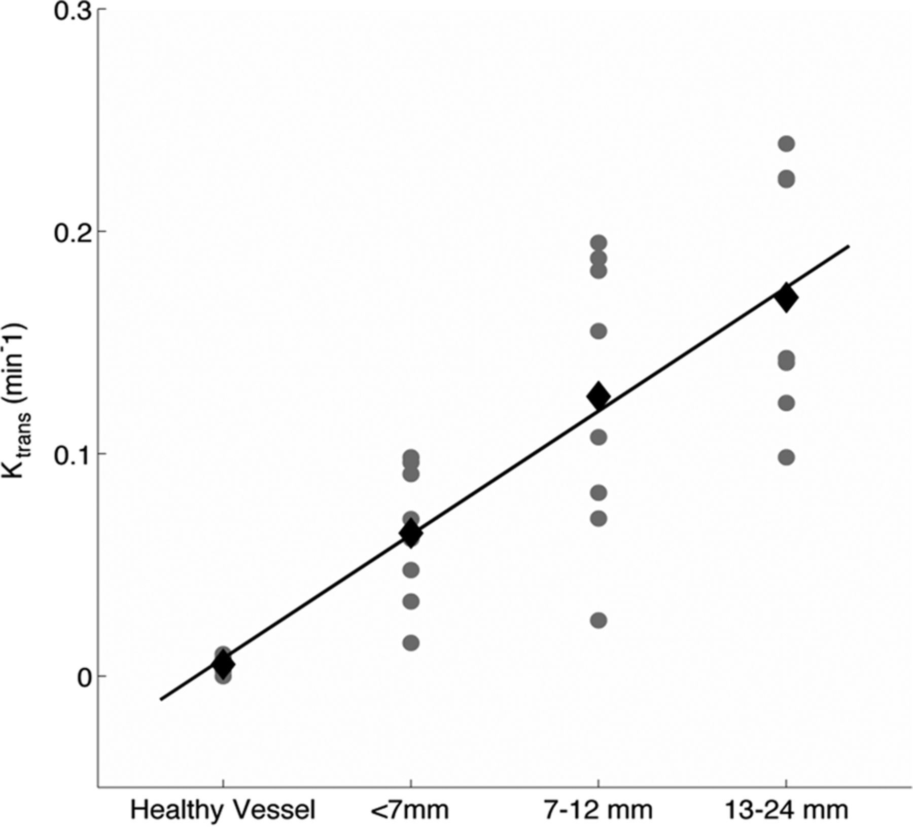
    Mean Ktrans and VL values (black diamonds) increase linearly with larger ISUIA-determined size/risk bins (<7, 7–12, and 13–24 mm) as shown in the scatterplot. Note that our cohort did not contain aneurysms in the >25-mm size/risk bin. The gray circles represent individual IA permeability values. The black diamonds are the mean for each size bin.

Tables

  • Figures
  • Results of univariate and multivariate logistic regression of demographic and imaging markers against size-independent risk paradigmsa

    Group A (High Risk = 7, Low Risk = 17)Group B (High Risk = 15, Low Risk = 9)Group AB (High Risk = 17, Low Risk = 7)
    P ValueCoefficientStandard ErrorP ValueCoefficientStandard ErrorP ValueCoefficientStandard Error
    Univariate analysis
        Sex.4092−0.79850.9675.52310.55960.8763.9395−0.06900.9090
        Age.4463−0.03020.0396.16870.05630.0409.9774−0.00110.0401
        HTNb (n = 19).999927.49103.32E + 05.25601.17871.0377.10731.72721.0724
        HLD (n = 17).32051.18561.1934.21151.16320.9309.35040.89100.9541
        Statins (n = 12).47840.64440.9090.91580.08960.8473.85110.16990.9052
        Tobacco
            Current use (n = 5).55260.62421.0511.999927.933.32E + 05.999927.49373.32E + 05
            Past use (n = 11).47840.64440.9090.91580.08960.8473.85110.16990.9052
        Alcoholc (n = 3).86540.22311.3166.2897−1.38631.3093.1620−1.18561.3276
        Imaging markers (n = 24)
            IA size.0182d0.2877d0.1222d.9583−0.00360.0695.39620.07640.0900
            IA neck.06160.57430.3072.6545−0.10610.2370.52230.18800.2938
            Ktrans.0280d19.5088d8.8789d.0243d23.6481d10.4975d.0286d36.5083d16.6795d
            Aspect ratio.10741.28140.7959.6270.20730.4266.45110.43850.5819
            VL.22864.52103.7548.41763.33954.1195.36324.48434.9321
    Multivariate analysis
        IA Size.03370.24480.1153.1534−0.15910.1114.4435−0.08170.1066
        Ktrans.133918.554412.3781.0231d33.9634d14.9545d.0346d43.0945d20.3981d
    • Note:—HTN indicates hypertension; HLD, hyperlipidemia.

    • ↵a Analysis performed on 24 saccular IAs.

    • ↵b All patients' hypertension was under control using medication.

    • ↵c Current use.

    • ↵d Significant (P < .05).

PreviousNext
Back to top

In this issue

American Journal of Neuroradiology: 36 (5)
American Journal of Neuroradiology
Vol. 36, Issue 5
1 May 2015
  • Table of Contents
  • Index by author
  • Complete Issue (PDF)
Advertisement
Print
Download PDF
Email Article

Thank you for your interest in spreading the word on American Journal of Neuroradiology.

NOTE: We only request your email address so that the person you are recommending the page to knows that you wanted them to see it, and that it is not junk mail. We do not capture any email address.

Enter multiple addresses on separate lines or separate them with commas.
Quantifying Intracranial Aneurysm Wall Permeability for Risk Assessment Using Dynamic Contrast-Enhanced MRI: A Pilot Study
(Your Name) has sent you a message from American Journal of Neuroradiology
(Your Name) thought you would like to see the American Journal of Neuroradiology web site.
CAPTCHA
This question is for testing whether or not you are a human visitor and to prevent automated spam submissions.
Cite this article
P. Vakil, S.A. Ansari, C.G. Cantrell, C.S. Eddleman, F.H. Dehkordi, J. Vranic, M.C. Hurley, H.H. Batjer, B.R. Bendok, T.J. Carroll
Quantifying Intracranial Aneurysm Wall Permeability for Risk Assessment Using Dynamic Contrast-Enhanced MRI: A Pilot Study
American Journal of Neuroradiology May 2015, 36 (5) 953-959; DOI: 10.3174/ajnr.A4225

Citation Manager Formats

  • BibTeX
  • Bookends
  • EasyBib
  • EndNote (tagged)
  • EndNote 8 (xml)
  • Medlars
  • Mendeley
  • Papers
  • RefWorks Tagged
  • Ref Manager
  • RIS
  • Zotero
0 Responses
Respond to this article
Share
Bookmark this article
Quantifying Intracranial Aneurysm Wall Permeability for Risk Assessment Using Dynamic Contrast-Enhanced MRI: A Pilot Study
P. Vakil, S.A. Ansari, C.G. Cantrell, C.S. Eddleman, F.H. Dehkordi, J. Vranic, M.C. Hurley, H.H. Batjer, B.R. Bendok, T.J. Carroll
American Journal of Neuroradiology May 2015, 36 (5) 953-959; DOI: 10.3174/ajnr.A4225
del.icio.us logo Twitter logo Facebook logo Mendeley logo
  • Tweet Widget
  • Facebook Like
  • Google Plus One
Purchase

Jump to section

  • Article
    • Abstract
    • ABBREVIATIONS:
    • Materials and Methods
    • Results
    • Discussion
    • Conclusions
    • Footnotes
    • REFERENCES
  • Figures & Data
  • Supplemental
  • Info & Metrics
  • Responses
  • References
  • PDF

Related Articles

  • PubMed
  • Google Scholar

Cited By...

  • Temporal Changes on Postgadolinium MR Vessel Wall Imaging Captures Enhancement Kinetics of Intracranial Atherosclerotic Plaques and Aneurysms
  • Quantitative analysis of unruptured intracranial aneurysm wall thickness and enhancement using 7T high resolution, black blood magnetic resonance imaging
  • Vessel Wall Imaging of Unruptured Intracranial Aneurysms: Ready for Prime Time? Not so Fast!
  • Complementary Roles of Dynamic Contrast-Enhanced MR Imaging and Postcontrast Vessel Wall Imaging in Detecting High-Risk Intracranial Aneurysms
  • Wall enhancement ratio and partial wall enhancement on MRI associated with the rupture of intracranial aneurysms
  • Quantifying Intracranial Plaque Permeability with Dynamic Contrast-Enhanced MRI: A Pilot Study
  • Crossref (44)
  • Google Scholar

This article has been cited by the following articles in journals that are participating in Crossref Cited-by Linking.

  • Hemodynamic Stress, Inflammation, and Intracranial Aneurysm Development and Rupture: A Systematic Review
    Francesco Signorelli, Sapir Sela, Loreto Gesualdo, Sophie Chevrel, Félix Tollet, Cyril Pailler-Mattei, Leonello Tacconi, Francis Turjman, Angelo Vacca, David B. Schul
    World Neurosurgery 2018 115
  • The biophysical role of hemodynamics in the pathogenesis of cerebral aneurysm formation and rupture
    Sauson Soldozy, Pedro Norat, Mazin Elsarrag, Ajay Chatrath, John S. Costello, Jennifer D. Sokolowski, Petr Tvrdik, M. Yashar S. Kalani, Min S. Park
    Neurosurgical Focus 2019 47 1
  • Wall enhancement ratio and partial wall enhancement on MRI associated with the rupture of intracranial aneurysms
    Guang-xian Wang, Li Wen, Sheng Lei, Qian Ran, Jin-bo Yin, Zi-li Gong, Dong Zhang
    Journal of NeuroInterventional Surgery 2018 10 6
  • Four-dimensional MRI flow examinations in cerebral and extracerebral vessels – ready for clinical routine?
    Susanne Schnell, Can Wu, Sameer A. Ansari
    Current Opinion in Neurology 2016 29 4
  • Imaging of cerebral aneurysms: a clinical perspective
    Nam K. Yoon, Scott McNally, Philipp Taussky, Min S. Park
    Neurovascular Imaging 2016 2 1
  • Current Perspectives in Imaging Modalities for the Assessment of Unruptured Intracranial Aneurysms: A Comparative Analysis and Review
    Nefize Turan, Robert A. Heider, Anil K. Roy, Brandon A. Miller, Mark E. Mullins, Daniel L. Barrow, Jonathan Grossberg, Gustavo Pradilla
    World Neurosurgery 2018 113
  • Clinical Significance of Circumferential Aneurysmal Wall Enhancement in Symptomatic Patients with Unruptured Intracranial Aneurysms: a High-resolution MRI Study
    Qichang Fu, Sheng Guan, Chao Liu, Keyan Wang, Jingliang Cheng
    Clinical Neuroradiology 2018 28 4
  • Vessel wall magnetic resonance imaging in intracranial aneurysms: Principles and emerging clinical applications
    Corrado Santarosa, Branden Cord, Andrew Koo, Pervinder Bhogal, Ajay Malhotra, Sam Payabvash, Frank J Minja, Charles C Matouk
    Interventional Neuroradiology 2020 26 2
  • Evaluation of the risk of rupture of intracranial aneurysms in patients with aneurysmal subarachnoid hemorrhage according to the PHASES score
    Belal Neyazi, I. Erol Sandalcioglu, Homajoun Maslehaty
    Neurosurgical Review 2019 42 2
  • Disturbed flow&#039;s impact on cellular changes indicative of vascular aneurysm initiation, expansion, and rupture: A pathological and methodological review
    Kevin Sunderland, Jingfeng Jiang, Feng Zhao
    Journal of Cellular Physiology 2022 237 1

More in this TOC Section

  • Factors Associated with Major Re-Recanalization following Second Coiling for Recanalized Aneurysms: A Multicenter Experience over 20 Years during Long-Term Follow-up
  • A Key Factor Shapes LS-DAVFs EVT Outcome
  • Optimizing Voxel Size in 3D Rotational Angiography
Show more NEUROINTERVENTION

Similar Articles

Advertisement

Indexed Content

  • Current Issue
  • Accepted Manuscripts
  • Article Preview
  • Past Issues
  • Editorials
  • Editor's Choice
  • Fellows' Journal Club
  • Letters to the Editor
  • Video Articles

Cases

  • Case Collection
  • Archive - Case of the Week
  • Archive - Case of the Month
  • Archive - Classic Case

More from AJNR

  • Trainee Corner
  • Imaging Protocols
  • MRI Safety Corner

Multimedia

  • AJNR Podcasts
  • AJNR Scantastics

Resources

  • Turnaround Time
  • Submit a Manuscript
  • Submit a Video Article
  • Submit an eLetter to the Editor/Response
  • Manuscript Submission Guidelines
  • Statistical Tips
  • Fast Publishing of Accepted Manuscripts
  • Graphical Abstract Preparation
  • Imaging Protocol Submission
  • Evidence-Based Medicine Level Guide
  • Publishing Checklists
  • Author Policies
  • Become a Reviewer/Academy of Reviewers
  • News and Updates

About Us

  • About AJNR
  • Editorial Board
  • Editorial Board Alumni
  • Alerts
  • Permissions
  • Not an AJNR Subscriber? Join Now
  • Advertise with Us
  • Librarian Resources
  • Feedback
  • Terms and Conditions
  • AJNR Editorial Board Alumni

American Society of Neuroradiology

  • Not an ASNR Member? Join Now

© 2025 by the American Society of Neuroradiology All rights, including for text and data mining, AI training, and similar technologies, are reserved.
Print ISSN: 0195-6108 Online ISSN: 1936-959X

Powered by HighWire