
Article Figures & Data
Figures
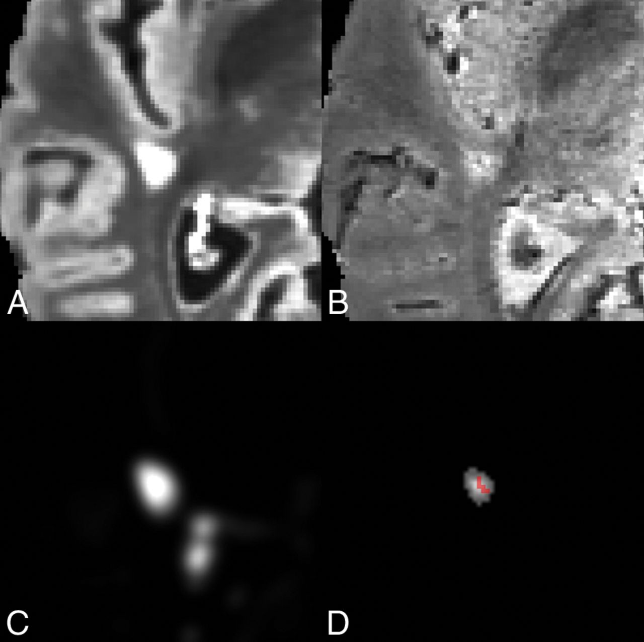
- Fig 1.
A, Axial slice of a lesion on T2-FLAIR volume. B, Axial slice of a lesion on T2*-EPI volume. C, MIMoSA lesion-probability map. D, Distance-to-lesion-boundary mask with the vesselness filter overlay. The lesion-level CVS probability following permutation was 0.975.
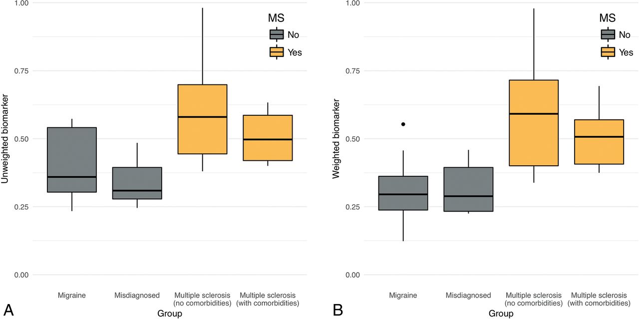
- Fig 2.
Boxplots of the patient-level central vein sign biomarker score by diagnostic group. The score can be interpreted as the proportion of lesions that are CVS+ according to the method described in this article. Groups shaded gray do not have an MS diagnosis, whereas groups shaded gold do. A, Boxplots for the unweighted biomarker. B, Boxplots for the noise-weighted biomarker. Points outside the boxplots represent outliers within their respective groups.
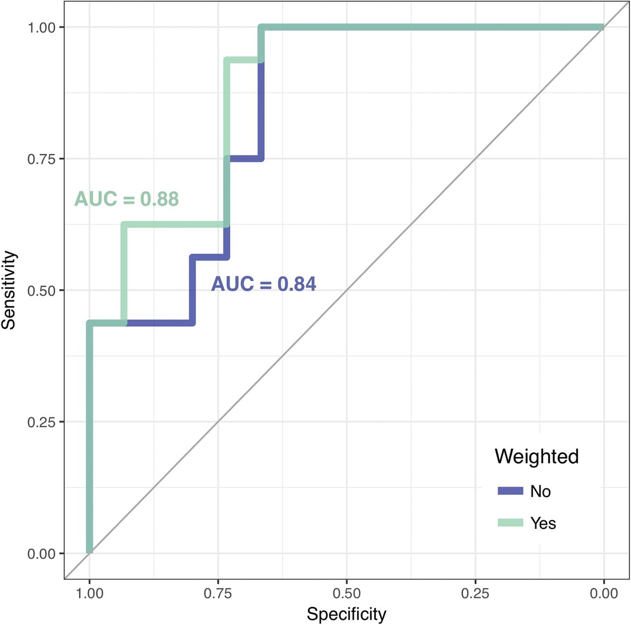
- Fig 3.
Receiver operating characteristic curves of MS diagnosis based on patient-level automated CVS biomarker scores. The receiver operating characteristic curve for the unweighted biomarker is shaded blue, and the receiver operating characteristic curve for the weighted biomarker is shaded green. The area under the curve (AUC) values for both curves are displayed in their respective colors.


Tables
Demographics MS (n = 10) Age (yr) 44 (16) Sex 9/10 Female Disease duration (yr) 9 (7) Disease subtype 10/10 RRMS MS with comorbidities (n = 10) Age (yr) 43 (9) Sex 9/10 Female Disease duration (yr) 9 (6) Disease subtype 10/10 RRMS Migraine (n = 10) Age (yr) 47 (13) Sex 10/10 Female Misdiagnosed with MS (n = 10) Age (yr) 53 (7) Sex 9/10 Female Note:—RRMS indicates relapsing-remitting MS.
↵a The numbers in parentheses are standard deviations.
Threshold Sensitivity Specificity Unweighted biomarker (ψi) 0.38 1.00 (0.75–1.00) 0.67 (0.42–0.84) 0.40 0.94 (0.70–1.00) 0.67 (0.42–0.84) 0.44 0.75 (0.50–0.90) 0.73 (0.47–0.89) 0.50 0.56 (0.25–0.65) 0.80 (0.58–0.94) Weighted biomarker (ψiw) 0.37 0.94 (0.70–1.00) 0.73 (0.42–0.84) 0.40 0.75 (0.50–0.90) 0.73 (0.42–0.84) 0.46 0.63 (0.40–0.80) 0.93 (0.68–1.00) 0.50 0.56 (0.35–0.75) 0.93 (0.74–1.00) ↵a Data in column 2 represent the given cutoff values; data in parentheses are the relevant 95% confidence intervals.


