
Article Figures & Data
Figures
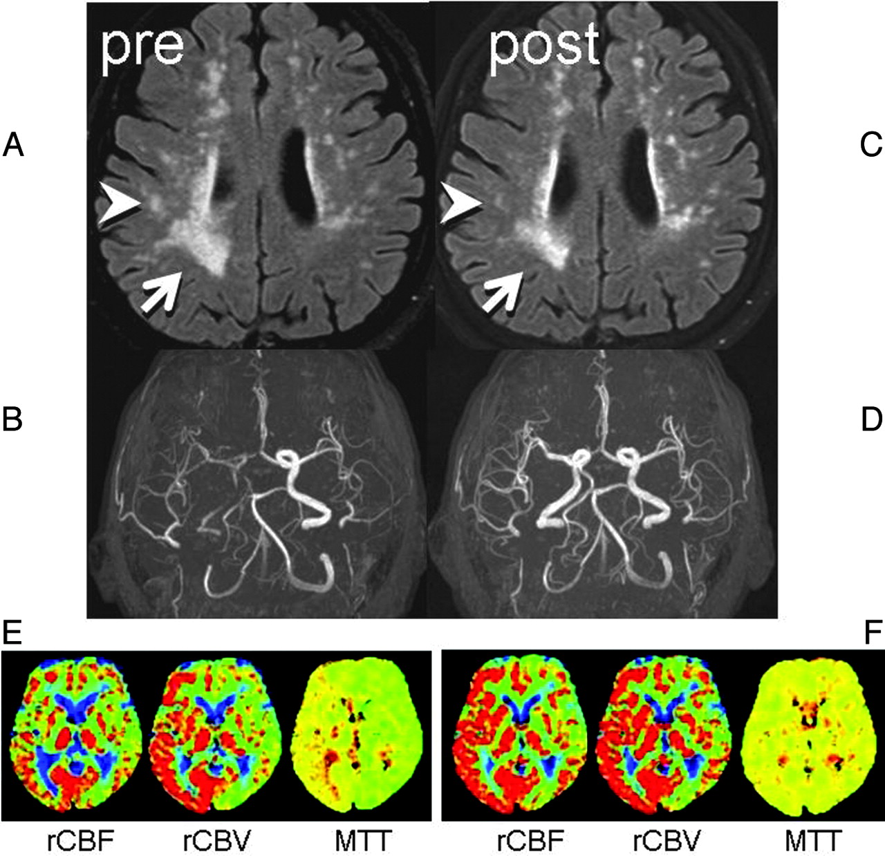
- Fig 1.
Two consecutive FLAIR images and MR angiography. A, Preprocedural FLAIR (delay time, 2200 ms; TR/TE, 8000/100 ms) demonstrates WMLs. B, Preprocedural MR angiography (TR/TE, 30/2.3 ms) shows poor visualization of the right anterior circulation branches. C, Postprocedural FLAIR a week later shows partial WML resolution in the right hemisphere (arrow/arrowhead). D, MR angiogram shows restoration of flow in the right ICA. E, Preprocedural perfusion-weighted images show MTT elongation at the right anterior circulation territory with compensatory vasodilation, indicated by increased cerebral blood volume. F, After the procedure, the MTT elongation is normalized. Increased cerebral blood flow at the same territory indicates postprocedural hyperperfusion, but the patient remained asymptomatic.
- Fig 2.
Image analysis on pre- and postprocedural images. A, Image coregistration was performed between the pre- and postprocedural FLAIR. B, Segmentation of the pixels with WMLs was then done. C, Subtraction images yielded pixels with/without reversal. Coregistration of these data onto the DWI/DTI (DWI: TR/TE, 6000/88 ms; b = 1000 s/mm2) and b = 0 imaging was performed to characterize the nature of the WMLs.


