Advertisement

Main menu

  • Home
  • Content
    • Current Issue
    • Accepted Manuscripts
    • Article Preview
    • Past Issue Archive
    • Video Articles
    • AJNR Case Collection
    • Case of the Week Archive
    • Case of the Month Archive
    • Classic Case Archive
  • Special Collections
    • AJNR Awards
    • Low-Field MRI
    • Alzheimer Disease
    • ASNR Foundation Special Collection
    • Photon-Counting CT
    • View All
  • Multimedia
    • AJNR Podcasts
    • AJNR SCANtastic
    • Trainee Corner
    • MRI Safety Corner
    • Imaging Protocols
  • For Authors
    • Submit a Manuscript
    • Submit a Video Article
    • Submit an eLetter to the Editor/Response
    • Manuscript Submission Guidelines
    • Statistical Tips
    • Fast Publishing of Accepted Manuscripts
    • Graphical Abstract Preparation
    • Imaging Protocol Submission
    • Author Policies
  • About Us
    • About AJNR
    • Editorial Board
    • Editorial Board Alumni
  • More
    • Become a Reviewer/Academy of Reviewers
    • Subscribers
    • Permissions
    • Alerts
    • Feedback
    • Advertisers
    • ASNR Home

User menu

  • Alerts
  • Log in

Search

  • Advanced search
American Journal of Neuroradiology

American Journal of Neuroradiology

ASHNR American Society of Functional Neuroradiology ASHNR American Society of Pediatric Neuroradiology ASSR
  • Alerts
  • Log in

Advanced Search

  • Home
  • Content
    • Current Issue
    • Accepted Manuscripts
    • Article Preview
    • Past Issue Archive
    • Video Articles
    • AJNR Case Collection
    • Case of the Week Archive
    • Case of the Month Archive
    • Classic Case Archive
  • Special Collections
    • AJNR Awards
    • Low-Field MRI
    • Alzheimer Disease
    • ASNR Foundation Special Collection
    • Photon-Counting CT
    • View All
  • Multimedia
    • AJNR Podcasts
    • AJNR SCANtastic
    • Trainee Corner
    • MRI Safety Corner
    • Imaging Protocols
  • For Authors
    • Submit a Manuscript
    • Submit a Video Article
    • Submit an eLetter to the Editor/Response
    • Manuscript Submission Guidelines
    • Statistical Tips
    • Fast Publishing of Accepted Manuscripts
    • Graphical Abstract Preparation
    • Imaging Protocol Submission
    • Author Policies
  • About Us
    • About AJNR
    • Editorial Board
    • Editorial Board Alumni
  • More
    • Become a Reviewer/Academy of Reviewers
    • Subscribers
    • Permissions
    • Alerts
    • Feedback
    • Advertisers
    • ASNR Home
  • Follow AJNR on Twitter
  • Visit AJNR on Facebook
  • Follow AJNR on Instagram
  • Join AJNR on LinkedIn
  • RSS Feeds

AJNR Awards, New Junior Editors, and more. Read the latest AJNR updates

Case ReportBrain

Cerebral White Matter Lesions May Be Partially Reversible in Patients with Carotid Artery Stenosis

K. Yamada, K. Sakai, K. Owada, K. Mineura and T. Nishimura
American Journal of Neuroradiology August 2010, 31 (7) 1350-1352; DOI: https://doi.org/10.3174/ajnr.A1873
K. Yamada
  • Find this author on Google Scholar
  • Find this author on PubMed
  • Search for this author on this site
K. Sakai
  • Find this author on Google Scholar
  • Find this author on PubMed
  • Search for this author on this site
K. Owada
  • Find this author on Google Scholar
  • Find this author on PubMed
  • Search for this author on this site
K. Mineura
  • Find this author on Google Scholar
  • Find this author on PubMed
  • Search for this author on this site
T. Nishimura
  • Find this author on Google Scholar
  • Find this author on PubMed
  • Search for this author on this site
  • Article
  • Figures & Data
  • Info & Metrics
  • Responses
  • References
  • PDF
Loading

Abstract

SUMMARY: Contrary to the common belief that age-related WMLs (also known as leukoaraiosis) are a progressive condition, a case of partial reversal of WMLs shortly after carotid artery stenting is described. A 75-year-old man presented with frequent TIAs, which were attributed to right ICA stenosis. He subsequently underwent successful carotid artery stenting. Follow-up MR imaging a week after the procedure showed improvement of WMLs in the right cerebral hemisphere. Pixel-by-pixel image analysis showed that the reversed WMLs tended to have higher λ1 and lower signal intensity on b = 0 images compared with nonreversed lesions, but by only approximately 10%.

Abbreviations

ADC
apparent diffusion coefficient
BBB
blood-brain barrier
ICA
internal carotid artery
DTI
diffusion tensor imaging
DWI
diffusion-weighted imaging
FLAIR
fluid-attenuated inversion recovery
λ1
axial diffusivity
λ2 and λ3
radial diffusivity
MTT
mean transit time
pre
before
post
after
rCBF
regional cerebral blood flow
rCBV
regional cerebral blood volume
TIA
transient ischemic attacks
WML
white matter lesion

Cerebral WMLs can often be detected on CT and MR imaging in elderly individuals. The term “leukoaraiosis” is often used to describe these WMLs.1 Epidemiologic studies have shown that these WMLs are correlated with age, hypertension, arteriosclerosis, cigarette smoking, and glucose intolerance.2,3 These predisposing factors are also found in patients with lacunar infarcts; thus, it has been proposed that WMLs and lacunar infarcts share a similar underlying vasculopathy, namely, subcortical occlusive small-vessel disease secondary to arteriolosclerosis. These age-related WMLs are considered to be a progressive process.3,4 In fact, to our knowledge, improvement of such ischemic lesions in a short period of time has not been well documented. We report herein a case with partial resolution of WMLs shortly after restoration of blood flow by carotid artery stenting. We conducted a retrospective image analysis on the pre- and postprocedural MR images to determine if there were any differences between these WMLs, hoping that this would cast some light on their pathophysiology.

Case Report

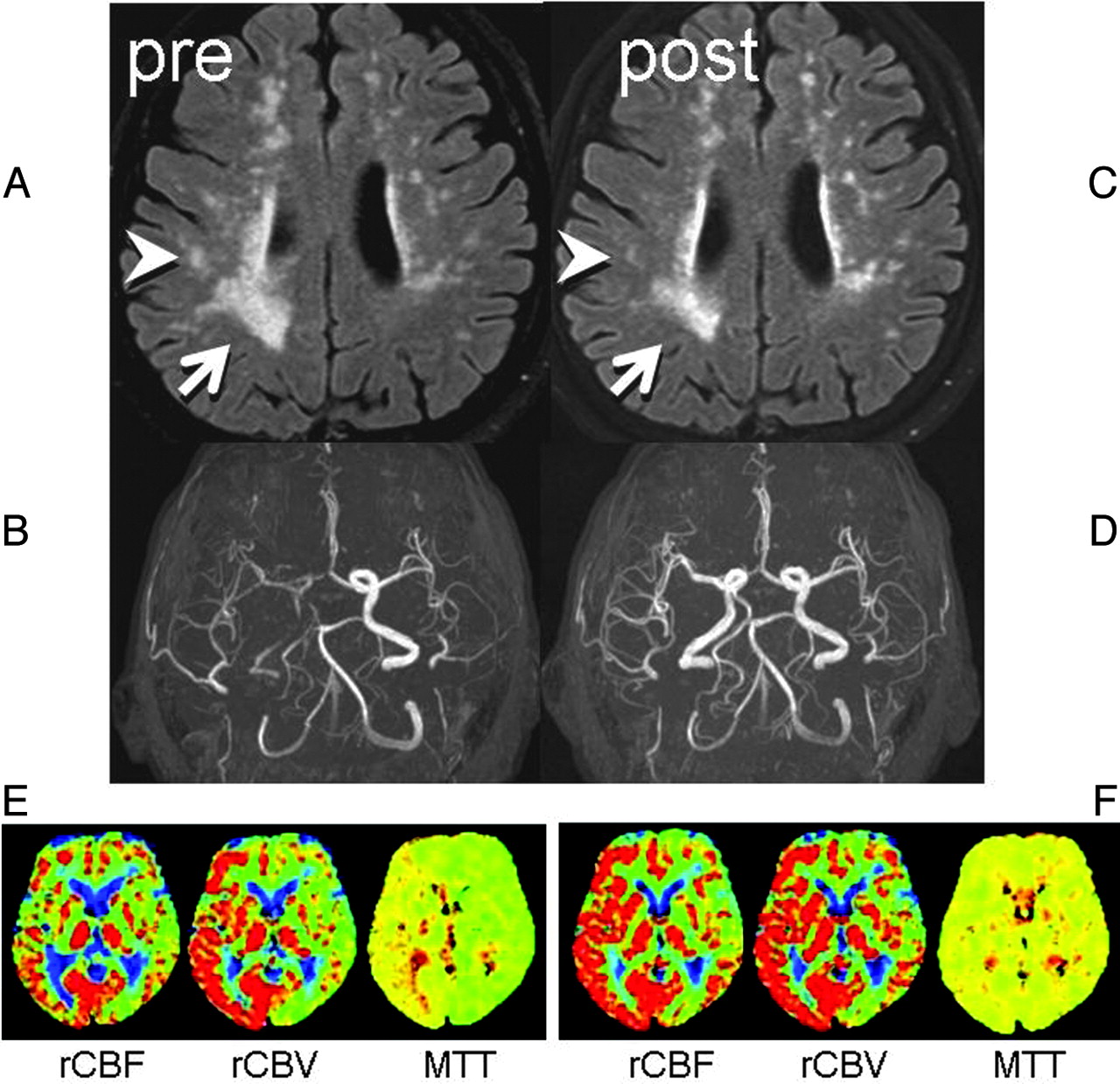
A 75-year-old man presented to our hospital with TIAs, characterized by intermittent left upper extremity weakness. DWI revealed no apparent acute infarction. Motion-sensitizing gradients of this DWI were applied to 15 different directions to enable DTI. Punctate WMLs were noted on FLAIR images at the centrum semiovale of both cerebral hemispheres, with right hemispheric laterality (Fig 1A). MR angiography revealed severe right ICA stenosis with distal flow compromise (Fig 1B).

Fig 1.
  • Download figure
  • Open in new tab
  • Download powerpoint
Fig 1.

Two consecutive FLAIR images and MR angiography. A, Preprocedural FLAIR (delay time, 2200 ms; TR/TE, 8000/100 ms) demonstrates WMLs. B, Preprocedural MR angiography (TR/TE, 30/2.3 ms) shows poor visualization of the right anterior circulation branches. C, Postprocedural FLAIR a week later shows partial WML resolution in the right hemisphere (arrow/arrowhead). D, MR angiogram shows restoration of flow in the right ICA. E, Preprocedural perfusion-weighted images show MTT elongation at the right anterior circulation territory with compensatory vasodilation, indicated by increased cerebral blood volume. F, After the procedure, the MTT elongation is normalized. Increased cerebral blood flow at the same territory indicates postprocedural hyperperfusion, but the patient remained asymptomatic.

Subsequent right carotid conventional angiography revealed pseudo-occlusion of the right ICA, with marked delay in contrast arrival at the right cerebral hemisphere. A successful stent-placement procedure was performed without any notable complications. Follow-up MR imaging 7 days after the procedure showed a patent right ICA and improvement of WMLs in the right cerebral hemisphere (Fig 1 C, -D). The patient remained asymptomatic at the recent 18-month follow-up.

To characterize the reversed WMLs, we performed a retrospective image analysis on the MR imaging data. First, affine registration of pre- and postprocedural FLAIR images was carried out, after which segmentation of the WMLs was performed (Fig 2A, -B). Subtraction images yielded pixels with/without reversal (Fig 2 C). We coregistered these data onto the DWI/DTI to characterize the nature of the WMLs. A t test was used for statistical analysis, and P values < .05 were considered significant.

Fig 2.
  • Download figure
  • Open in new tab
  • Download powerpoint
Fig 2.

Image analysis on pre- and postprocedural images. A, Image coregistration was performed between the pre- and postprocedural FLAIR. B, Segmentation of the pixels with WMLs was then done. C, Subtraction images yielded pixels with/without reversal. Coregistration of these data onto the DWI/DTI (DWI: TR/TE, 6000/88 ms; b = 1000 s/mm2) and b = 0 imaging was performed to characterize the nature of the WMLs.

This image analysis revealed that the reversed WMLs had 10% higher λ1 and 11% lower signal intensity on b = 0 images compared with nonreversed WMLs. The ADC and λ2 and λ3 were not significantly different.

Discussion

Age-related WMLs are usually considered a progressive condition.4 A population-based study that analyzed 2 MR images separated by 5 years showed worsening in 28% of participants.3 The previously proposed mechanisms for progression included intermittent changes in cerebral perfusion pressure leading to incomplete infarction,5,6 leakage of toxic plasma components due to altered BBB permeability,7–9 and microembolic events.10 Each of these mechanisms would induce a progressive, but not reversible, condition. The present case with WML reversal must, therefore, have been induced by a different etiology.

There are a few clinical conditions in which transient brain edema is known to occur, which include hyperammonemia and posterior reversible encephalopathy syndrome. From the imaging perspective, the present case most closely resembles hyperammonemia.11 The previous report dealing with hyperammonemia illustrated reversal of punctate WMLs in a series of patients with liver cirrhosis.11 These patients were treated with liver transplantation, after which the authors noted WML reversal. Hyperammonemia causes increased permeability through the BBB, which was considered to be the source of transient edema.

WMLsT Test
ReversedNonreversed
ADC × 10(−6)1.151.280.127
λ1 × 10(−6)2.53>2.290.001 >
λ2 or λ3 × 10(−6)0.580.580.336
b = 0292<3280.001 >

Comparison between reversed and nonreversed WMLs

A case report described reversal of WMLs after brain infarction.12 This elderly patient had an acute brain infarction involving the occipital lobe. A follow-up MR imaging performed a year later revealed partial regression of WMLs. The authors of this report also speculated that this was due to transient disturbance of the BBB, causing patchy white matter edema.

To investigate the underlying pathophysiology of WML reversal in the present case, we performed a retrospective image analysis. This revealed that the λ1 and signal intensity on b = 0 images were different, but by only 10%. This finding may indicate that image-based differentiation between reversible and nonreversible WMLs may be challenging. A difference in λ1 may indicate that nonreversed WMLs have axonal fragmentation or reduced axonal flow, but future pathologic correlation studies are needed to discover the true underlying mechanisms.

Aside from the pathophysiology, a few other clinical questions may arise. For instance, how often do we expect to see such reversal of WMLs after blood flow restoration and does this reversal indicate a successful treatment result? These issues would have sufficient impact on our clinical practice; thus, we have started a study to elucidate these issues.

Acknowledgments

We thank Drs Sachiko Yuen, Kentaro Akazawa, and Hiroyasu Sasajima for valuable comments on this study.

References

  1. 1.↵
    1. Hachinski VC,
    2. Potter P,
    3. Merskey H
    . Leukoaraiosis. Arch Neurol 1987;4:21–23
  2. 2.↵
    1. Wiszniewska M,
    2. Devuyst G,
    3. Bogousslavsky J,
    4. et al
    . What is the significance of leukoaraiosis in patients with acute ischemic stroke? Arch Neurol 2000;57:967–73
    CrossRefPubMed
  3. 3.↵
    1. Longstreth WT Jr.,
    2. Arnold AM,
    3. Beauchamp NJ Jr.,
    4. et al
    . Incidence, manifestations, and predictors of worsening white matter on serial cranial magnetic resonance imaging in the elderly. Stroke 2005;36:56–61
    Abstract/FREE Full Text
  4. 4.↵
    1. Streifler JY,
    2. Eliasziw M,
    3. Benavente OR,
    4. et al
    . Development and progression of leukoaraiosis in patients with brain ischemia and carotid artery disease. Stroke 2003;34:1913–16
    Abstract/FREE Full Text
  5. 5.↵
    1. Brun A,
    2. Englund EA
    . White matter disorder in dementia of the Alzheimer type: a pathoanatomical study. Ann Neurol 1986;19:253–62
    CrossRefPubMed
  6. 6.↵
    1. Yamada K,
    2. Nagakane Y,
    3. Sasajima H,
    4. et al
    . Incidental acute infarcts identified on diffusion-weighted images: a university hospital-based study. AJNR Am J Neuroradiol 2008;29:937–40
    Abstract/FREE Full Text
  7. 7.↵
    1. Nakaji K,
    2. Ihara M,
    3. Takahashi C,
    4. et al
    . Matrix metalloproteinase-2 plays a critical role in the pathogenesis of white matter lesions after chronic cerebral hypoperfusion in rodents. Stroke 2006;37:2816–23
    Abstract/FREE Full Text
  8. 8.↵
    1. Caplan LR
    . Dilatative arteriopathy: what is known and not known. Ann Neurol 2005;57:469–71
    CrossRefPubMed
  9. 9.↵
    1. Wardlaw JM,
    2. Sandercock PA,
    3. Dennis MS,
    4. et al
    . Is breakdown of the blood brain barrier responsible for lacunar stroke, leukoaraiosis, and dementia? Stroke 2003;34:806–12
    Abstract/FREE Full Text
  10. 10.↵
    1. Tejada J,
    2. Diez-Tejedor E,
    3. Hernandez-Echebarria L,
    4. et al
    . Does a relationship exist between carotid stenosis and lacunar infarction? Stroke 2003;34:1404–09
    Abstract/FREE Full Text
  11. 11.↵
    1. Rovira A,
    2. Mínguez B,
    3. Aymerich FX,
    4. et al
    . Decreased white matter lesion volume and improved cognitive function after liver transplantation. Hepatology 2007;465:1485–90
  12. 12.↵
    1. Moriya Y,
    2. Kozaki K,
    3. Nagai K,
    4. et al
    . Attenuation of brain white matter hyperintensities after cerebral infarction. [Letter] AJNR Am J Neuroradiol 2009;30:E43
    CrossRefPubMed
  • Received July 22, 2009.
  • Accepted after revision August 5, 2009.
  • Copyright © American Society of Neuroradiology
View Abstract
PreviousNext
Back to top

In this issue

American Journal of Neuroradiology: 31 (7)
American Journal of Neuroradiology
Vol. 31, Issue 7
1 Aug 2010
  • Table of Contents
  • Index by author
Advertisement
Print
Download PDF
Email Article

Thank you for your interest in spreading the word on American Journal of Neuroradiology.

NOTE: We only request your email address so that the person you are recommending the page to knows that you wanted them to see it, and that it is not junk mail. We do not capture any email address.

Enter multiple addresses on separate lines or separate them with commas.
Cerebral White Matter Lesions May Be Partially Reversible in Patients with Carotid Artery Stenosis
(Your Name) has sent you a message from American Journal of Neuroradiology
(Your Name) thought you would like to see the American Journal of Neuroradiology web site.
CAPTCHA
This question is for testing whether or not you are a human visitor and to prevent automated spam submissions.
Cite this article
K. Yamada, K. Sakai, K. Owada, K. Mineura, T. Nishimura
Cerebral White Matter Lesions May Be Partially Reversible in Patients with Carotid Artery Stenosis
American Journal of Neuroradiology Aug 2010, 31 (7) 1350-1352; DOI: 10.3174/ajnr.A1873

Citation Manager Formats

  • BibTeX
  • Bookends
  • EasyBib
  • EndNote (tagged)
  • EndNote 8 (xml)
  • Medlars
  • Mendeley
  • Papers
  • RefWorks Tagged
  • Ref Manager
  • RIS
  • Zotero
0 Responses
Respond to this article
Share
Bookmark this article
Cerebral White Matter Lesions May Be Partially Reversible in Patients with Carotid Artery Stenosis
K. Yamada, K. Sakai, K. Owada, K. Mineura, T. Nishimura
American Journal of Neuroradiology Aug 2010, 31 (7) 1350-1352; DOI: 10.3174/ajnr.A1873
del.icio.us logo Twitter logo Facebook logo Mendeley logo
  • Tweet Widget
  • Facebook Like
  • Google Plus One
Purchase

Jump to section

  • Article
    • Abstract
    • Abbreviations
    • Case Report
    • Discussion
    • Acknowledgments
    • References
  • Figures & Data
  • Info & Metrics
  • Responses
  • References
  • PDF

Related Articles

  • No related articles found.
  • PubMed
  • Google Scholar

Cited By...

  • Automatic Spatial Estimation of White Matter Hyperintensities Evolution in Brain MRI using Disease Evolution Predictor Deep Neural Networks
  • Enzyme replacement therapy and white matter hyperintensity progression in Fabry disease
  • Crossref (22)
  • Google Scholar

This article has been cited by the following articles in journals that are participating in Crossref Cited-by Linking.

  • Age‐related changes in cerebrovascular health and their effects on neural function and cognition: A comprehensive review
    Benjamin Zimmerman, Bart Rypma, Gabriele Gratton, Monica Fabiani
    Psychophysiology 2021 58 7
  • White Matter Hyperintensity in Ischemic Stroke Patients: It May Regress Over Time
    A-Hyun Cho, Hyeong-Ryul Kim, Woojun Kim, Dong Won Yang
    Journal of Stroke 2015 17 1
  • Advances in chronic cerebral circulation insufficiency
    Da Zhou, Ran Meng, Si‐Jie Li, Jing‐Yuan Ya, Jia‐Yue Ding, Shu‐Ling Shang, Yu‐Chuan Ding, Xun‐Ming Ji
    CNS Neuroscience & Therapeutics 2018 24 1
  • Dynamic Progression of White Matter Hyperintensities in Alzheimer’s Disease and Normal Aging: Results from the Sunnybrook Dementia Study
    Joel Ramirez, Alicia A. McNeely, Courtney Berezuk, Fuqiang Gao, Sandra E. Black
    Frontiers in Aging Neuroscience 2016 8
  • Carotid Stenosis: A Risk Factor for Cerebral White-matter Disease
    Nagaendran Kandiah, Orlanda Goh, Elijah Mak, Maznah Marmin, Amanda Ng
    Journal of Stroke and Cerebrovascular Diseases 2014 23 1
  • Automatic spatial estimation of white matter hyperintensities evolution in brain MRI using disease evolution predictor deep neural networks
    Muhammad Febrian Rachmadi, Maria del C. Valdés-Hernández, Stephen Makin, Joanna Wardlaw, Taku Komura
    Medical Image Analysis 2020 63
  • Reversibility of White Matter Hyperintensity by Revascularization Surgery in Moyamoya Disease
    Katsuya Komatsu, Takeshi Mikami, Shouhei Noshiro, Kei Miyata, Masahiko Wanibuchi, Nobuhiro Mikuni
    Journal of Stroke and Cerebrovascular Diseases 2016 25 6
  • Characterizing the Penumbras of White Matter Hyperintensities and Their Associations With Cognitive Function in Patients With Subcortical Vascular Mild Cognitive Impairment
    Xiaowei Wu, Xin Ge, Jing Du, Yao Wang, Yawen Sun, Xu Han, Weina Ding, Mengqiu Cao, Qun Xu, Yan Zhou
    Frontiers in Neurology 2019 10
  • Association Between the Volume of Carotid Artery Plaque and Its Subcomponents and the Volume of White Matter Lesions in Patients Selected for Endarterectomy
    Luca Saba, Eytan Raz, Roberto Grassi, Pier Luigi Di Paolo, Aniello Iacomino, Roberto Montisci, Mario Piga
    American Journal of Roentgenology 2013 201 5
  • Unraveling Specific Brain Microstructural Damage in Moyamoya Disease Using Diffusion Magnetic Resonance Imaging and Positron Emission Tomography
    Shoko Hara, Masaaki Hori, Ryo Ueda, Shihori Hayashi, Motoki Inaji, Yoji Tanaka, Taketoshi Maehara, Kenji Ishii, Shigeki Aoki, Tadashi Nariai
    Journal of Stroke and Cerebrovascular Diseases 2019 28 4

More in this TOC Section

  • Fast Contrast-Enhanced 4D MRA and 4D Flow MRI Using Constrained Reconstruction (HYPRFlow): Potential Applications for Brain Arteriovenous Malformations
  • Multimodal CT Provides Improved Performance for Lacunar Infarct Detection
  • Optimal MRI Sequence for Identifying Occlusion Location in Acute Stroke: Which Value of Time-Resolved Contrast-Enhanced MRA?
Show more BRAIN

Similar Articles

Advertisement

Indexed Content

  • Current Issue
  • Accepted Manuscripts
  • Article Preview
  • Past Issues
  • Editorials
  • Editor's Choice
  • Fellows' Journal Club
  • Letters to the Editor
  • Video Articles

Cases

  • Case Collection
  • Archive - Case of the Week
  • Archive - Case of the Month
  • Archive - Classic Case

More from AJNR

  • Trainee Corner
  • Imaging Protocols
  • MRI Safety Corner
  • Book Reviews

Multimedia

  • AJNR Podcasts
  • AJNR Scantastics

Resources

  • Turnaround Time
  • Submit a Manuscript
  • Submit a Video Article
  • Submit an eLetter to the Editor/Response
  • Manuscript Submission Guidelines
  • Statistical Tips
  • Fast Publishing of Accepted Manuscripts
  • Graphical Abstract Preparation
  • Imaging Protocol Submission
  • Evidence-Based Medicine Level Guide
  • Publishing Checklists
  • Author Policies
  • Become a Reviewer/Academy of Reviewers
  • News and Updates

About Us

  • About AJNR
  • Editorial Board
  • Editorial Board Alumni
  • Alerts
  • Permissions
  • Not an AJNR Subscriber? Join Now
  • Advertise with Us
  • Librarian Resources
  • Feedback
  • Terms and Conditions
  • AJNR Editorial Board Alumni

American Society of Neuroradiology

  • Not an ASNR Member? Join Now

© 2025 by the American Society of Neuroradiology All rights, including for text and data mining, AI training, and similar technologies, are reserved.
Print ISSN: 0195-6108 Online ISSN: 1936-959X

Powered by HighWire