Advertisement

Main menu

  • Home
  • Content
    • Current Issue
    • Accepted Manuscripts
    • Article Preview
    • Past Issue Archive
    • Video Articles
    • AJNR Case Collection
    • Case of the Week Archive
    • Case of the Month Archive
    • Classic Case Archive
  • Special Collections
    • AJNR Awards
    • Low-Field MRI
    • Alzheimer Disease
    • ASNR Foundation Special Collection
    • Photon-Counting CT
    • View All
  • Multimedia
    • AJNR Podcasts
    • AJNR SCANtastic
    • Trainee Corner
    • MRI Safety Corner
    • Imaging Protocols
  • For Authors
    • Submit a Manuscript
    • Submit a Video Article
    • Submit an eLetter to the Editor/Response
    • Manuscript Submission Guidelines
    • Statistical Tips
    • Fast Publishing of Accepted Manuscripts
    • Graphical Abstract Preparation
    • Imaging Protocol Submission
    • Author Policies
  • About Us
    • About AJNR
    • Editorial Board
    • Editorial Board Alumni
  • More
    • Become a Reviewer/Academy of Reviewers
    • Subscribers
    • Permissions
    • Alerts
    • Feedback
    • Advertisers
    • ASNR Home

User menu

  • Alerts
  • Log in

Search

  • Advanced search
American Journal of Neuroradiology

American Journal of Neuroradiology

ASHNR American Society of Functional Neuroradiology ASHNR American Society of Pediatric Neuroradiology ASSR
  • Alerts
  • Log in

Advanced Search

  • Home
  • Content
    • Current Issue
    • Accepted Manuscripts
    • Article Preview
    • Past Issue Archive
    • Video Articles
    • AJNR Case Collection
    • Case of the Week Archive
    • Case of the Month Archive
    • Classic Case Archive
  • Special Collections
    • AJNR Awards
    • Low-Field MRI
    • Alzheimer Disease
    • ASNR Foundation Special Collection
    • Photon-Counting CT
    • View All
  • Multimedia
    • AJNR Podcasts
    • AJNR SCANtastic
    • Trainee Corner
    • MRI Safety Corner
    • Imaging Protocols
  • For Authors
    • Submit a Manuscript
    • Submit a Video Article
    • Submit an eLetter to the Editor/Response
    • Manuscript Submission Guidelines
    • Statistical Tips
    • Fast Publishing of Accepted Manuscripts
    • Graphical Abstract Preparation
    • Imaging Protocol Submission
    • Author Policies
  • About Us
    • About AJNR
    • Editorial Board
    • Editorial Board Alumni
  • More
    • Become a Reviewer/Academy of Reviewers
    • Subscribers
    • Permissions
    • Alerts
    • Feedback
    • Advertisers
    • ASNR Home
  • Follow AJNR on Twitter
  • Visit AJNR on Facebook
  • Follow AJNR on Instagram
  • Join AJNR on LinkedIn
  • RSS Feeds

AJNR Awards, New Junior Editors, and more. Read the latest AJNR updates

Case ReportBrain

MR Spectroscopy Detects Lipid Peaks in Cerebrotendinous Xanthomatosis

E.K. Embiruçu, M.C.G. Otaduy, A.K. Taneja, C.C. Leite, F. Kok and L.T. Lucato
American Journal of Neuroradiology August 2010, 31 (7) 1347-1349; DOI: https://doi.org/10.3174/ajnr.A1885
E.K. Embiruçu
  • Find this author on Google Scholar
  • Find this author on PubMed
  • Search for this author on this site
M.C.G. Otaduy
  • Find this author on Google Scholar
  • Find this author on PubMed
  • Search for this author on this site
A.K. Taneja
  • Find this author on Google Scholar
  • Find this author on PubMed
  • Search for this author on this site
C.C. Leite
  • Find this author on Google Scholar
  • Find this author on PubMed
  • Search for this author on this site
F. Kok
  • Find this author on Google Scholar
  • Find this author on PubMed
  • Search for this author on this site
L.T. Lucato
  • Find this author on Google Scholar
  • Find this author on PubMed
  • Search for this author on this site
  • Article
  • Figures & Data
  • Info & Metrics
  • Responses
  • References
  • PDF
Loading

Abstract

SUMMARY: CTX is a rare lipid-storage disease. Novel MRS findings from 3 patients, using a short TE, were the presence of lipid peaks at 0.9 and 1.3 ppm in the depth of the cerebellar hemisphere; this might represent an additional marker of disease that is CNS-specific and noninvasive. A decrease in NAA concentration was also detected and attributed to neuroaxonal damage. One patient presented an increase in mIns concentration, pointing to gliosis and astrocytic proliferation.

Abbreviations

a.u.
arbitrary unit
Cho
choline-containing compounds
CNS
central nervous system
Cr
creatine/phosphocreatine
CTX
cerebrotendinous xanthomatosis
Lip
lipid
Lip09
lipid signals at 0.9 ppm
Lip13
lipid signals at 1.3 ppm
mIns
myo-inositol
MRS
MR spectroscopy
NAA
N-acetylaspartate
PRESS
point-resolved spectroscopy sequence
STEAM
short echo time stimulated echo acquisition mode
VOI
volume of interest

CTX is a rare metabolic disorder whose main clinical manifestations are juvenile cataracts, chronic diarrhea, and tendinous xanthomas.1–5 It is caused by mutations in the CYP27A1 gene, which codes for sterol 27-hydroxylase, an enzyme essential for bile acid synthesis.2,4 This defect leads to abnormally high levels of cholestanol in various tissues—the biochemical hallmark of CTX.1–5 Treatment is oral administration of chenodeoxycholic acid, which inhibits cholestanol synthesis.5

The most distinctive MR imaging abnormalities are bilateral T2 hyperintensity in the dentate nuclei and adjacent cerebellar white matter.1,6 A previous MRS study disclosed a reduction of the NAA peak and the presence of a lactate peak.6 We studied 3 patients with CTX, attempting to assess possible novel MRS findings, focusing on short-TE acquisitions.

Case Reports

Case 1

A 22-year-old man, African-Brazilian, a second sibling of first-cousin parents, had normal developmental milestones until 3 years of age, when his motor and cognitive abilities declined. During childhood, he had diagnosed bilateral cataracts and started to present chronic intermittent diarrhea. Clinical examination disclosed bilateral achillean xanthomas. He was severely mentally retarded, dysarthric, with trunk and limb ataxia, spastic gait, and distal limb atrophy. His serum cholestanol level was elevated (118.7 μg/mL; reference value, <6.6 μg/mL). MRS, at 1.5T, was performed on the depth of cerebellar hemisphere, encompassing T2-hyperintense white matter (Fig 1), by using a single-voxel STEAM sequence (TR/TE/mixing time, 1500/30/13.7 ms; 128 acquisitions; and VOI size, 8 cm3). Spectral analysis was performed by using LCModel software (Stephen Provencher, Oakville, Ontario, Canada)7; it demonstrated a decreased NAA level and abnormal lipid peaks at 0.9 and 1.3 ppm (Fig 2 and Table).

Fig 1.
  • Download figure
  • Open in new tab
  • Download powerpoint
Fig 1.

Axial T2-weighted image demonstrates hyperintense lesions encompassing both dentate nuclei and surrounding white matter; this image also shows region-of-interest placement for MRS.

Fig 2.
  • Download figure
  • Open in new tab
  • Download powerpoint
Fig 2.

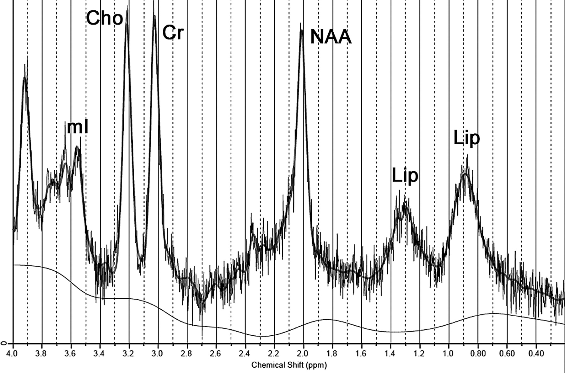
Spectrum obtained from case 1 (1.5T; STEAM; TR/TE, 1500/30 ms) demonstrates lipid peaks at 0.9 and 1.3 ppm. Notice also a slight decrease in the NAA peak.

View this table:
  • View inline
  • View popup

Short TE (30–35 ms) single-voxel MRS data: cerebellar tissue and absolute concentrationsa

Case 2

A 47-year-old man, who was the third of 6 siblings of nonconsanguineous parents from Okinawa Island, Japan, had an infancy-onset progressive decline of motor and cognitive abilities. He had no history of chronic diarrhea or lens opacities. Clinical examination disclosed achillean, plantar, and elbow xanthomas. He was profoundly demented, wheelchair-bound, anarthric, drooling, and totally dependent for daily activities. He had limb spasticity and ataxia. His serum cholestanol level was only slightly elevated (9.1 μg/mL).

Cerebellar MRS was performed twice, 3 years apart, one time at initial diagnosis (1.5T) and the other during follow-up (3T). Parameters of first examination were similar to those of case 1; his last MRS examination was performed with a single-voxel PRESS sequence (TR/TE, 1871/35 ms; 96 acquisitions; and VOI size, 8 cm3). The same spectral analysis disclosed, in both examinations, markedly decreased NAA levels, high mIns concentrations, and the same abnormal lipid peaks (Fig 3 and Table).

Fig 3.
  • Download figure
  • Open in new tab
  • Download powerpoint
Fig 3.

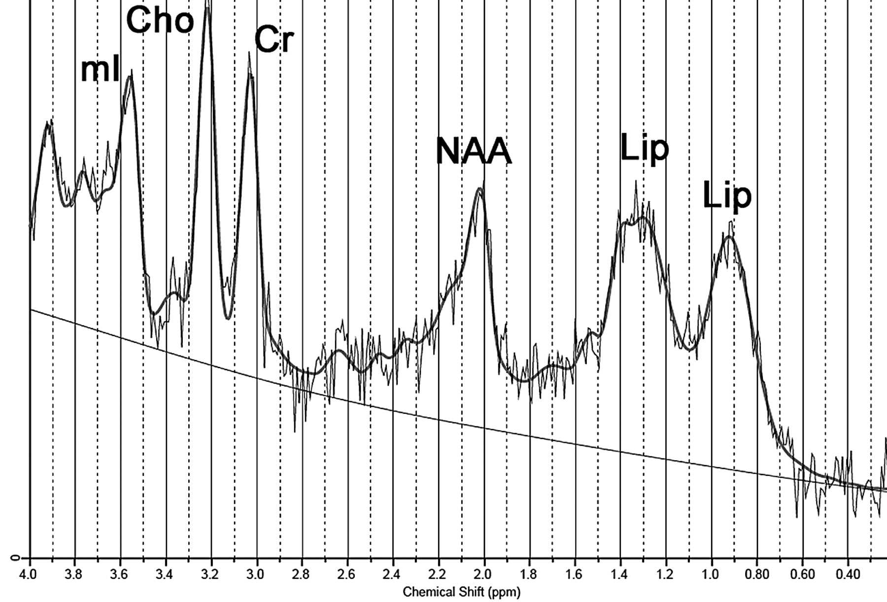
Spectrum obtained from case 2 (3T; PRESS; TR/TE, 1871/35 ms) demonstrates lipid peaks at 0.9 and 1.3 ppm. Notice also a marked decrease in the NAA peak and an increase in the mIns peak.

Case 3

A 35-year-old man, who is the fifth sibling of case 2, had normal developmental milestones. He started presenting progressive spastic gait and muscle cramps at age 28. He finished high school but, since early childhood, had chronic intermittent diarrhea and bilateral achillean xanthomas. Neurologic examination disclosed no cognitive abnormality, mild dysarthria, and spastic gait. Mild lower limb distal atrophy was also detected. His serum cholestanol level was elevated (61.5 μg/mL).

Cerebellar MRS, performed at 3T, in the same way as in case 2, showed a slight decrease in NAA level and also the same abnormal lipid peaks (Fig 4 and Table).

Fig 4.
  • Download figure
  • Open in new tab
  • Download powerpoint
Fig 4.

Spectrum obtained from case 3 (3T; PRESS; TR/TE, 1871/35 ms) demonstrates lipid peaks at 0.9 and 1.3 ppm. Notice also a slight trend toward a decrease in the NAA peak.

Discussion

Neuropathologic findings in CTX include demyelination and gliosis in the cerebellar white matter and corticospinal tracts, with multiple dispersed lipid crystal clefts, the consequence of accumulation of cholesterol and cholestanol in affected tissues.8

In this study, MRS showed a decrease in NAA concentration, which was similar to that previously reported6; the decrease can be interpreted as the expression of neuronal damage,6 because deposition of cholestanol seems to be neurotoxic.8 The magnitude of decrease in our sample roughly paralleled clinical impairment; case 2 was the most affected.

Case 2 also presented an increase in mIns levels. If one assumes that mIns represents a glial marker,9 the increase points to the well-known gliosis and astrocytic proliferation.8

A previously unreported finding was the presence of abnormal lipid peaks detected at 0.9 and 1.3 ppm. One MRS study available in patients with CTX used a long TE (272 ms),6 probably precluding identification of these lipid peaks, which usually have short T2s and do not appear in long-TE spectra.10

Lipid peaks are not detected in normal tissue. Although it is speculative, their presence in CTX can have 2 explanations. On one hand, they can represent membrane breakdown and can precede histologic necrosis, as seen in other diseases.9 On the other hand, they can be the surrogate markers of the major lipid storage that occurs in this disease.8

Some aspects reinforce the hypothesis that at least part of these peaks represent abnormal lipid accumulation. In serum samples from patients with CTX, by using high-resolution nuclear MRS, peaks assigned to cholestanol were found at 0.645 and 0.817 ppm.11 Also, besides the more commonly found abnormal lipid peaks detected at 0.9 and 1.3 ppm, which correspond to methyl (CH3) and the methylene group (CH2) respectively,12 other lipid protons resonate in these areas, as is the case for cholesterol.11

The apparent lack of correlation between the 1.3 and 0.9 ppm peaks may point to a more complex pattern of lipid deposit. If both represented the same single lipid molecule in all patients, a constant ratio between both peaks would be expected, which is not the case. Some similar findings have already emerged from MRS data of other diseases, which correspond with lipid accumulation in brain tissue, such as Sjögren-Larsson syndrome.10

This case report has limitations. The sample size is small, though this is a rare disease. There are no controls for the 3T spectra, but we believe that all the assumptions made for the 1.5T examinations are also valid for the 3T ones, because MRS examinations from case 2 performed on both MR imaging scanners presented no significant differences. Another point is that lipid-peak detection does not depend heavily on the existence of a control group because these peaks are not present in significant amounts in normal tissue. Finally, we cannot preclude the possibility that lactate may have contributed to the observed resonance at 1.3 ppm because lactate and lipids are known to overlap in this region. However, if there is any lactate contribution, it is probably negligible because long-TE spectra acquired in our patients revealed a small lactate peak in only 2 of them.

In conclusion, our cases suggest that lipid peaks detected at short-TE MRS in the cerebellum of patients with CTX might represent an additional marker of the disease, which is CNS-specific and noninvasive, with a potential role in monitoring therapy response.

References

  1. 1.↵
    1. Barkhof F,
    2. Verrips A,
    3. Wesseling P,
    4. et al
    . Cerebrotendinous xanthomatosis: the spectrum of imaging findings and the correlation with neuropathologic findings. Radiology 2000;217:869–76
    PubMed
  2. 2.↵
    1. Verrips A,
    2. Hoefsloot LH,
    3. Steenbergen GC,
    4. et al
    . Clinical and molecular genetic characteristics of patients with cerebrotendinous xanthomatosis. Brain 2000;123:908–19
    Abstract/FREE Full Text
  3. 3.↵
    1. Federico A,
    2. Dotti MT
    . Cerebrotendinous xanthomatosis: clinical manifestations, diagnostic criteria, pathogenesis, and therapy. J Child Neurol 2003;18:633–38
    Abstract/FREE Full Text
  4. 4.↵
    1. Gallus GN,
    2. Dotti MT,
    3. Federico A
    . Clinical and molecular diagnosis of cerebrotendinous xanthomatosis with a review of the mutations in the CYP27A1 gene. Neurol Sci 2006;27:143–49
    CrossRefPubMed
  5. 5.↵
    1. Berginer VM,
    2. Gross B,
    3. Morad K,
    4. et al
    . Chronic diarrhea and juvenile cataracts: think cerebrotendinous xanthomatosis and treat. Pediatrics 2009;123:143–47
    Abstract/FREE Full Text
  6. 6.↵
    1. De Stefano N,
    2. Dotti MT,
    3. Mortilla M,
    4. et al
    . Magnetic resonance imaging and spectroscopic changes in brains of patients with cerebrotendinous xanthomatosis. Brain 2001;124:121–31
    Abstract/FREE Full Text
  7. 7.↵
    1. Provencher SW
    . Estimation of metabolite concentrations from localized in vivo proton NMR spectra. Magn Reson Med 1993;30:672–79
    CrossRefPubMed
  8. 8.↵
    1. Pilo de la Fuente B,
    2. Ruiz I,
    3. Lopez de Munain A,
    4. et al
    . Cerebrotendinous xanthomatosis: neuropathological findings. J Neurol 2008;255:839–42
    CrossRefPubMed
  9. 9.↵
    1. Gillard JH,
    2. Waldman A,
    3. Barker P
    . Clinical MR neuroimaging: diffusion, perfusion, and spectroscopy. Cambridge, UK: Cambridge University Press; 2005:7–26
  10. 10.↵
    1. Willemsen MA,
    2. Van Der Graaf M,
    3. Van Der Knaap MS,
    4. et al
    . MR imaging and proton MR spectroscopic studies in Sjogren-Larsson syndrome: characterization of the leukoencephalopathy. AJNR Am J Neuroradiol 2004;25:649–57
    Abstract/FREE Full Text
  11. 11.↵
    1. Oostendorp M,
    2. Engelke UF,
    3. Willemsen MA,
    4. et al
    . Diagnosing inborn errors of lipid metabolism with proton nuclear magnetic resonance spectroscopy. Clin Chem 2006;52:1395–405
    Abstract/FREE Full Text
  12. 12.↵
    1. Maheshwari SR,
    2. Fatterpekar GM,
    3. Castillo M,
    4. et al
    . Proton MR spectroscopy of the brain. Semin Ultrasound CT MR 2000;21:434–51
    CrossRefPubMed
  • Received July 17, 2009.
  • Accepted after revision August 17, 2009.
  • Copyright © American Society of Neuroradiology
View Abstract
PreviousNext
Back to top

In this issue

American Journal of Neuroradiology: 31 (7)
American Journal of Neuroradiology
Vol. 31, Issue 7
1 Aug 2010
  • Table of Contents
  • Index by author
Advertisement
Print
Download PDF
Email Article

Thank you for your interest in spreading the word on American Journal of Neuroradiology.

NOTE: We only request your email address so that the person you are recommending the page to knows that you wanted them to see it, and that it is not junk mail. We do not capture any email address.

Enter multiple addresses on separate lines or separate them with commas.
MR Spectroscopy Detects Lipid Peaks in Cerebrotendinous Xanthomatosis
(Your Name) has sent you a message from American Journal of Neuroradiology
(Your Name) thought you would like to see the American Journal of Neuroradiology web site.
CAPTCHA
This question is for testing whether or not you are a human visitor and to prevent automated spam submissions.
Cite this article
E.K. Embiruçu, M.C.G. Otaduy, A.K. Taneja, C.C. Leite, F. Kok, L.T. Lucato
MR Spectroscopy Detects Lipid Peaks in Cerebrotendinous Xanthomatosis
American Journal of Neuroradiology Aug 2010, 31 (7) 1347-1349; DOI: 10.3174/ajnr.A1885

Citation Manager Formats

  • BibTeX
  • Bookends
  • EasyBib
  • EndNote (tagged)
  • EndNote 8 (xml)
  • Medlars
  • Mendeley
  • Papers
  • RefWorks Tagged
  • Ref Manager
  • RIS
  • Zotero
0 Responses
Respond to this article
Share
Bookmark this article
MR Spectroscopy Detects Lipid Peaks in Cerebrotendinous Xanthomatosis
E.K. Embiruçu, M.C.G. Otaduy, A.K. Taneja, C.C. Leite, F. Kok, L.T. Lucato
American Journal of Neuroradiology Aug 2010, 31 (7) 1347-1349; DOI: 10.3174/ajnr.A1885
del.icio.us logo Twitter logo Facebook logo Mendeley logo
  • Tweet Widget
  • Facebook Like
  • Google Plus One
Purchase

Jump to section

  • Article
    • Abstract
    • Abbreviations
    • Case Reports
    • Discussion
    • References
  • Figures & Data
  • Info & Metrics
  • Responses
  • References
  • PDF

Related Articles

  • No related articles found.
  • PubMed
  • Google Scholar

Cited By...

  • Teaching NeuroImages: Cerebrotendinous xanthomatosis: A rare treatable adult-onset lipid storage disease
  • Teaching NeuroImages: Cerebrotendinous xanthomatosis: A rare treatable adult-onset lipid storage disease
  • Crossref (21)
  • Google Scholar

This article has been cited by the following articles in journals that are participating in Crossref Cited-by Linking.

  • Cerebrotendinous xanthomatosis: a comprehensive review of pathogenesis, clinical manifestations, diagnosis, and management
    Shuke Nie, Guiqin Chen, Xuebing Cao, Yunjian Zhang
    Orphanet Journal of Rare Diseases 2014 9 1
  • Epidemiology, diagnosis, and treatment of cerebrotendinous xanthomatosis (CTX)
    Gerald Salen, Robert D. Steiner
    Journal of Inherited Metabolic Disease 2017 40 6
  • Adult Leukodystrophies: A Step-by-Step Diagnostic Approach
    Lucas Lopes Resende, Anderson Rodrigues Brandão de Paiva, Fernando Kok, Claudia da Costa Leite, Leandro Tavares Lucato
    RadioGraphics 2019 39 1
  • Cerebrotendinous Xanthomatosis: Molecular Pathogenesis, Clinical Spectrum, Diagnosis, and Disease-Modifying Treatments
    Shingo Koyama, Yoshiki Sekijima, Masatsune Ogura, Mika Hori, Kota Matsuki, Takashi Miida, Mariko Harada-Shiba
    Journal of Atherosclerosis and Thrombosis 2021 28 9
  • Magnetic Resonance Imaging Biomarkers in Patients with Progressive Ataxia: Current Status and Future Direction
    Stuart Currie, Marios Hadjivassiliou, Ian J Craven, Iain D Wilkinson, Paul D Griffiths, Nigel Hoggard
    The Cerebellum 2013 12 2
  • Cerebrotendinous xanthomatosis without neurological involvement
    B. M. L. Stelten, F. J. Raal, A. D. Marais, N. P. Riksen, J. E. Roeters van Lennep, P.B Duell, M. van der Graaf, L. A. J. Kluijtmans, R. A. Wevers, A. Verrips
    Journal of Internal Medicine 2021 290 5
  • Cerebral lipid accumulation in Chanarin–Dorfman Syndrome
    Marleen C.D.G. Huigen, Marinette van der Graaf, Eva Morava, A. Carin M. Dassel, Maurice A.M. van Steensel, Marieke M.B. Seyger, Ron A. Wevers, Michèl A. Willemsen
    Molecular Genetics and Metabolism 2015 114 1
  • JIMD Reports, Volume 28
    Carlos R. Ferreira, Molly H. Silber, Taeun Chang, Jonathan G. Murnick, Brian Kirmse
    2015 28
  • A case of Sjögren-Larsson syndrome with minimal MR imaging findings facilitated by proton spectroscopy
    Yasuhiko Tachibana, Noriko Aida, Keisuke Enomoto, Mizue Iai, Kenji Kurosawa
    Pediatric Radiology 2012 42 3
  • Neurologic Aspects of Systemic Disease Part III
    Jean-Philippe Neau, Gaëlle Godeneche, Stéphane Mathis, Gérard Guillet
    2014 121

More in this TOC Section

  • Fast Contrast-Enhanced 4D MRA and 4D Flow MRI Using Constrained Reconstruction (HYPRFlow): Potential Applications for Brain Arteriovenous Malformations
  • Multimodal CT Provides Improved Performance for Lacunar Infarct Detection
  • Optimal MRI Sequence for Identifying Occlusion Location in Acute Stroke: Which Value of Time-Resolved Contrast-Enhanced MRA?
Show more BRAIN

Similar Articles

Advertisement

Indexed Content

  • Current Issue
  • Accepted Manuscripts
  • Article Preview
  • Past Issues
  • Editorials
  • Editor's Choice
  • Fellows' Journal Club
  • Letters to the Editor
  • Video Articles

Cases

  • Case Collection
  • Archive - Case of the Week
  • Archive - Case of the Month
  • Archive - Classic Case

More from AJNR

  • Trainee Corner
  • Imaging Protocols
  • MRI Safety Corner
  • Book Reviews

Multimedia

  • AJNR Podcasts
  • AJNR Scantastics

Resources

  • Turnaround Time
  • Submit a Manuscript
  • Submit a Video Article
  • Submit an eLetter to the Editor/Response
  • Manuscript Submission Guidelines
  • Statistical Tips
  • Fast Publishing of Accepted Manuscripts
  • Graphical Abstract Preparation
  • Imaging Protocol Submission
  • Evidence-Based Medicine Level Guide
  • Publishing Checklists
  • Author Policies
  • Become a Reviewer/Academy of Reviewers
  • News and Updates

About Us

  • About AJNR
  • Editorial Board
  • Editorial Board Alumni
  • Alerts
  • Permissions
  • Not an AJNR Subscriber? Join Now
  • Advertise with Us
  • Librarian Resources
  • Feedback
  • Terms and Conditions
  • AJNR Editorial Board Alumni

American Society of Neuroradiology

  • Not an ASNR Member? Join Now

© 2025 by the American Society of Neuroradiology All rights, including for text and data mining, AI training, and similar technologies, are reserved.
Print ISSN: 0195-6108 Online ISSN: 1936-959X

Powered by HighWire