Advertisement

Main menu

  • Home
  • Content
    • Current Issue
    • Accepted Manuscripts
    • Article Preview
    • Past Issue Archive
    • Video Articles
    • AJNR Case Collection
    • Case of the Week Archive
    • Case of the Month Archive
    • Classic Case Archive
  • Special Collections
    • AJNR Awards
    • Low-Field MRI
    • Alzheimer Disease
    • ASNR Foundation Special Collection
    • Photon-Counting CT
    • View All
  • Multimedia
    • AJNR Podcasts
    • AJNR SCANtastic
    • Trainee Corner
    • MRI Safety Corner
    • Imaging Protocols
  • For Authors
    • Submit a Manuscript
    • Submit a Video Article
    • Submit an eLetter to the Editor/Response
    • Manuscript Submission Guidelines
    • Statistical Tips
    • Fast Publishing of Accepted Manuscripts
    • Graphical Abstract Preparation
    • Imaging Protocol Submission
    • Author Policies
  • About Us
    • About AJNR
    • Editorial Board
    • Editorial Board Alumni
  • More
    • Become a Reviewer/Academy of Reviewers
    • Subscribers
    • Permissions
    • Alerts
    • Feedback
    • Advertisers
    • ASNR Home

User menu

  • Alerts
  • Log in

Search

  • Advanced search
American Journal of Neuroradiology

American Journal of Neuroradiology

ASHNR American Society of Functional Neuroradiology ASHNR American Society of Pediatric Neuroradiology ASSR
  • Alerts
  • Log in

Advanced Search

  • Home
  • Content
    • Current Issue
    • Accepted Manuscripts
    • Article Preview
    • Past Issue Archive
    • Video Articles
    • AJNR Case Collection
    • Case of the Week Archive
    • Case of the Month Archive
    • Classic Case Archive
  • Special Collections
    • AJNR Awards
    • Low-Field MRI
    • Alzheimer Disease
    • ASNR Foundation Special Collection
    • Photon-Counting CT
    • View All
  • Multimedia
    • AJNR Podcasts
    • AJNR SCANtastic
    • Trainee Corner
    • MRI Safety Corner
    • Imaging Protocols
  • For Authors
    • Submit a Manuscript
    • Submit a Video Article
    • Submit an eLetter to the Editor/Response
    • Manuscript Submission Guidelines
    • Statistical Tips
    • Fast Publishing of Accepted Manuscripts
    • Graphical Abstract Preparation
    • Imaging Protocol Submission
    • Author Policies
  • About Us
    • About AJNR
    • Editorial Board
    • Editorial Board Alumni
  • More
    • Become a Reviewer/Academy of Reviewers
    • Subscribers
    • Permissions
    • Alerts
    • Feedback
    • Advertisers
    • ASNR Home
  • Follow AJNR on Twitter
  • Visit AJNR on Facebook
  • Follow AJNR on Instagram
  • Join AJNR on LinkedIn
  • RSS Feeds

AJNR Awards, New Junior Editors, and more. Read the latest AJNR updates

Research ArticleAdult Brain
Open Access

Deep Learning Segmentation of the Nucleus Basalis of Meynert on 3T MRI

D.J. Doss, G.W. Johnson, S. Narasimhan, J.S. Shless, J.W. Jiang, H.F.J. González, D.L. Paulo, A. Lucas, K.A. Davis, C. Chang, V.L. Morgan, C. Constantinidis, B.M. Dawant and D.J. Englot
American Journal of Neuroradiology August 2023, DOI: https://doi.org/10.3174/ajnr.A7950
D.J. Doss
aFrom the Department of Biomedical Engineering (D.J.D., G.W.J., S.N., H.F.J.G., C. Chang., V.L.M., C. Constantinidis, D.J.E.)
bInstitute of Imaging Science (D.J.D., G.W.J., S.N., J.S.S., J.W.J., H.F.J.G., C. Chang, V.L.M., B.M.D., D.J.E.)
fVanderbilt Institute for Surgery and Engineering (D.J.D., G.W.J., S.N., H.F.J.G., C. Chang, V.L.M., B.M.D., D.J.E.), Nashville, Tennessee
  • Find this author on Google Scholar
  • Find this author on PubMed
  • Search for this author on this site
  • ORCID record for D.J. Doss
G.W. Johnson
aFrom the Department of Biomedical Engineering (D.J.D., G.W.J., S.N., H.F.J.G., C. Chang., V.L.M., C. Constantinidis, D.J.E.)
bInstitute of Imaging Science (D.J.D., G.W.J., S.N., J.S.S., J.W.J., H.F.J.G., C. Chang, V.L.M., B.M.D., D.J.E.)
fVanderbilt Institute for Surgery and Engineering (D.J.D., G.W.J., S.N., H.F.J.G., C. Chang, V.L.M., B.M.D., D.J.E.), Nashville, Tennessee
  • Find this author on Google Scholar
  • Find this author on PubMed
  • Search for this author on this site
  • ORCID record for G.W. Johnson
S. Narasimhan
aFrom the Department of Biomedical Engineering (D.J.D., G.W.J., S.N., H.F.J.G., C. Chang., V.L.M., C. Constantinidis, D.J.E.)
bInstitute of Imaging Science (D.J.D., G.W.J., S.N., J.S.S., J.W.J., H.F.J.G., C. Chang, V.L.M., B.M.D., D.J.E.)
fVanderbilt Institute for Surgery and Engineering (D.J.D., G.W.J., S.N., H.F.J.G., C. Chang, V.L.M., B.M.D., D.J.E.), Nashville, Tennessee
gDepartments of Neurological Surgery (S.N., J.S.S., J.W.J., D.L.P., V.L.M., D.J.E.)
  • Find this author on Google Scholar
  • Find this author on PubMed
  • Search for this author on this site
  • ORCID record for S. Narasimhan
J.S. Shless
bInstitute of Imaging Science (D.J.D., G.W.J., S.N., J.S.S., J.W.J., H.F.J.G., C. Chang, V.L.M., B.M.D., D.J.E.)
gDepartments of Neurological Surgery (S.N., J.S.S., J.W.J., D.L.P., V.L.M., D.J.E.)
  • Find this author on Google Scholar
  • Find this author on PubMed
  • Search for this author on this site
  • ORCID record for J.S. Shless
J.W. Jiang
bInstitute of Imaging Science (D.J.D., G.W.J., S.N., J.S.S., J.W.J., H.F.J.G., C. Chang, V.L.M., B.M.D., D.J.E.)
gDepartments of Neurological Surgery (S.N., J.S.S., J.W.J., D.L.P., V.L.M., D.J.E.)
  • Find this author on Google Scholar
  • Find this author on PubMed
  • Search for this author on this site
H.F.J. González
aFrom the Department of Biomedical Engineering (D.J.D., G.W.J., S.N., H.F.J.G., C. Chang., V.L.M., C. Constantinidis, D.J.E.)
bInstitute of Imaging Science (D.J.D., G.W.J., S.N., J.S.S., J.W.J., H.F.J.G., C. Chang, V.L.M., B.M.D., D.J.E.)
fVanderbilt Institute for Surgery and Engineering (D.J.D., G.W.J., S.N., H.F.J.G., C. Chang, V.L.M., B.M.D., D.J.E.), Nashville, Tennessee
  • Find this author on Google Scholar
  • Find this author on PubMed
  • Search for this author on this site
  • ORCID record for H.F.J. González
D.L. Paulo
gDepartments of Neurological Surgery (S.N., J.S.S., J.W.J., D.L.P., V.L.M., D.J.E.)
  • Find this author on Google Scholar
  • Find this author on PubMed
  • Search for this author on this site
  • ORCID record for D.L. Paulo
A. Lucas
kDepartments of Bioengineering (A.L.)
  • Find this author on Google Scholar
  • Find this author on PubMed
  • Search for this author on this site
  • ORCID record for A. Lucas
K.A. Davis
lNeuroscience (K.A.D.)
mCenter for Neuroengineering and Therapeutics (K.A.D.)
nDepartment of Neurology (K.A.D.), University of Pennsylvania, Philadelphia, Pennsylvania
  • Find this author on Google Scholar
  • Find this author on PubMed
  • Search for this author on this site
  • ORCID record for K.A. Davis
C. Chang
aFrom the Department of Biomedical Engineering (D.J.D., G.W.J., S.N., H.F.J.G., C. Chang., V.L.M., C. Constantinidis, D.J.E.)
bInstitute of Imaging Science (D.J.D., G.W.J., S.N., J.S.S., J.W.J., H.F.J.G., C. Chang, V.L.M., B.M.D., D.J.E.)
cDepartments of Electrical and Computer Engineering (C. Chang, B.M.D., D.J.E.)
dComputer Science (C. Chang)
fVanderbilt Institute for Surgery and Engineering (D.J.D., G.W.J., S.N., H.F.J.G., C. Chang, V.L.M., B.M.D., D.J.E.), Nashville, Tennessee
  • Find this author on Google Scholar
  • Find this author on PubMed
  • Search for this author on this site
  • ORCID record for C. Chang
V.L. Morgan
aFrom the Department of Biomedical Engineering (D.J.D., G.W.J., S.N., H.F.J.G., C. Chang., V.L.M., C. Constantinidis, D.J.E.)
bInstitute of Imaging Science (D.J.D., G.W.J., S.N., J.S.S., J.W.J., H.F.J.G., C. Chang, V.L.M., B.M.D., D.J.E.)
fVanderbilt Institute for Surgery and Engineering (D.J.D., G.W.J., S.N., H.F.J.G., C. Chang, V.L.M., B.M.D., D.J.E.), Nashville, Tennessee
gDepartments of Neurological Surgery (S.N., J.S.S., J.W.J., D.L.P., V.L.M., D.J.E.)
hNeurology (V.L.M.)
iRadiological Sciences (V.L.M., D.J.E.)
  • Find this author on Google Scholar
  • Find this author on PubMed
  • Search for this author on this site
  • ORCID record for V.L. Morgan
C. Constantinidis
aFrom the Department of Biomedical Engineering (D.J.D., G.W.J., S.N., H.F.J.G., C. Chang., V.L.M., C. Constantinidis, D.J.E.)
eNeuroscience (C. Constantinidis), Vanderbilt University, Nashville, Tennessee
jOphthalmology and Visual Sciences (C. Constantinidis), Vanderbilt University Medical Center, Nashville, Tennessee
  • Find this author on Google Scholar
  • Find this author on PubMed
  • Search for this author on this site
  • ORCID record for C. Constantinidis
B.M. Dawant
bInstitute of Imaging Science (D.J.D., G.W.J., S.N., J.S.S., J.W.J., H.F.J.G., C. Chang, V.L.M., B.M.D., D.J.E.)
cDepartments of Electrical and Computer Engineering (C. Chang, B.M.D., D.J.E.)
fVanderbilt Institute for Surgery and Engineering (D.J.D., G.W.J., S.N., H.F.J.G., C. Chang, V.L.M., B.M.D., D.J.E.), Nashville, Tennessee
  • Find this author on Google Scholar
  • Find this author on PubMed
  • Search for this author on this site
  • ORCID record for B.M. Dawant
D.J. Englot
aFrom the Department of Biomedical Engineering (D.J.D., G.W.J., S.N., H.F.J.G., C. Chang., V.L.M., C. Constantinidis, D.J.E.)
bInstitute of Imaging Science (D.J.D., G.W.J., S.N., J.S.S., J.W.J., H.F.J.G., C. Chang, V.L.M., B.M.D., D.J.E.)
cDepartments of Electrical and Computer Engineering (C. Chang, B.M.D., D.J.E.)
fVanderbilt Institute for Surgery and Engineering (D.J.D., G.W.J., S.N., H.F.J.G., C. Chang, V.L.M., B.M.D., D.J.E.), Nashville, Tennessee
gDepartments of Neurological Surgery (S.N., J.S.S., J.W.J., D.L.P., V.L.M., D.J.E.)
iRadiological Sciences (V.L.M., D.J.E.)
  • Find this author on Google Scholar
  • Find this author on PubMed
  • Search for this author on this site
  • ORCID record for D.J. Englot
  • Article
  • Figures & Data
  • Supplemental
  • Info & Metrics
  • Responses
  • References
  • PDF
Loading

Abstract

BACKGROUND AND PURPOSE: The nucleus basalis of Meynert is a key subcortical structure that is important in arousal and cognition and has been explored as a deep brain stimulation target but is difficult to study due to its small size, variability among patients, and lack of contrast on 3T MR imaging. Thus, our goal was to establish and evaluate a deep learning network for automatic, accurate, and patient-specific segmentations with 3T MR imaging.

MATERIALS AND METHODS: Patient-specific segmentations can be produced manually; however, the nucleus basalis of Meynert is difficult to accurately segment on 3T MR imaging, with 7T being preferred. Thus, paired 3T and 7T MR imaging data sets of 21 healthy subjects were obtained. A test data set of 6 subjects was completely withheld. The nucleus was expertly segmented on 7T, providing accurate labels for the paired 3T MR imaging. An external data set of 14 patients with temporal lobe epilepsy was used to test the model on brains with neurologic disorders. A 3D-Unet convolutional neural network was constructed, and a 5-fold cross-validation was performed.

RESULTS: The novel segmentation model demonstrated significantly improved Dice coefficients over the standard probabilistic atlas for both healthy subjects (mean, 0.68 [SD, 0.10] versus 0.45 [SD, 0.11], P = .002, t test) and patients (0.64 [SD, 0.10] versus 0.37 [SD, 0.22], P < .001). Additionally, the model demonstrated significantly decreased centroid distance in patients (1.18 [SD, 0.43] mm, 3.09 [SD, 2.56] mm, P = .007).

CONCLUSIONS: We developed the first model, to our knowledge, for automatic and accurate patient-specific segmentation of the nucleus basalis of Meynert. This model may enable further study into the nucleus, impacting new treatments such as deep brain stimulation.

ABBREVIATIONS:

DBS
deep brain stimulation
DnSeg
deep nuclei segmentation network
NBM
nucleus basalis of Meynert
TLE
temporal lobe epilepsy

The nucleus basalis of Meynert (NBM) is a basal forebrain nucleus and is one of the major sources of cholinergic signal in the brain.1,2 It has been implicated as abnormal in several disorders with cognitive decline such as Parkinson disease dementia, Alzheimer disease, and temporal lobe epilepsy (TLE).3,4 These abnormal changes include prediction of cognitive impairment in Parkinson disease, neuronal loss and atrophy in Alzheimer disease, and abnormal functional connectivity in TLE.3⇓⇓⇓-7 Furthermore, there has recently been increased interest in studying the functional and structural network abnormalities involving NBM connections in these and other disease states.3,6,7 Finally, several studies have started investigating neurostimulation of the NBM to improve cognitive outcomes in Alzheimer disease using deep brain stimulation (DBS).8,9

While the NBM is thought to be important in several disease states, study into it has been limited because it exhibits patient-specific changes and no accurate patient-specific segmentation exists.10 Patient-specific changes may be accounted for with manual segmentation, but the NBM is difficult to visualize on commonly available 3T T1-weighted MR imaging. More accurate manual segmentations can be performed on high-resolution 7T MR imaging; however, these high-resolution scans are rare (Online Supplemental Data). To overcome this limitation, previous groups have developed a probabilistic atlas of the NBM, derived from postmortem histologic staining of 10 healthy subject’s brains.11 The segmented histologic slices were then aligned to the Montreal Neurological Institute 152 atlas space to provide a probabilistic atlas of the NBM. The probabilistic atlas has gained popularity and is the most used atlas of the NBM.12 While the probabilistic atlas provides an accurate segmentation for most cases, it does not capture patient-specific anatomic variability (Fig 1). Considering that the anatomy of the NBM varies among patients and changes throughout the disease states of interest, this limitation substantially impacts nearly all studies of the NBM.3⇓⇓⇓-7,13⇓⇓-16 Furthermore, recent studies have demonstrated that the probabilistic atlas is unable to capture the patient-specific differences, and atlas innovation is needed to address this limitation.17

FIG 1.
  • Download figure
  • Open in new tab
  • Download powerpoint
FIG 1.

Comparison of the probabilistic and manual labels on a similar coronal section of the Montreal Neurological Institute 152 atlas space brain and a subject’s 7T brain. The left NBM is shown in blue, and the right NBM is shown in red.

Deep learning methods can learn subtle features for medical imaging segmentation that are beyond human perception.18 Thus, to expand study of the NBM, we propose a novel deep learning—based method for patient-specific NBM segmentation using only 3T MR imaging.

MATERIALS AND METHODS

Data Set

We trained the network using only 3T MR imaging and both 7T and 3T MR imaging scans of the same healthy subjects. Data from a total of 21 healthy subjects were available from 2 separate institutions (Vanderbilt University and the University of Amsterdam).19⇓-21 A total of 6 healthy subjects from this data set were completely withheld as a test data set.

An additional external data set of 14 paired 3T and 7T MR images of patients with TLE was obtained. Half of the patients had lesional changes on MR imaging, and the other half had nonlesional changes. This data set was withheld from the training and validation process and was used only for testing of the deep learning network on brains with neurologic disorders. The demographics of both data sets can be seen in the Table.

View this table:
  • View inline
  • View popup

Data set demographicsa

Manual Segmentation

Although the anatomic borders of the NBM cannot be easily discerned on 3T MR imaging, 7T MR imaging enables enhanced contrast in the NBM. According to the landmarks described in the literature, the anatomic borders of the NBM were identified for manual segmentation.1,22 The left and right NBMs were segmented for each subject and patient using our in-house CRAnial Vault Explorer (CRAVE) software (https://www.sciencedirect.com/science/article/abs/pii/S1361841510001015).23 The segmentations of the NBM were completed by 2 authors and were verified by 2 neurological surgeons.

Data Preprocessing

The 7T and 3T paired images were converted to Right Anterior Superior (RAS) orientation, were skull-stripped, and were rigidly registered to the same patient’s 7T scan with SPM8 (https://www.fil.ion.ucl.ac.uk/spm/software/spm8/).24,25 The registration accuracy was then individually verified for every patient (Fig 2).

FIG 2.
  • Download figure
  • Open in new tab
  • Download powerpoint
FIG 2.

Flow chart describing how the 7T NBM segmentation is used for training the network to segment the NBM on 3T MR imaging.

Data Augmentation

To enhance model generalization and promote learning of anatomic variability, we augmented the healthy subject data set from 21 to 210 samples. Augmentations were performed with torchIO (https://torchio.readthedocs.io/) and included random rigid affine transformations, random elastic transformations, and random bias field additions, examples of which can be seen in the Online Supplemental Data.26

Volume Reduction

Considering the large amount of video RAM required to train a deep learning network using the whole brain, the size of the images was reduced for ease of training. Given that the mamillary bodies are the posterior anatomic borders of the NBM, we used the patient-specific segmentation package implemented in FreeSurfer ScLimbic (surfer.nmr.mgh.harvard.edu/fswiki/ScLimbic) to segment the mamillary bodies.15 The mamillary bodies were used as the center of a 64 × 64 × 64 mm region, which includes the entire NBM in all cases. The final network performs this step automatically, allowing for an end-to-end network that requires only 3T MR imaging for accurate segmentation.

Convolutional Neural Network

The modified 3D U-Net architecture for biomedical image segmentation was used.27 A complete visualization of the architecture can be seen in the Online Supplemental Data. The network was trained using the soft Dice loss function.28

The data were divided into train, test, and validation sets. The data sets were split on the subject level. The train data set consisted of 12 subjects (120 scans), the validation data set consisted of 3 subjects (30 scans), and the test data set consisted of 6 subjects (60 scans). The test data set was completely held out from model selection and only used for the evaluation after the final model was selected.

A 5-fold cross-validation was used on the train and validation data sets. The average Dice performance from each fold on the validation data set was used as the performance measure for model selection. Hyperparameter tuning during model selection was performed.29 The final network comprised an ensemble of 5 trained and optimized models.

Evaluation Metrics

Evaluation of the deep nuclei segmentation network (DnSeg) encompassed methods to assess volumetric overlap of the NBM and localization of the NBM. The Dice coefficient, mean surface distance, and centroid distance were calculated between the predicted and ground truth segmentation, defined as the overlap of both rater segmentations.13,14,30

The performance of DnSeg was compared against that of the probabilistic atlas. The probabilistic atlas was thresholded such that all voxels with a >50% probability of being the NBM were assigned as NBM. The probabilistic atlas was registered from Montreal Neurological Institute 152 atlas space to patient-specific space using SPM8 (https://www.fil.ion.ucl.ac.uk/spm/software/spm8/). The same metrics were then computed on the probabilistic atlas segmentation of the NBM.

RESULTS

All results were computed in the patient’s 3T MR imaging space on nonaugmented data with 1-mm isotropic slices. The results were all computed on the test data sets of 6 healthy subjects, which were held out from the training and validation of the DnSeg. Results were also computed on held-out external data sets of 14 subjects with diagnosed TLE that were not used in development of the DnSeg.

Dice Coefficient

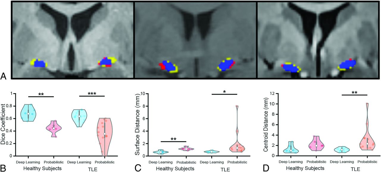
The Dice coefficient was computed between the predicted and ground truth–segmented NBM.30 The Dice coefficient is known to underestimate the performance of small, complex structures, for example, segmentation studies of other small structures such as the ventral tegmental area and the interposed nuclei report Dice coefficients of 0.52 and 0.69, respectively.13⇓⇓-16,31 The Dice coefficient of the NBM computed from the DnSeg (mean, 0.68 [SD, 0.10]) significantly outperformed that of the probabilistic atlas (0.45 [SD, 0.11]) in the completely withheld data sets of 6 healthy subjects (paired t test, P = .001), as seen in Fig 3B. Additionally, the Dice coefficient was significantly increased in DnSeg (mean, 0.64 [SD, 0.10]) compared with the probabilistic atlas Dice coefficient (mean, 0.37 [SD, 0.22) for the TLE data set (paired t test, P < .001). The probabilistic atlas demonstrated substantial variability in the Dice coefficient for the TLE data set, likely due to anatomic changes that have been noted in the NBM for TLE.

FIG 3.
  • Download figure
  • Open in new tab
  • Download powerpoint
FIG 3.

Example of favorable (A, right), average (A, middle), and poor (A, left) performance. Blue is the correct prediction of NBM, red is the overpredicted NBM (false-positive), and yellow is the ground truth label that was not predicted (false-negative). The Dice coefficient of the favorable example (right) was 0.82, the Dice coefficient of the average example (middle) was 0.63, and the Dice coefficient of the poor example (left) was 0.55. B, The Dice coefficient. C, The mean surface distance. D, The centroid distance of DnSeg versus the probabilistic atlas for both healthy subjects and patients with TLE. The healthy subjects included the nonaugmented held-out test data set (n = 6). The patients with TLE were also not augmented and were held out until final analysis (n = 14). Paired t test: single asterisk indicates P < .05; double asterisks, P < .01; triple asterisks, P < .001.

Mean Surface Distance

The mean surface distance between the predicted surface and the ground truth surface of the NBM segmentation was calculated for the test data set and the TLE data set (Fig 3C). For healthy subjects, DnSeg demonstrated significantly lower mean surface distance (mean, 0.65 [SD, 0.24] mm) computed with the probabilistic atlas (mean, 1.18 [SD, 0.35] mm) in the completely withheld data set of 6 healthy subjects (paired t test, P = .002). Furthermore, the mean surface distance was significantly lower with DnSeg (0.69 [SD, 0.15] mm) compared with the probabilistic atlas (mean, 1.91 [SD, 2.03] mm) in the TLE data set (paired t test P = .027). The variability of the probabilistic atlas on the TLE data set was far more exaggerated than that of the healthy subject data set, as seen with the 95% confidence intervals of 1.2 and 0.2 mm, respectively.

Centroid Distance

The centroid distance represents the error in localization of the NBM that can occur due to patient-specific anatomic variability. It is most relevant for DBS applications because it could contribute to a targeting error. The centroid distance performance of DnSeg (mean, 1.35 [SD, 0.86] mm) was not significantly different from the probabilistic atlas (mean, 2.19 [SD, 1.36] mm) in the completely withheld data sets of 6 healthy subjects (paired t test, P = .014), as can be seen in Fig 3D. However, the centroid distance of DnSeg (mean, 1.18 [SD, 0.43] mm) was significantly lower than that of the probabilistic atlas (mean, 3.09 [SD, 2.56] mm) in the TLE data set (paired t test, P = .007).

Qualitative Comparison

Because localization and overlap are difficult to quantify for small structures, the results were also analyzed qualitatively. An evaluation of NBM prediction of DnSeg compared with the ground truth manual segmentation was completed qualitatively by visual inspection (Fig 3A). The poor-performance example demonstrates that even the poor-performing examples perform relatively well. Additionally, it is seen that the NBM shape differs slightly from patient to patient and that DnSeg successfully accounts for heterogeneous patient anatomy (Online Supplemental Data).

DISCUSSION

The purpose of this work was to enable study of the NBM by accounting for patient-specific differences. DnSeg may have widespread impact across several diseases, perhaps enabling new treatment modalities targeting the NBM.

DnSeg Can Distinguish Subject-Specific Anatomic Differences

The NBM has anatomic variability among healthy subjects and has been shown to change in size in patients with Parkinson disease, Alzheimer disease, and TLE.32⇓⇓⇓⇓⇓⇓⇓-40 This variability presents a substantial challenge in studying the NBM and a barrier for possible treatment innovations such as DBS. Manual segmentation to capture these differences would be preferable; however, the NBM cannot be accurately visualized on 3T MR imaging. Thus, current research is limited by the non-patient-specific atlas that does not capture variability in the NBM, as seen by the wide 95% confidence intervals for patients with TLE (Fig 3). DnSeg was able to capture patient-specific differences in the NBM and accurately segment it using only 3T T1-weighted MR imaging. The results of DnSeg in patients with TLE were comparable with those of healthy subjects, indicating that it can accurately segment the NBM despite anatomic changes (Fig 3B–D), demonstrating that DnSeg can capture pathologic changes in the NBM in at least 1 disease state.

DBS Targeting

DBS targeting for the NBM has been explored in several studies with mixed results.8,9 One possible factor in these studies is DBS targeting accuracy. In this investigation, DnSeg localized the NBM with far more accuracy than the probabilistic atlas, which can have a substantial impact on targeting (Fig 3). The impact of this distance can be seen in Fig 4, where an electrode planned with DnSeg seems to target the NBM accurately, while an electrode planned with the center of the 50% thresholded probabilistic atlas targets the border of the true NBM.

FIG 4.
  • Download figure
  • Open in new tab
  • Download powerpoint
FIG 4.

Increased accuracy of NBM electrode targeting with DnSeg. The orange electrode was placed in the center of the NBM segmentation generated by the probabilistic atlas. The blue electrode was placed in the center of the segmentation generated by DnSeg. D, Both electrode positions compared with the ground truth segmentation mesh. The ground truth NBM (manually segmented on 7T MR imaging) is shown in red. The outline of the ground truth NBM as well as the lead trajectories can be seen in A, B, and C. The sample DBS planning for this figure was generated using CRAnial Vault Explorer.23

In addition to this practical application, DnSeg also has 10× faster run-time than standard registration methods required for applying a Montreal Neurological Institute 152 atlas space to patient-specific space. With a modern CPU, it takes approximately 1 minute per scan if skull-stripping is not required and approximately 2 minutes if skull stripping is required. The pipeline does not require the patient data to be registered to any standard space; thus, it is much faster than other methods that require a registration step.

Limitations

Despite these benefits of the deep learning pipeline, there are some limitations. Most notably, the expert segmentations could not be verified with a postmortem histologic analysis. While the segmentations were verified by 2 neurological surgeons, a histologic analysis would be ideal. Furthermore, the current analysis validates only DnSeg in one disease state. The NBM is both of interest and has pathologic changes in several disease states. Thus, it would be preferred to validate DnSeg in other disease states. However, paired 7T and 3T MR imaging scans of the same patients needed to achieve this goal are rare. Furthermore, DnSeg is limited by the small sample size used for training. Although this limitation is common among studies of paired 3T and 7T MR imaging, it has the potential to limit the generalizability of DnSeg. Aggressive augmentation was used to attenuate this limitation and increase generalizability.

CONCLUSIONS

In this work, we have presented an accurate, patient-specific method of segmenting the NBM using only 3T T1-weighted MR imaging. The NBM has been implicated in Alzheimer disease, Parkinson disease dementia, and TLE.3,6,7 It has been shown to change in neuronal density and gray matter volume across age and disease states.32⇓⇓⇓⇓⇓⇓⇓-40 With the use of an expertly segmented NBM and aggressive data augmentation, we have trained a deep learning network to capture anatomic differences when segmenting the NBM. We have presented evidence that DnSeg is a powerful and accurate NBM segmentation model.

Methodologically, this work represents an innovative approach to the segmentation of regions with little contrast enhancement. The NBM represents a small region of the brain with little contrast on commonly used 3T MR imaging but with high importance to several disease states. Paired 3T and 7T MR imaging of the same subjects is a useful approach to providing accurate labels for training a deep learning network to segment structures that cannot be accurately visualized on 3T imaging. However, this approach is intrinsically limited by the rarity of paired 3T-7T data sets.

DnSeg, therefore, opens the possibility of further study of the NBM. The NBM has long been considered a region of key interest, but study of the NBM in vivo has been difficult, in part because of the lack of an adaptive, patient-specific atlas. The presented model is available (https://github.com/DerekDoss/DnSeg) and may greatly assist in novel studies of the NBM.

Acknowledgments

The authors would like to thank Laurie Cutting, PhD, for providing paired 7T-3T MR imaging scans of healthy subjects. The authors would also like to thank William (Bill) Rodriguez, MEd for his assistance with CRAVE and data set curation. A preprint of this article was originally released on biorxiv.

Footnotes

  • This work was supported by the National Institutes of Health (grant Nos. T32GM007347, T32EB001628-17, T32EB021937, F31NS106735, F31NS120401, R01NS112252, R01NS108445, R01NS110130, and R01NS116504).

  • Disclosure forms provided by the authors are available with the full text and PDF of this article at www.ajnr.org.

Indicates open access to non-subscribers at www.ajnr.org

References

  1. 1.↵
    1. Liu AK,
    2. Chang RC,
    3. Pearce RK, et al
    . Nucleus basalis of Meynert revisited: anatomy, history and differential involvement in Alzheimer’s and Parkinson’s disease. Acta Neuropathol 2015;129:527–40 doi:10.1007/s00401-015-1392-5 pmid:25633602
    CrossRefPubMed
  2. 2.↵
    1. Mesulam MM,
    2. Mufson EJ,
    3. Levey AI, et al
    . Cholinergic innervation of cortex by the basal forebrain: cytochemistry and cortical connections of the septal area, diagonal band nuclei, nucleus basalis (substantia innominata), and hypothalamus in the rhesus monkey. J Comp Neurol 1983;214:170–97 doi:10.1002/cne.902140206 pmid:6841683
    CrossRefPubMed
  3. 3.↵
    1. Gonzalez HFJ,
    2. Narasimhan S,
    3. Johnson GW, et al
    . Role of the nucleus basalis as a key network node in temporal lobe epilepsy. Neurology 2021;96:e1334–46 doi:10.1212/WNL.0000000000011523 pmid:33441453
    CrossRefPubMed
  4. 4.↵
    1. Schulz J,
    2. Pagano G,
    3. Fernández Bonfante JA, et al
    . Nucleus basalis of Meynert degeneration precedes and predicts cognitive impairment in Parkinson’s disease. Brain 2018;141:1501–16 doi:10.1093/brain/awy072 pmid:29701787
    CrossRefPubMed
  5. 5.↵
    1. Whitehouse PJ,
    2. Price DL,
    3. Clark AW, et al
    . Alzheimer disease: evidence for selective loss of cholinergic neurons in the nucleus basalis. Ann Neurol 1981;10:122–26 doi:10.1002/ana.410100203 pmid:7283399
    CrossRefPubMed
  6. 6.↵
    1. Schumacher J,
    2. Thomas AJ,
    3. Peraza LR, et al
    . Functional connectivity of the nucleus basalis of Meynert in Lewy body dementia and Alzheimer’s disease. Int Psychogeriatr 2021;33:89–94 doi:10.1017/S1041610220003944 pmid:33413710
    CrossRefPubMed
  7. 7.↵
    1. Oswal A,
    2. Gratwicke J,
    3. Akram H, et al
    . Cortical connectivity of the nucleus basalis of Meynert in Parkinson’s disease and Lewy body dementias. Brain 2021;144:781–88 doi:10.1093/brain/awaa411 pmid:33521808
    CrossRefPubMed
  8. 8.↵
    1. Chen YS,
    2. Shu K,
    3. Kang HC
    . Deep brain stimulation in Alzheimer’s disease: targeting the nucleus basalis of Meynert. J Alzheimers Dis 2021;80:53–70 doi:10.3233/JAD-201141 pmid:33492288
    CrossRefPubMed
  9. 9.↵
    1. Kuhn J,
    2. Hardenacke K,
    3. Lenartz D, et al
    . Deep brain stimulation of the nucleus basalis of Meynert in Alzheimer’s dementia. Mol Psychiatry 2015;20:353–60 doi:10.1038/mp.2014.32 pmid:24798585
    CrossRefPubMed
  10. 10.↵
    1. Wilson H,
    2. de Natale ER,
    3. Politis M
    . Nucleus basalis of Meynert degeneration predicts cognitive impairment in Parkinson’s disease. Handb Clin Neurol 2021;179:189–205 doi:10.1016/B978-0-12-819975-6.00010-8 pmid:34225962
    CrossRefPubMed
  11. 11.↵
    1. Zaborszky L,
    2. Hoemke L,
    3. Mohlberg H, et al
    . Stereotaxic probabilistic maps of the magnocellular cell groups in human basal forebrain. Neuroimage 2008;42:1127–41 doi:10.1016/j.neuroimage.2008.05.055 pmid:18585468
    CrossRefPubMed
  12. 12.↵
    1. Eickhoff SB,
    2. Stephan KE,
    3. Mohlberg H, et al
    . A new SPM toolbox for combining probabilistic cytoarchitectonic maps and functional imaging data. Neuroimage 2005;25:1325–35 doi:10.1016/j.neuroimage.2004.12.034 pmid:15850749
    CrossRefPubMed
  13. 13.↵
    1. Kim J,
    2. Duchin Y,
    3. Shamir RR, et al
    . Automatic localization of the subthalamic nucleus on patient-specific clinical MRI by incorporating 7 T MRI and machine learning: application in deep brain stimulation. Hum Brain Mapp 2019;40:679–98 doi:10.1002/hbm.24404 pmid:30379376
    CrossRefPubMed
  14. 14.↵
    1. Kim J,
    2. Patriat R,
    3. Kaplan J, et al
    . Deep cerebellar nuclei segmentation via semi-supervised deep context-aware learning from 7T diffusion MRI. IEEE Access 2020;8:101550–68 doi:10.1109/access.2020.2998537 pmid:32656051
    CrossRefPubMed
  15. 15.↵
    1. Greve DN,
    2. Billot B,
    3. Cordero D, et al
    . A deep learning toolbox for automatic segmentation of subcortical limbic structures from MRI images. Neuroimage 2021;244:118610 doi:10.1016/j.neuroimage.2021.118610 pmid:34571161
    CrossRefPubMed
  16. 16.↵
    1. Billot B,
    2. Bocchetta M,
    3. Todd E, et al
    . Automated segmentation of the hypothalamus and associated subunits in brain MRI. Neuroimage 2020;223:117287 doi:10.1016/j.neuroimage.2020.117287 pmid:32853816
    CrossRefPubMed
  17. 17.↵
    1. Wang Y,
    2. Zhan M,
    3. Roebroeck A, et al
    . Inconsistencies in atlas-based volumetric measures of the human nucleus basalis of Meynert: a need for high-resolution alternatives. Neuroimage 2022;259:119421 doi:10.1016/j.neuroimage.2022.119421
    CrossRef
  18. 18.↵
    1. Niazi MK,
    2. Parwani AV,
    3. Gurcan MN
    . Digital pathology and artificial intelligence. Lancet Oncol 2019;20:e253–61 doi:10.1016/S1470-2045(19)30154-8 pmid:31044723
    CrossRefPubMed
  19. 19.↵
    1. Isaacs BR,
    2. Mulder MJ,
    3. Groot JM, et al
    . 3 versus 7 Tesla magnetic resonance imaging for parcellations of subcortical brain structures in clinical settings. PLoS One 2020;15:e0236208 doi:10.1371/journal.pone.0236208 pmid:33232325
    CrossRefPubMed
  20. 20.↵
    1. Liu Y,
    2. D’Haese PF,
    3. Newton AT, et al
    . Generation of human thalamus atlases from 7T data and application to intrathalamic nuclei segmentation in clinical 3T T1-weighted images. Magn Reson Imaging 2020;65:114–28 doi:10.1016/j.mri.2019.09.004 pmid:31629074
    CrossRefPubMed
  21. 21.↵
    1. Ramadass K,
    2. Rheault F,
    3. Cai LY, et al
    . Ultra-high-resolution mapping of cortical layers 3T-guided 7T MRI. Proc SPIE Int Soc Opt Eng 2022;12032:120321G doi:10.1117/12.2611857 pmid:36303575
    CrossRefPubMed
  22. 22.↵
    1. Jethwa K,
    2. Aphiwatthanasumet K,
    3. Mougin O, et al
    . Phase enhanced PSIR T1-weighted imaging improves contrast resolution of the nucleus basalis of Meynert at 7T: a preliminary study. Magn Reson Imaging 2019;61:296–99 doi:10.1016/j.mri.2019.06.004 pmid:31202788
    CrossRefPubMed
  23. 23.↵
    1. D’Haese PF,
    2. Pallavaram S,
    3. Li R, et al
    . CranialVault and its CRAVE tools: a clinical computer assistance system for deep brain stimulation (DBS) therapy. Med Image Anal 2012;16:744–53 doi:10.1016/j.media.2010.07.009 pmid:20732828
    CrossRefPubMed
  24. 24.↵
    1. Brett M,
    2. Markiewicz CJ,
    3. Hanke M, et al
    . nipy/nibabel: 4.0.0rc0. 2022/6/2022 doi:10.5281/ZENODO.6617127
    CrossRef
  25. 25.↵
    1. Iglesias JE,
    2. Liu CY,
    3. Thompson PM, et al
    . Robust brain extraction across datasets and comparison with publicly available methods. IEEE Trans Med Imaging 2011;30:1617–34 doi:10.1109/TMI.2011.2138152 pmid:21880566
    CrossRefPubMed
  26. 26.↵
    1. Pérez-García F,
    2. Sparks R,
    3. Ourselin S
    . TorchIO: a Python library for efficient loading, preprocessing, augmentation and patch-based sampling of medical images in deep learning. Comput Methods Programs Biomed 2021;208:106236 doi:10.1016/j.cmpb.2021.106236 pmid:34311413
    CrossRefPubMed
  27. 27.↵
    1. Navab N,
    2. Hornegger J,
    3. Wells WM, et al.
    1. Ronneberger O,
    2. Fischer P,
    3. Brox T
    . U-net: Convolutional networks for biomedical image segmentation. In: Navab N, Hornegger J, Wells WM, et al., eds. Medical Image Computing and Computer-Assisted Intervention–MICCAI 2015. Springer; 2015: 234–41
  28. 28.↵
    1. Taha AA,
    2. Hanbury A
    . Metrics for evaluating 3D medical image segmentation: analysis, selection, and tool. BMC Med Imaging 2015;15:29 doi:10.1186/s12880-015-0068-x pmid:26263899
    CrossRefPubMed
  29. 29.↵
    1. Bergstra J,
    2. Yamins D,
    3. Cox D
    . Making a science of model search: hyperparameter optimization in hundreds of dimensions for vision architectures. Proceedings of the 30th International Conference on Machine Learning, 2013. https://proceedings.mlr.press/v28/bergstra13.html. Accessed November 11, 2022
  30. 30.↵
    1. Dice LR
    . Measures of the amount of ecologic association between species. Ecology 1945;26:297–302 doi:10.2307/1932409
    CrossRef
  31. 31.↵
    1. Trutti AC,
    2. Fontanesi L,
    3. Mulder MJ, et al
    . A probabilistic atlas of the human ventral tegmental area (VTA) based on 7 Tesla MRI data. Brain Struct Funct 2021;226:1155–67 doi:10.1007/s00429-021-02231-w pmid:33580320
    CrossRefPubMed
  32. 32.↵
    1. Cantero JL,
    2. Zaborszky L,
    3. Atienza M
    . Volume loss of the nucleus basalis of Meynert is associated with atrophy of innervated regions in mild cognitive impairment. Cereb Cortex 2017;27:3881–89 doi:10.1093/cercor/bhw195 pmid:27371762
    CrossRefPubMed
  33. 33.↵
    1. Grothe M,
    2. Heinsen H,
    3. Teipel SJ
    . Atrophy of the cholinergic basal forebrain over the adult age range and in early stages of Alzheimer’s disease. Biol Psychiatry 2012;71:805–13 doi:10.1016/j.biopsych.2011.06.019 pmid:21816388
    CrossRefPubMed
  34. 34.↵
    1. Kilimann I,
    2. Grothe M,
    3. Heinsen H, et al
    . Subregional basal forebrain atrophy in Alzheimer's disease: a multicenter study. J Alzheimers Dis 2014;40:687–700 doi:10.3233/JAD-132345 pmid:24503619
    CrossRefPubMed
  35. 35.↵
    1. Kim HJ,
    2. Lee JE,
    3. Shin SJ, et al
    . Analysis of the substantia innominata volume in patients with Parkinson’s disease with dementia, dementia with Lewy bodies, and Alzheimer’s disease. J Mov Disord 2011;4:68–72 doi:10.14802/jmd.11014 pmid:24868398
    CrossRefPubMed
  36. 36.↵
    1. Lammers F,
    2. Borchers F,
    3. Feinkohl I, et al
    ; BioCog consortium. Basal forebrain cholinergic system volume is associated with general cognitive ability in the elderly. Neuropsychologia 2018;119:145–56 doi:10.1016/j.neuropsychologia.2018.08.005 pmid:30096414
    CrossRefPubMed
  37. 37.↵
    1. Ray NJ,
    2. Bradburn S,
    3. Murgatroyd C, et al
    . In vivo cholinergic basal forebrain atrophy predicts cognitive decline in de novo Parkinson’s disease. Brain 2018;141:165–76 doi:10.1093/brain/awx310 pmid:29228203
    CrossRefPubMed
  38. 38.↵
    1. Rong S,
    2. Li Y,
    3. Li B, et al
    . Meynert nucleus-related cortical thinning in Parkinson’s disease with mild cognitive impairment. Quant Imaging Med Surg 2021;11:1554–66 doi:10.21037/qims-20-444 pmid:33816191
    CrossRefPubMed
  39. 39.↵
    1. Scheef L,
    2. Grothe MJ,
    3. Koppara A, et al
    . Subregional volume reduction of the cholinergic forebrain in subjective cognitive decline (SCD). Neuroimage Clin 2019;21:101612 doi:10.1016/j.nicl.2018.101612 pmid:30555006
    CrossRefPubMed
  40. 40.↵
    1. Wolf D,
    2. Grothe M,
    3. Fischer FU, et al
    . Association of basal forebrain volumes and cognition in normal aging. Neuropsychologia 2014;53:54–63 doi:10.1016/j.neuropsychologia.2013.11.002 pmid:24269297
    CrossRefPubMed
  • Received November 11, 2022.
  • Accepted after revision June 25, 2023.
  • © 2023 by American Journal of Neuroradiology
PreviousNext
Back to top
Advertisement
Print
Download PDF
Email Article

Thank you for your interest in spreading the word on American Journal of Neuroradiology.

NOTE: We only request your email address so that the person you are recommending the page to knows that you wanted them to see it, and that it is not junk mail. We do not capture any email address.

Enter multiple addresses on separate lines or separate them with commas.
Deep Learning Segmentation of the Nucleus Basalis of Meynert on 3T MRI
(Your Name) has sent you a message from American Journal of Neuroradiology
(Your Name) thought you would like to see the American Journal of Neuroradiology web site.
CAPTCHA
This question is for testing whether or not you are a human visitor and to prevent automated spam submissions.
Cite this article
D.J. Doss, G.W. Johnson, S. Narasimhan, J.S. Shless, J.W. Jiang, H.F.J. González, D.L. Paulo, A. Lucas, K.A. Davis, C. Chang, V.L. Morgan, C. Constantinidis, B.M. Dawant, D.J. Englot
Deep Learning Segmentation of the Nucleus Basalis of Meynert on 3T MRI
American Journal of Neuroradiology Aug 2023, DOI: 10.3174/ajnr.A7950

Citation Manager Formats

  • BibTeX
  • Bookends
  • EasyBib
  • EndNote (tagged)
  • EndNote 8 (xml)
  • Medlars
  • Mendeley
  • Papers
  • RefWorks Tagged
  • Ref Manager
  • RIS
  • Zotero
0 Responses
Respond to this article
Share
Bookmark this article
Deep Learning Segmentation of the Nucleus Basalis of Meynert on 3T MRI
D.J. Doss, G.W. Johnson, S. Narasimhan, J.S. Shless, J.W. Jiang, H.F.J. González, D.L. Paulo, A. Lucas, K.A. Davis, C. Chang, V.L. Morgan, C. Constantinidis, B.M. Dawant, D.J. Englot
American Journal of Neuroradiology Aug 2023, DOI: 10.3174/ajnr.A7950
del.icio.us logo Twitter logo Facebook logo Mendeley logo
  • Tweet Widget
  • Facebook Like
  • Google Plus One
Purchase

Jump to section

  • Article
    • Abstract
    • ABBREVIATIONS:
    • MATERIALS AND METHODS
    • RESULTS
    • DISCUSSION
    • CONCLUSIONS
    • Acknowledgments
    • Footnotes
    • References
  • Figures & Data
  • Supplemental
  • Info & Metrics
  • Responses
  • References
  • PDF

Related Articles

  • PubMed
  • Google Scholar

Cited By...

  • Deep Learning-Based Algorithm for Automatic Quantification of Nigrosome-1 and Parkinsonism Classification Using Susceptibility Map-Weighted MRI
  • Multimodal state-dependent connectivity analysis of arousal and autonomic centers in the brainstem and basal forebrain
  • Crossref (5)
  • Google Scholar

This article has been cited by the following articles in journals that are participating in Crossref Cited-by Linking.

  • Deep Learning–Based Algorithm for Automatic Quantification of Nigrosome-1 and Parkinsonism Classification Using Susceptibility Map–Weighted MRI
    Pae Sun Suh, Hwan Heo, Chong Hyun Suh, MyeongOh Lee, Soohwa Song, Donghoon Shin, Sungyang Jo, Sun Ju Chung, Hwon Heo, Woo Hyun Shim, Ho Sung Kim, Sang Joon Kim, Eung Yeop Kim
    American Journal of Neuroradiology 2025 46 5
  • Effects of strategic white matter hyperintensities of cholinergic pathways on basal forebrain volume in patients with amyloid-negative neurocognitive disorders
    Ye Eun Kim, Jae-Sung Lim, Chong Hyun Suh, Hwon Heo, Jee Hoon Roh, E-nae Cheong, Yoojin Lee, Jae Woo Kim, Jae-Hong Lee
    Alzheimer's Research & Therapy 2024 16 1
  • Network signatures define consciousness state during focal seizures
    Derek J. Doss, Graham W. Johnson, Ghassan S. Makhoul, Rohan V. Rashingkar, Jared S. Shless, Camden E. Bibro, Danika L. Paulo, Abhijeet Gummadavelli, Tyler J. Ball, Shilpa B. Reddy, Robert P. Naftel, Kevin F. Haas, Benoit M. Dawant, Christos Constantinidis, Shawniqua Williams Roberson, Sarah K. Bick, Victoria L. Morgan, Dario J. Englot
    Epilepsia 2024 65 9
  • An individualized tractography pipeline for the nucleus basalis of Meynert lateral tract
    Rachel A. Crockett, Kevin B. Wilkins, Michael M. Zeineh, Jennifer A. McNab, Jaimie M. Henderson, Vivek P. Buch, Helen M. Brontë-Stewart
    Imaging Neuroscience 2024 2
  • Fully automated segmentation and volumetric measurement of ocular adnexal lymphoma by deep learning-based self-configuring nnU-net on multi-sequence MRI: a multi-center study
    Guorong Wang, Bingbing Yang, Xiaoxia Qu, Jian Guo, Yongheng Luo, Xiaoquan Xu, Feiyun Wu, Xiaoxue Fan, Yang Hou, Song Tian, Sicong Huang, Junfang Xian
    Neuroradiology 2024 66 10

More in this TOC Section

  • Diagnostic Neuroradiology of Monoclonal Antibodies
  • Clinical Outcomes After Chiari I Decompression
  • Segmentation of Brain Metastases with BLAST
Show more Adult Brain

Similar Articles

Advertisement

Indexed Content

  • Current Issue
  • Accepted Manuscripts
  • Article Preview
  • Past Issues
  • Editorials
  • Editor's Choice
  • Fellows' Journal Club
  • Letters to the Editor
  • Video Articles

Cases

  • Case Collection
  • Archive - Case of the Week
  • Archive - Case of the Month
  • Archive - Classic Case

More from AJNR

  • Trainee Corner
  • Imaging Protocols
  • MRI Safety Corner
  • Book Reviews

Multimedia

  • AJNR Podcasts
  • AJNR Scantastics

Resources

  • Turnaround Time
  • Submit a Manuscript
  • Submit a Video Article
  • Submit an eLetter to the Editor/Response
  • Manuscript Submission Guidelines
  • Statistical Tips
  • Fast Publishing of Accepted Manuscripts
  • Graphical Abstract Preparation
  • Imaging Protocol Submission
  • Evidence-Based Medicine Level Guide
  • Publishing Checklists
  • Author Policies
  • Become a Reviewer/Academy of Reviewers
  • News and Updates

About Us

  • About AJNR
  • Editorial Board
  • Editorial Board Alumni
  • Alerts
  • Permissions
  • Not an AJNR Subscriber? Join Now
  • Advertise with Us
  • Librarian Resources
  • Feedback
  • Terms and Conditions
  • AJNR Editorial Board Alumni

American Society of Neuroradiology

  • Not an ASNR Member? Join Now

© 2025 by the American Society of Neuroradiology All rights, including for text and data mining, AI training, and similar technologies, are reserved.
Print ISSN: 0195-6108 Online ISSN: 1936-959X

Powered by HighWire