Advertisement

Main menu

  • Home
  • Content
    • Current Issue
    • Accepted Manuscripts
    • Article Preview
    • Past Issue Archive
    • Video Articles
    • AJNR Case Collection
    • Case of the Week Archive
    • Case of the Month Archive
    • Classic Case Archive
  • Special Collections
    • AJNR Awards
    • Low-Field MRI
    • Alzheimer Disease
    • ASNR Foundation Special Collection
    • Photon-Counting CT
    • View All
  • Multimedia
    • AJNR Podcasts
    • AJNR SCANtastic
    • Trainee Corner
    • MRI Safety Corner
    • Imaging Protocols
  • For Authors
    • Submit a Manuscript
    • Submit a Video Article
    • Submit an eLetter to the Editor/Response
    • Manuscript Submission Guidelines
    • Statistical Tips
    • Fast Publishing of Accepted Manuscripts
    • Graphical Abstract Preparation
    • Imaging Protocol Submission
    • Author Policies
  • About Us
    • About AJNR
    • Editorial Board
    • Editorial Board Alumni
  • More
    • Become a Reviewer/Academy of Reviewers
    • Subscribers
    • Permissions
    • Alerts
    • Feedback
    • Advertisers
    • ASNR Home

User menu

  • Alerts
  • Log in

Search

  • Advanced search
American Journal of Neuroradiology

American Journal of Neuroradiology

ASHNR American Society of Functional Neuroradiology ASHNR American Society of Pediatric Neuroradiology ASSR
  • Alerts
  • Log in

Advanced Search

  • Home
  • Content
    • Current Issue
    • Accepted Manuscripts
    • Article Preview
    • Past Issue Archive
    • Video Articles
    • AJNR Case Collection
    • Case of the Week Archive
    • Case of the Month Archive
    • Classic Case Archive
  • Special Collections
    • AJNR Awards
    • Low-Field MRI
    • Alzheimer Disease
    • ASNR Foundation Special Collection
    • Photon-Counting CT
    • View All
  • Multimedia
    • AJNR Podcasts
    • AJNR SCANtastic
    • Trainee Corner
    • MRI Safety Corner
    • Imaging Protocols
  • For Authors
    • Submit a Manuscript
    • Submit a Video Article
    • Submit an eLetter to the Editor/Response
    • Manuscript Submission Guidelines
    • Statistical Tips
    • Fast Publishing of Accepted Manuscripts
    • Graphical Abstract Preparation
    • Imaging Protocol Submission
    • Author Policies
  • About Us
    • About AJNR
    • Editorial Board
    • Editorial Board Alumni
  • More
    • Become a Reviewer/Academy of Reviewers
    • Subscribers
    • Permissions
    • Alerts
    • Feedback
    • Advertisers
    • ASNR Home
  • Follow AJNR on Twitter
  • Visit AJNR on Facebook
  • Follow AJNR on Instagram
  • Join AJNR on LinkedIn
  • RSS Feeds

AJNR Awards, New Junior Editors, and more. Read the latest AJNR updates

Research ArticleUltra-High-Field MRI/Imaging of Epilepsy/Demyelinating Diseases/Inflammation/Infection

7T MRI for Cushing Disease: A Single-Institution Experience and Literature Review

Ian T. Mark, Kirk Welker, Dana Erickson, Derek R. Johnson, Girish Bathla, Steven Messina, Paul J. Farnsworth and Jamie Van Gompel
American Journal of Neuroradiology July 2024, 45 (7) 971-976; DOI: https://doi.org/10.3174/ajnr.A8209
Ian T. Mark
aFrom the Department of Radiology (I.T.M., K.W., D.R.J., G.B., S.M., P.J.F.), Mayo Clinic, Rochester, Minnesota
  • Find this author on Google Scholar
  • Find this author on PubMed
  • Search for this author on this site
  • ORCID record for Ian T. Mark
Kirk Welker
aFrom the Department of Radiology (I.T.M., K.W., D.R.J., G.B., S.M., P.J.F.), Mayo Clinic, Rochester, Minnesota
  • Find this author on Google Scholar
  • Find this author on PubMed
  • Search for this author on this site
  • ORCID record for Kirk Welker
Dana Erickson
bDepartment of Endocrinology (D.E.), Mayo Clinic, Rochester, Minnesota
  • Find this author on Google Scholar
  • Find this author on PubMed
  • Search for this author on this site
Derek R. Johnson
aFrom the Department of Radiology (I.T.M., K.W., D.R.J., G.B., S.M., P.J.F.), Mayo Clinic, Rochester, Minnesota
  • Find this author on Google Scholar
  • Find this author on PubMed
  • Search for this author on this site
  • ORCID record for Derek R. Johnson
Girish Bathla
aFrom the Department of Radiology (I.T.M., K.W., D.R.J., G.B., S.M., P.J.F.), Mayo Clinic, Rochester, Minnesota
  • Find this author on Google Scholar
  • Find this author on PubMed
  • Search for this author on this site
  • ORCID record for Girish Bathla
Steven Messina
aFrom the Department of Radiology (I.T.M., K.W., D.R.J., G.B., S.M., P.J.F.), Mayo Clinic, Rochester, Minnesota
  • Find this author on Google Scholar
  • Find this author on PubMed
  • Search for this author on this site
  • ORCID record for Steven Messina
Paul J. Farnsworth
aFrom the Department of Radiology (I.T.M., K.W., D.R.J., G.B., S.M., P.J.F.), Mayo Clinic, Rochester, Minnesota
  • Find this author on Google Scholar
  • Find this author on PubMed
  • Search for this author on this site
  • ORCID record for Paul J. Farnsworth
Jamie Van Gompel
cDepartment of Neurosurgery (J.V.G.), Mayo Clinic, Rochester, Minnesota
  • Find this author on Google Scholar
  • Find this author on PubMed
  • Search for this author on this site
  • ORCID record for Jamie Van Gompel
  • Article
  • Figures & Data
  • Supplemental
  • Info & Metrics
  • Responses
  • References
  • PDF
Loading

Article Figures & Data

Figures

  • FIG 1.
    • Download figure
    • Open in new tab
    • Download powerpoint
    FIG 1.

    Three different types of 7T postcontrast T1-weighted imaging in a patient with a left-sided pituitary adenoma (arrows): (left to right) DCE, 2D TSE, and 3D SPACE with 1.5-, 1.5-, and 0.7-mm slice thickness, respectively.

  • FIG 2.
    • Download figure
    • Open in new tab
    • Download powerpoint
    FIG 2.

    An example of 7T MR imaging showing a nondiscrete lesion (arrows) in the left paramidline pituitary gland on postcontrast MPRAGE T1-weighted imaging at 0.33 mm that was a pathology-confirmed Cushing adenoma.

  • FIG 3.
    • Download figure
    • Open in new tab
    • Download powerpoint
    FIG 3.

    Flow chart of the search and selection of studies.
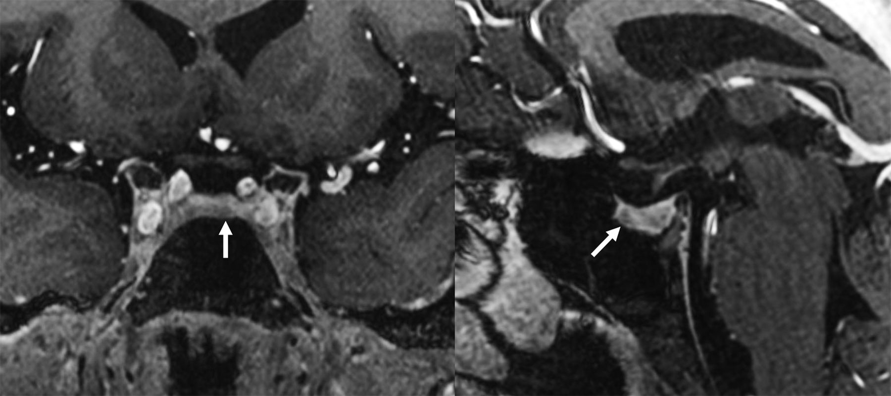
  • FIG 4.
    • Download figure
    • Open in new tab
    • Download powerpoint
    FIG 4.

    7T postcontrast T1-weighted imaging in a patient with a right-sided pituitary adenoma (arrows). The left image is a 3D MPRAGE sequence, while the right image is a 2D TSE sequence. The MPRAGE image demonstrates greater contrast between the normally enhancing pituitary gland compared with the hypoenhancing adenoma.

PreviousNext
Back to top

In this issue

American Journal of Neuroradiology: 45 (7)
American Journal of Neuroradiology
Vol. 45, Issue 7
1 Jul 2024
  • Table of Contents
  • Index by author
  • Complete Issue (PDF)
Advertisement
Print
Download PDF
Email Article

Thank you for your interest in spreading the word on American Journal of Neuroradiology.

NOTE: We only request your email address so that the person you are recommending the page to knows that you wanted them to see it, and that it is not junk mail. We do not capture any email address.

Enter multiple addresses on separate lines or separate them with commas.
7T MRI for Cushing Disease: A Single-Institution Experience and Literature Review
(Your Name) has sent you a message from American Journal of Neuroradiology
(Your Name) thought you would like to see the American Journal of Neuroradiology web site.
CAPTCHA
This question is for testing whether or not you are a human visitor and to prevent automated spam submissions.
Cite this article
Ian T. Mark, Kirk Welker, Dana Erickson, Derek R. Johnson, Girish Bathla, Steven Messina, Paul J. Farnsworth, Jamie Van Gompel
7T MRI for Cushing Disease: A Single-Institution Experience and Literature Review
American Journal of Neuroradiology Jul 2024, 45 (7) 971-976; DOI: 10.3174/ajnr.A8209

Citation Manager Formats

  • BibTeX
  • Bookends
  • EasyBib
  • EndNote (tagged)
  • EndNote 8 (xml)
  • Medlars
  • Mendeley
  • Papers
  • RefWorks Tagged
  • Ref Manager
  • RIS
  • Zotero
0 Responses
Respond to this article
Share
Bookmark this article
7T MRI for Cushing Disease: Review and Experience
Ian T. Mark, Kirk Welker, Dana Erickson, Derek R. Johnson, Girish Bathla, Steven Messina, Paul J. Farnsworth, Jamie Van Gompel
American Journal of Neuroradiology Jul 2024, 45 (7) 971-976; DOI: 10.3174/ajnr.A8209
del.icio.us logo Twitter logo Facebook logo Mendeley logo
  • Tweet Widget
  • Facebook Like
  • Google Plus One
Purchase

Jump to section

  • Article
    • Abstract
    • ABBREVIATIONS:
    • MATERIALS AND METHODS
    • RESULTS
    • DISCUSSION
    • CONCLUSIONS
    • Footnotes
    • References
  • Figures & Data
  • Supplemental
  • Info & Metrics
  • Responses
  • References
  • PDF

Related Articles

  • PubMed
  • Google Scholar

Cited By...

  • Contrast-Enhanced CISS/FIESTA Imaging for Increased Conspicuity of Pituitary Microadenomas in Cushing Disease
  • Prevalence of Rathke Cleft and Other Incidental Pituitary Gland Findings on Contrast-Enhanced 3D Fat-Saturated T1 MPRAGE at 7T MRI
  • Crossref
  • Google Scholar

This article has not yet been cited by articles in journals that are participating in Crossref Cited-by Linking.

More in this TOC Section

Ultra-High-Field MRI/Imaging of Epilepsy/Demyelinating Diseases/Inflammation/Infection

  • DL Image Reconstruction in T2-Weighted TSE at 7T
  • Automated Lesion Segmentation Software in MS
  • Interpretation Errors in Adults with MRE
Show more Ultra-High-Field MRI/Imaging of Epilepsy/Demyelinating Diseases/Inflammation/Infection

Head and Neck Imaging

  • Hydrops Herniation into the Semicircular Canals
  • ASL Sensitivity for Head and Neck Paraganglioma
  • Post SRS Peritumoral Hyperintense Signal of VSs
Show more Head and Neck Imaging

Similar Articles

Advertisement

Indexed Content

  • Current Issue
  • Accepted Manuscripts
  • Article Preview
  • Past Issues
  • Editorials
  • Editor's Choice
  • Fellows' Journal Club
  • Letters to the Editor
  • Video Articles

Cases

  • Case Collection
  • Archive - Case of the Week
  • Archive - Case of the Month
  • Archive - Classic Case

More from AJNR

  • Trainee Corner
  • Imaging Protocols
  • MRI Safety Corner
  • Book Reviews

Multimedia

  • AJNR Podcasts
  • AJNR Scantastics

Resources

  • Turnaround Time
  • Submit a Manuscript
  • Submit a Video Article
  • Submit an eLetter to the Editor/Response
  • Manuscript Submission Guidelines
  • Statistical Tips
  • Fast Publishing of Accepted Manuscripts
  • Graphical Abstract Preparation
  • Imaging Protocol Submission
  • Evidence-Based Medicine Level Guide
  • Publishing Checklists
  • Author Policies
  • Become a Reviewer/Academy of Reviewers
  • News and Updates

About Us

  • About AJNR
  • Editorial Board
  • Editorial Board Alumni
  • Alerts
  • Permissions
  • Not an AJNR Subscriber? Join Now
  • Advertise with Us
  • Librarian Resources
  • Feedback
  • Terms and Conditions
  • AJNR Editorial Board Alumni

American Society of Neuroradiology

  • Not an ASNR Member? Join Now

© 2025 by the American Society of Neuroradiology All rights, including for text and data mining, AI training, and similar technologies, are reserved.
Print ISSN: 0195-6108 Online ISSN: 1936-959X

Powered by HighWire