Advertisement

Main menu

  • Home
  • Content
    • Current Issue
    • Accepted Manuscripts
    • Article Preview
    • Past Issue Archive
    • Video Articles
    • AJNR Case Collection
    • Case of the Week Archive
    • Case of the Month Archive
    • Classic Case Archive
  • Special Collections
    • AJNR Awards
    • Low-Field MRI
    • Alzheimer Disease
    • ASNR Foundation Special Collection
    • Photon-Counting CT
    • View All
  • Multimedia
    • AJNR Podcasts
    • AJNR SCANtastic
    • Trainee Corner
    • MRI Safety Corner
    • Imaging Protocols
  • For Authors
    • Submit a Manuscript
    • Submit a Video Article
    • Submit an eLetter to the Editor/Response
    • Manuscript Submission Guidelines
    • Statistical Tips
    • Fast Publishing of Accepted Manuscripts
    • Graphical Abstract Preparation
    • Imaging Protocol Submission
    • Author Policies
  • About Us
    • About AJNR
    • Editorial Board
    • Editorial Board Alumni
  • More
    • Become a Reviewer/Academy of Reviewers
    • Subscribers
    • Permissions
    • Alerts
    • Feedback
    • Advertisers
    • ASNR Home

User menu

  • Alerts
  • Log in

Search

  • Advanced search
American Journal of Neuroradiology

American Journal of Neuroradiology

ASHNR American Society of Functional Neuroradiology ASHNR American Society of Pediatric Neuroradiology ASSR
  • Alerts
  • Log in

Advanced Search

  • Home
  • Content
    • Current Issue
    • Accepted Manuscripts
    • Article Preview
    • Past Issue Archive
    • Video Articles
    • AJNR Case Collection
    • Case of the Week Archive
    • Case of the Month Archive
    • Classic Case Archive
  • Special Collections
    • AJNR Awards
    • Low-Field MRI
    • Alzheimer Disease
    • ASNR Foundation Special Collection
    • Photon-Counting CT
    • View All
  • Multimedia
    • AJNR Podcasts
    • AJNR SCANtastic
    • Trainee Corner
    • MRI Safety Corner
    • Imaging Protocols
  • For Authors
    • Submit a Manuscript
    • Submit a Video Article
    • Submit an eLetter to the Editor/Response
    • Manuscript Submission Guidelines
    • Statistical Tips
    • Fast Publishing of Accepted Manuscripts
    • Graphical Abstract Preparation
    • Imaging Protocol Submission
    • Author Policies
  • About Us
    • About AJNR
    • Editorial Board
    • Editorial Board Alumni
  • More
    • Become a Reviewer/Academy of Reviewers
    • Subscribers
    • Permissions
    • Alerts
    • Feedback
    • Advertisers
    • ASNR Home
  • Follow AJNR on Twitter
  • Visit AJNR on Facebook
  • Follow AJNR on Instagram
  • Join AJNR on LinkedIn
  • RSS Feeds

AJNR Awards, New Junior Editors, and more. Read the latest AJNR updates

Research ArticleUltra-High-Field MRI/Imaging of Epilepsy/Demyelinating Diseases/Inflammation/Infection

Cross-Sectional Validation of an Automated Lesion Segmentation Software in Multiple Sclerosis: Comparison with Radiologist Assessments

Maria Vittoria Spampinato, Heather R. Collins, Hannah Wells, William Dennis, Jordan H. Chamberlin, Emily Ye, Justin A. Chetta, Maria Gisele Matheus, Seth T. Stalcup and Donna R. Roberts
American Journal of Neuroradiology July 2025, 46 (7) 1510-1516; DOI: https://doi.org/10.3174/ajnr.A8655
Maria Vittoria Spampinato
aFrom the Department of Radiology (M.V.S., H.R.C., W.D., J.H.C, J.A.C., M.G.M., S.T.S., D.R.R.), Medical University of South Carolina, Charleston, South Carolina
  • Find this author on Google Scholar
  • Find this author on PubMed
  • Search for this author on this site
  • ORCID record for Maria Vittoria Spampinato
Heather R. Collins
aFrom the Department of Radiology (M.V.S., H.R.C., W.D., J.H.C, J.A.C., M.G.M., S.T.S., D.R.R.), Medical University of South Carolina, Charleston, South Carolina
  • Find this author on Google Scholar
  • Find this author on PubMed
  • Search for this author on this site
  • ORCID record for Heather R. Collins
Hannah Wells
bCollege of Medicine (H.W., E.Y.), Medical University of South Carolina, Charleston, South Carolina
  • Find this author on Google Scholar
  • Find this author on PubMed
  • Search for this author on this site
William Dennis
aFrom the Department of Radiology (M.V.S., H.R.C., W.D., J.H.C, J.A.C., M.G.M., S.T.S., D.R.R.), Medical University of South Carolina, Charleston, South Carolina
  • Find this author on Google Scholar
  • Find this author on PubMed
  • Search for this author on this site
Jordan H. Chamberlin
aFrom the Department of Radiology (M.V.S., H.R.C., W.D., J.H.C, J.A.C., M.G.M., S.T.S., D.R.R.), Medical University of South Carolina, Charleston, South Carolina
  • Find this author on Google Scholar
  • Find this author on PubMed
  • Search for this author on this site
  • ORCID record for Jordan H. Chamberlin
Emily Ye
bCollege of Medicine (H.W., E.Y.), Medical University of South Carolina, Charleston, South Carolina
  • Find this author on Google Scholar
  • Find this author on PubMed
  • Search for this author on this site
  • ORCID record for Emily Ye
Justin A. Chetta
aFrom the Department of Radiology (M.V.S., H.R.C., W.D., J.H.C, J.A.C., M.G.M., S.T.S., D.R.R.), Medical University of South Carolina, Charleston, South Carolina
  • Find this author on Google Scholar
  • Find this author on PubMed
  • Search for this author on this site
Maria Gisele Matheus
aFrom the Department of Radiology (M.V.S., H.R.C., W.D., J.H.C, J.A.C., M.G.M., S.T.S., D.R.R.), Medical University of South Carolina, Charleston, South Carolina
  • Find this author on Google Scholar
  • Find this author on PubMed
  • Search for this author on this site
  • ORCID record for Maria Gisele Matheus
Seth T. Stalcup
aFrom the Department of Radiology (M.V.S., H.R.C., W.D., J.H.C, J.A.C., M.G.M., S.T.S., D.R.R.), Medical University of South Carolina, Charleston, South Carolina
  • Find this author on Google Scholar
  • Find this author on PubMed
  • Search for this author on this site
  • ORCID record for Seth T. Stalcup
Donna R. Roberts
bCollege of Medicine (H.W., E.Y.), Medical University of South Carolina, Charleston, South Carolina
  • Find this author on Google Scholar
  • Find this author on PubMed
  • Search for this author on this site
  • ORCID record for Donna R. Roberts
  • Article
  • Figures & Data
  • Supplemental
  • Info & Metrics
  • Responses
  • References
  • PDF
Loading

This article requires a subscription to view the full text. If you have a subscription you may use the login form below to view the article. Access to this article can also be purchased.

Graphical Abstract

Figure
  • Download figure
  • Open in new tab
  • Download powerpoint

Abstract

BACKGROUND AND PURPOSE: MRI is widely used to assess disease burden in MS. This study aimed to evaluate the effectiveness of a commercially available k-nearest neighbors (kNN) network software in quantifying white matter lesion (WML) burden in MS. We compared the software’s WML quantification to expert radiologists’ assessments.

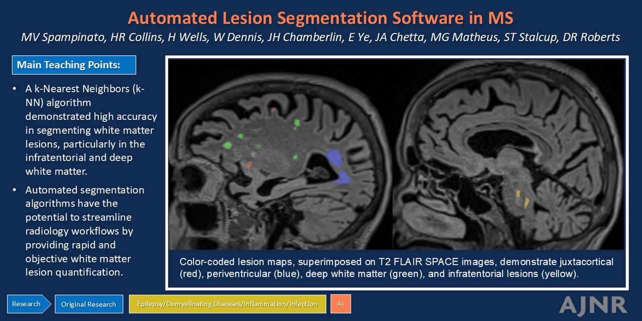
MATERIALS AND METHODS: We retrospectively reviewed brain MRI examinations of adult patients with MS and of adult patients without MS and with a normal brain MRI referred from the neurology clinic. MR images were processed by using an AI-powered, cloud-based kNN software, which generated a DICOM lesion distribution map and a report of WML count and volume in 4 brain regions (periventricular, deep, juxtacortical, and infratentorial white matter). Two blinded radiologists performed semiquantitative assessments of WM lesion load and lesion segmentation accuracy. Additionally, 4 blinded neuroradiologists independently reviewed the data to determine if MRI findings supported an MS diagnosis. The associations between radiologist-rated WML load and kNN model WML volume and count were evaluated with Spearman rank order correlation coefficient (rho) because these variables were not normally distributed. Results were considered significant when P < .05.

RESULTS: The study included 32 patients with MS (35.4 years ±9.1) and 19 patients without MS (33.5 years ±12.1). The kNN software demonstrated 94.1% and 84.3% accuracy in differentiating MS from non-MS subjects based respectively on WML count and WML volume, compared with radiologists’ accuracy of 90.2% to 94.1%. Lesion segmentation was more accurate for the deep WM and infratentorial regions than for the juxtacortical region (both P < .001).

CONCLUSIONS: kNN-derived WML volume and WML count provide valuable quantitative metrics of disease burden in MS. AI-powered postprocessing software may enhance the interpretation of brain MRIs in MS patients.

ABBREVIATIONS:

3D DIR
3D double-inversion recovery
AUC
area under the curve
EDSS
Expanded Disability Status Scale
FN
false-negative
FP
false-positive
ICC
intraclass correlation coefficient
kNN
k-nearest neighbors
MP2RAGE
magnetization prepared 2 rapid acquisition gradient echoes
ROC
receiver operating characteristic
SPACE
sampling perfection with application-optimized contrasts by using a different flip angle evolution
WMH
white matter hyperintensity
WML
white matter lesion
  • © 2025 by American Journal of Neuroradiology
View Full Text

Log in using your username and password

Forgot your user name or password?

Log in through your institution

You may be able to gain access using your login credentials for your institution. Contact your library if you do not have a username and password.
PreviousNext
Back to top

In this issue

American Journal of Neuroradiology: 46 (7)
American Journal of Neuroradiology
Vol. 46, Issue 7
1 Jul 2025
  • Table of Contents
  • Index by author
  • Complete Issue (PDF)
Advertisement
Print
Download PDF
Email Article

Thank you for your interest in spreading the word on American Journal of Neuroradiology.

NOTE: We only request your email address so that the person you are recommending the page to knows that you wanted them to see it, and that it is not junk mail. We do not capture any email address.

Enter multiple addresses on separate lines or separate them with commas.
Cross-Sectional Validation of an Automated Lesion Segmentation Software in Multiple Sclerosis: Comparison with Radiologist Assessments
(Your Name) has sent you a message from American Journal of Neuroradiology
(Your Name) thought you would like to see the American Journal of Neuroradiology web site.
CAPTCHA
This question is for testing whether or not you are a human visitor and to prevent automated spam submissions.
Cite this article
Maria Vittoria Spampinato, Heather R. Collins, Hannah Wells, William Dennis, Jordan H. Chamberlin, Emily Ye, Justin A. Chetta, Maria Gisele Matheus, Seth T. Stalcup, Donna R. Roberts
Cross-Sectional Validation of an Automated Lesion Segmentation Software in Multiple Sclerosis: Comparison with Radiologist Assessments
American Journal of Neuroradiology Jul 2025, 46 (7) 1510-1516; DOI: 10.3174/ajnr.A8655

Citation Manager Formats

  • BibTeX
  • Bookends
  • EasyBib
  • EndNote (tagged)
  • EndNote 8 (xml)
  • Medlars
  • Mendeley
  • Papers
  • RefWorks Tagged
  • Ref Manager
  • RIS
  • Zotero
0 Responses
Respond to this article
Share
Bookmark this article
Automated Lesion Segmentation Software in MS
Maria Vittoria Spampinato, Heather R. Collins, Hannah Wells, William Dennis, Jordan H. Chamberlin, Emily Ye, Justin A. Chetta, Maria Gisele Matheus, Seth T. Stalcup, Donna R. Roberts
American Journal of Neuroradiology Jul 2025, 46 (7) 1510-1516; DOI: 10.3174/ajnr.A8655
del.icio.us logo Twitter logo Facebook logo Mendeley logo
  • Tweet Widget
  • Facebook Like
  • Google Plus One
Purchase

Jump to section

  • Article
    • Graphical Abstract
    • Abstract
    • ABBREVIATIONS:
    • MATERIALS AND METHODS
    • RESULTS
    • DISCUSSION
    • CONCLUSIONS
    • Footnotes
    • References
  • Figures & Data
  • Supplemental
  • Info & Metrics
  • Responses
  • References
  • PDF

Related Articles

  • PubMed
  • Google Scholar

Cited By...

  • No citing articles found.
  • Crossref (1)
  • Google Scholar

This article has been cited by the following articles in journals that are participating in Crossref Cited-by Linking.

  • New approaches to lesion assessment in multiple sclerosis
    Paolo Preziosa, Massimo Filippi, Maria A. Rocca
    Current Opinion in Neurology 2025 38 4

More in this TOC Section

  • DL Image Reconstruction in T2-Weighted TSE at 7T
  • Interpretation Errors in Adults with MRE
Show more Ultra-High-Field MRI/Imaging of Epilepsy/Demyelinating Diseases/Inflammation/Infection

Similar Articles

Advertisement

Indexed Content

  • Current Issue
  • Accepted Manuscripts
  • Article Preview
  • Past Issues
  • Editorials
  • Editor's Choice
  • Fellows' Journal Club
  • Letters to the Editor
  • Video Articles

Cases

  • Case Collection
  • Archive - Case of the Week
  • Archive - Case of the Month
  • Archive - Classic Case

More from AJNR

  • Trainee Corner
  • Imaging Protocols
  • MRI Safety Corner
  • Book Reviews

Multimedia

  • AJNR Podcasts
  • AJNR Scantastics

Resources

  • Turnaround Time
  • Submit a Manuscript
  • Submit a Video Article
  • Submit an eLetter to the Editor/Response
  • Manuscript Submission Guidelines
  • Statistical Tips
  • Fast Publishing of Accepted Manuscripts
  • Graphical Abstract Preparation
  • Imaging Protocol Submission
  • Evidence-Based Medicine Level Guide
  • Publishing Checklists
  • Author Policies
  • Become a Reviewer/Academy of Reviewers
  • News and Updates

About Us

  • About AJNR
  • Editorial Board
  • Editorial Board Alumni
  • Alerts
  • Permissions
  • Not an AJNR Subscriber? Join Now
  • Advertise with Us
  • Librarian Resources
  • Feedback
  • Terms and Conditions
  • AJNR Editorial Board Alumni

American Society of Neuroradiology

  • Not an ASNR Member? Join Now

© 2025 by the American Society of Neuroradiology All rights, including for text and data mining, AI training, and similar technologies, are reserved.
Print ISSN: 0195-6108 Online ISSN: 1936-959X

Powered by HighWire