Advertisement

Main menu

  • Home
  • Content
    • Current Issue
    • Accepted Manuscripts
    • Article Preview
    • Past Issue Archive
    • Video Articles
    • AJNR Case Collection
    • Case of the Week Archive
    • Case of the Month Archive
    • Classic Case Archive
  • Special Collections
    • AJNR Awards
    • Low-Field MRI
    • Alzheimer Disease
    • ASNR Foundation Special Collection
    • Photon-Counting CT
    • View All
  • Multimedia
    • AJNR Podcasts
    • AJNR SCANtastic
    • Trainee Corner
    • MRI Safety Corner
    • Imaging Protocols
  • For Authors
    • Submit a Manuscript
    • Submit a Video Article
    • Submit an eLetter to the Editor/Response
    • Manuscript Submission Guidelines
    • Statistical Tips
    • Fast Publishing of Accepted Manuscripts
    • Graphical Abstract Preparation
    • Imaging Protocol Submission
    • Author Policies
  • About Us
    • About AJNR
    • Editorial Board
    • Editorial Board Alumni
  • More
    • Become a Reviewer/Academy of Reviewers
    • Subscribers
    • Permissions
    • Alerts
    • Feedback
    • Advertisers
    • ASNR Home

User menu

  • Alerts
  • Log in

Search

  • Advanced search
American Journal of Neuroradiology

American Journal of Neuroradiology

ASHNR American Society of Functional Neuroradiology ASHNR American Society of Pediatric Neuroradiology ASSR
  • Alerts
  • Log in

Advanced Search

  • Home
  • Content
    • Current Issue
    • Accepted Manuscripts
    • Article Preview
    • Past Issue Archive
    • Video Articles
    • AJNR Case Collection
    • Case of the Week Archive
    • Case of the Month Archive
    • Classic Case Archive
  • Special Collections
    • AJNR Awards
    • Low-Field MRI
    • Alzheimer Disease
    • ASNR Foundation Special Collection
    • Photon-Counting CT
    • View All
  • Multimedia
    • AJNR Podcasts
    • AJNR SCANtastic
    • Trainee Corner
    • MRI Safety Corner
    • Imaging Protocols
  • For Authors
    • Submit a Manuscript
    • Submit a Video Article
    • Submit an eLetter to the Editor/Response
    • Manuscript Submission Guidelines
    • Statistical Tips
    • Fast Publishing of Accepted Manuscripts
    • Graphical Abstract Preparation
    • Imaging Protocol Submission
    • Author Policies
  • About Us
    • About AJNR
    • Editorial Board
    • Editorial Board Alumni
  • More
    • Become a Reviewer/Academy of Reviewers
    • Subscribers
    • Permissions
    • Alerts
    • Feedback
    • Advertisers
    • ASNR Home
  • Follow AJNR on Twitter
  • Visit AJNR on Facebook
  • Follow AJNR on Instagram
  • Join AJNR on LinkedIn
  • RSS Feeds

AJNR Awards, New Junior Editors, and more. Read the latest AJNR updates

Research ArticlePediatric Neuroimaging
Open Access

Incidental Thalamic Lesions Identified on Brain MRI in Pediatric and Young Adult Patients: Imaging Features and Natural History

Vinicius de Padua V. Alves, Marguerite M. Care and James L. Leach
American Journal of Neuroradiology February 2024, 45 (2) 211-217; DOI: https://doi.org/10.3174/ajnr.A8090
Vinicius de Padua V. Alves
aFrom the Department of Radiology (V.d.P.V.A., M.M.C., J.L.L.), Cincinnati Children’s Hospital Medical Center, Cincinnati, Ohio
  • Find this author on Google Scholar
  • Find this author on PubMed
  • Search for this author on this site
  • ORCID record for Vinicius de Padua V. Alves
Marguerite M. Care
aFrom the Department of Radiology (V.d.P.V.A., M.M.C., J.L.L.), Cincinnati Children’s Hospital Medical Center, Cincinnati, Ohio
bDepartment of Radiology (M.M.C., J.L.L.), University of Cincinnati College of Medicine, Cincinnati, Ohio
  • Find this author on Google Scholar
  • Find this author on PubMed
  • Search for this author on this site
  • ORCID record for Marguerite M. Care
James L. Leach
aFrom the Department of Radiology (V.d.P.V.A., M.M.C., J.L.L.), Cincinnati Children’s Hospital Medical Center, Cincinnati, Ohio
bDepartment of Radiology (M.M.C., J.L.L.), University of Cincinnati College of Medicine, Cincinnati, Ohio
  • Find this author on Google Scholar
  • Find this author on PubMed
  • Search for this author on this site
  • ORCID record for James L. Leach
  • Article
  • Figures & Data
  • Info & Metrics
  • Responses
  • References
  • PDF
Loading

Article Figures & Data

Figures

  • Tables
  • FIG 1.
    • Download figure
    • Open in new tab
    • Download powerpoint
    FIG 1.

    Flow chart of study participant selection process. Conditions on right denote exclusion criteria.

  • FIG 2.
    • Download figure
    • Open in new tab
    • Download powerpoint
    FIG 2.

    Example cases of incidental thalamic lesions identified in this study. A, Axial T2-weighted images of a 13-year-old adolescent girl with headache show a focal thalamic lesion (arrow) within the anterior lateral right thalamus on baseline MR imaging. The lesion was stable on the 3-month follow-up and resolved at 22-month follow-up. B, Axial T2-weighted images of a 9-year-old girl with headache show ill-defined thalamic signal (arrow) in the posterior right thalamus on baseline MR imaging. At 1-year follow-up, the lesion was slightly less defined and smaller. At 9-year follow-up, the lesion was smaller and less defined. This patient had 12 follow-up scans for this lesion over a 9-year period. C, Axial T2 FLAIR images of a 5-year-old girl after a single seizure episode. Baseline MR imaging shows a small focal signal abnormality in the right thalamus (arrow). This lesion slightly enlarged at 1-year follow-up and resolved at 18-month follow-up imaging.
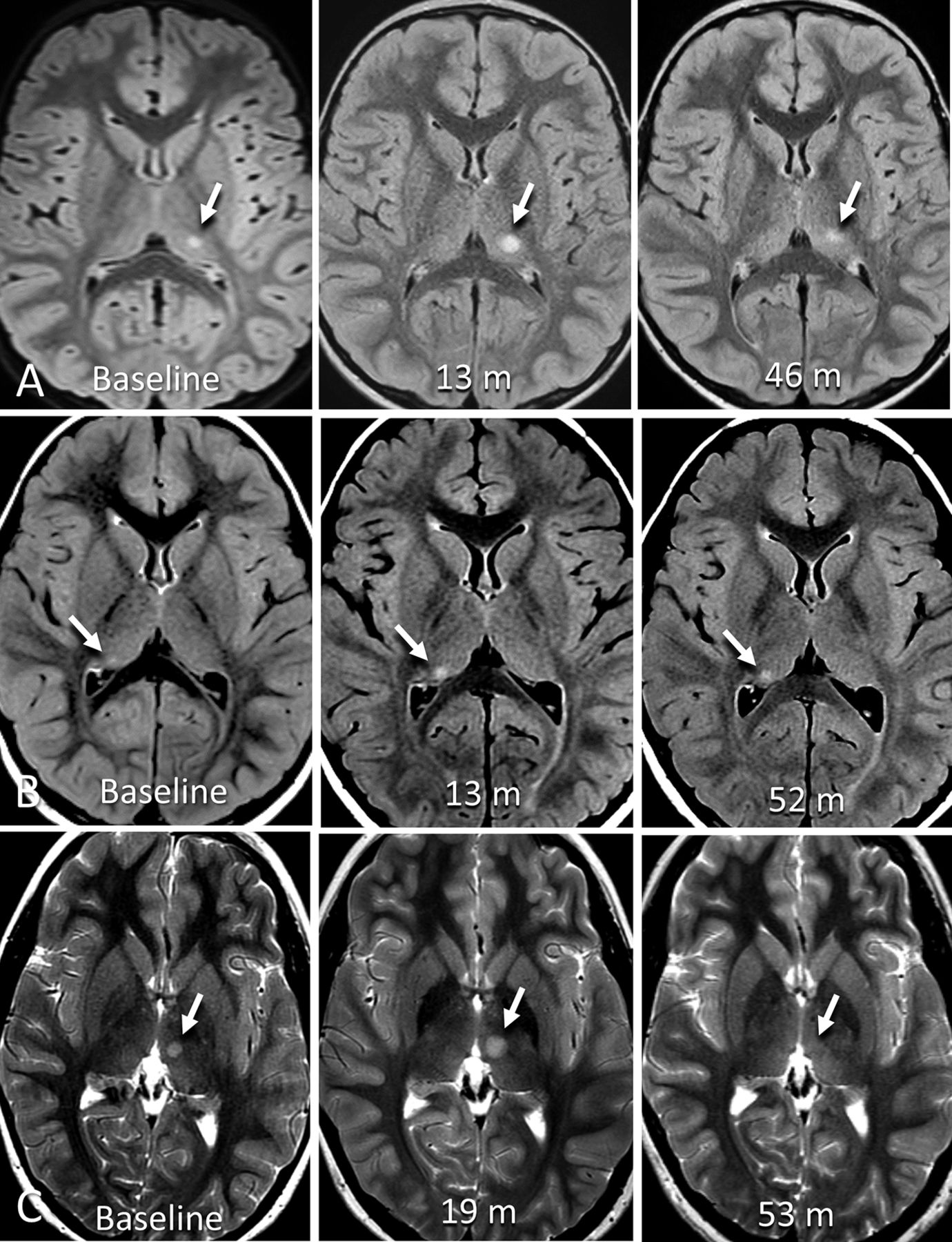
  • FIG 3.
    • Download figure
    • Open in new tab
    • Download powerpoint
    FIG 3.

    Example cases of enlarging incidental thalamic lesions identified in this study. A, Axial T2 FLAIR images of a 6-year-old boy with headache show a focal thalamic lesion (arrow) within the posterior left thalamus on baseline MR imaging. At 13 month follow-up, the lesion was enlarged. At 46-month follow-up, the lesion was more ill-defined and slightly decreased posteriorly. B, Axial T2 FLAIR images part of routine follow-up of a 4-year-old boy with history of right cerebellar complex developmental venous anomaly. Baseline MR imaging shows a small focus of increased signal in the posterior right thalamus, enlarged at 13-month follow-up, then stable 52 months after baseline MRI. C, Axial T2-weighted images of an 8-year-old girl with history of head trauma and headache showing a focal lesion within the left medial thalamus (7 × 5 mm). There was slow interval enlargement over 5 MR imaging studies for 19 months, at which point the lesion was classified as presumed low-grade glioma, and was treated with a total of 50.4 Gy fractionated radiation therapy over 8 weeks. Following therapy, there was a decrease in the site over subsequent 34 months.
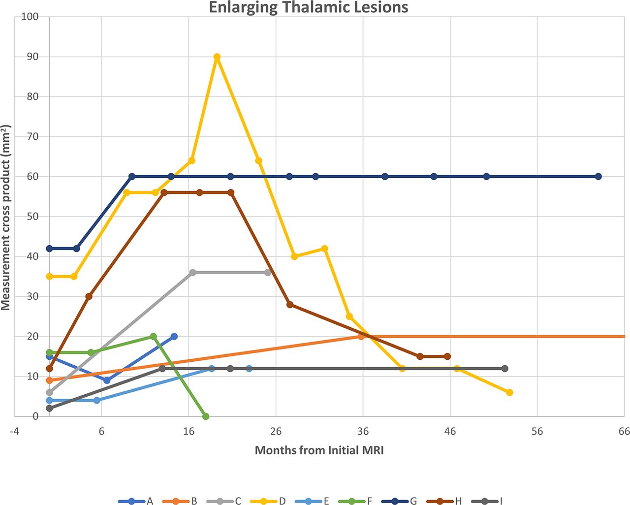
  • FIG 4.
    • Download figure
    • Open in new tab
    • Download powerpoint
    FIG 4.

    Growth trajectories of 9 thalamic lesions that enlarged at any time during follow-up MR imaging. Patient D, an 8-year-old girl with history of head trauma and headache, was treated with radiation therapy 19 months after initial lesion identification after growth identified with a subsequent decrease in size. Patient B, a 14-year-old adolescent boy with history of fetal alcohol syndrome and tethered cord, had additional stable follow-up examinations at 138 and 190 months (not shown).

Tables

  • Figures
    • View popup
    Table 1:

    Clinical scenario for baseline MR imaging examinations (n = 171)

    MR Imaging Examination Indicationn%
    Migraine headache5331
    Seizures/epilepsy4023.4
    NOS headache2011.7
    Othera1810.5
    Behavioral/developmentalb137.6
    Head injury84.7
    Movement disorder52.9
    Ophthalmologic42.3
    Psychiatric42.3
    Hearing loss31.8
    Idiopathic intracranial hypertension31.8
    • Note:— NOS indicates not otherwise specified.

    • ↵a Includes precocious puberty, hypopituitarism, central sleep apnea or congenital central hypoventilation, focal neurologic findings, Fanconi anemia, lymphoma, Li-Fraumeni syndrome screening, unspecified neck pain, Chiari syndrome, syncope, prior stroke, and vertigo.

    • ↵b Includes attention deficit/hyperactivity disorder, autism spectrum disorder, fine and gross motor delay, intermittent explosive disorder, and language delay.

    • View popup
    Table 2:

    Thalamic lesion signal characteristics on baseline MR imaging (n = 180 lesions)

    SignalPDT1T2T2 FLAIRDWIADCGRE/SWI
    Hypointense0.026.50.00.01.80.00.0
    Isointense42.816.021.019.019.81.816.5
    Hyperintense52.41.777.379.911.153.155.3
    Not visible9.555.81.11.168.546.329.8
    • View popup
    Table 3:

    MR imaging follow-up recommendations available on baseline imaging reports (n = 171 patients)a

    Noneb1 Monthc2 Months3 Months4 Months6 Months9 Months12 MonthsNOSd
    347762441376
    • Note:— NOS indicates not otherwise specified.

    • ↵a Values are absolute numbers.

    • ↵b Study report did not provide recommendations for follow-up imaging.

    • ↵c In the case an interval range of follow-up was given, the smallest interval was considered.

    • ↵d Imaging follow-up was recommended but no specified time interval.

PreviousNext
Back to top

In this issue

American Journal of Neuroradiology: 45 (2)
American Journal of Neuroradiology
Vol. 45, Issue 2
1 Feb 2024
  • Table of Contents
  • Index by author
  • Complete Issue (PDF)
Advertisement
Print
Download PDF
Email Article

Thank you for your interest in spreading the word on American Journal of Neuroradiology.

NOTE: We only request your email address so that the person you are recommending the page to knows that you wanted them to see it, and that it is not junk mail. We do not capture any email address.

Enter multiple addresses on separate lines or separate them with commas.
Incidental Thalamic Lesions Identified on Brain MRI in Pediatric and Young Adult Patients: Imaging Features and Natural History
(Your Name) has sent you a message from American Journal of Neuroradiology
(Your Name) thought you would like to see the American Journal of Neuroradiology web site.
CAPTCHA
This question is for testing whether or not you are a human visitor and to prevent automated spam submissions.
Cite this article
Vinicius de Padua V. Alves, Marguerite M. Care, James L. Leach
Incidental Thalamic Lesions Identified on Brain MRI in Pediatric and Young Adult Patients: Imaging Features and Natural History
American Journal of Neuroradiology Feb 2024, 45 (2) 211-217; DOI: 10.3174/ajnr.A8090

Citation Manager Formats

  • BibTeX
  • Bookends
  • EasyBib
  • EndNote (tagged)
  • EndNote 8 (xml)
  • Medlars
  • Mendeley
  • Papers
  • RefWorks Tagged
  • Ref Manager
  • RIS
  • Zotero
0 Responses
Respond to this article
Share
Bookmark this article
Incidental Thalamic Lesions on MRI in the Young
Vinicius de Padua V. Alves, Marguerite M. Care, James L. Leach
American Journal of Neuroradiology Feb 2024, 45 (2) 211-217; DOI: 10.3174/ajnr.A8090
del.icio.us logo Twitter logo Facebook logo Mendeley logo
  • Tweet Widget
  • Facebook Like
  • Google Plus One
Purchase

Jump to section

  • Article
    • Abstract
    • ABBREVIATIONS:
    • MATERIALS AND METHODS
    • RESULTS
    • DISCUSSION
    • CONCLUSIONS
    • Footnotes
    • REFERENCES
  • Figures & Data
  • Info & Metrics
  • Responses
  • References
  • PDF

Related Articles

  • No related articles found.
  • PubMed
  • Google Scholar

Cited By...

  • No citing articles found.
  • Crossref
  • Google Scholar

This article has not yet been cited by articles in journals that are participating in Crossref Cited-by Linking.

More in this TOC Section

  • Clinical SVR of Fetal Brain MRI
  • FRACTURE MR in Congenital Vertebral Anomalies
  • Comparing MRI Perfusion in Pediatric Brain Tumors
Show more Pediatric Neuroimaging

Similar Articles

Advertisement

Indexed Content

  • Current Issue
  • Accepted Manuscripts
  • Article Preview
  • Past Issues
  • Editorials
  • Editor's Choice
  • Fellows' Journal Club
  • Letters to the Editor
  • Video Articles

Cases

  • Case Collection
  • Archive - Case of the Week
  • Archive - Case of the Month
  • Archive - Classic Case

More from AJNR

  • Trainee Corner
  • Imaging Protocols
  • MRI Safety Corner
  • Book Reviews

Multimedia

  • AJNR Podcasts
  • AJNR Scantastics

Resources

  • Turnaround Time
  • Submit a Manuscript
  • Submit a Video Article
  • Submit an eLetter to the Editor/Response
  • Manuscript Submission Guidelines
  • Statistical Tips
  • Fast Publishing of Accepted Manuscripts
  • Graphical Abstract Preparation
  • Imaging Protocol Submission
  • Evidence-Based Medicine Level Guide
  • Publishing Checklists
  • Author Policies
  • Become a Reviewer/Academy of Reviewers
  • News and Updates

About Us

  • About AJNR
  • Editorial Board
  • Editorial Board Alumni
  • Alerts
  • Permissions
  • Not an AJNR Subscriber? Join Now
  • Advertise with Us
  • Librarian Resources
  • Feedback
  • Terms and Conditions
  • AJNR Editorial Board Alumni

American Society of Neuroradiology

  • Not an ASNR Member? Join Now

© 2025 by the American Society of Neuroradiology All rights, including for text and data mining, AI training, and similar technologies, are reserved.
Print ISSN: 0195-6108 Online ISSN: 1936-959X

Powered by HighWire