
Article Figures & Data
Figures
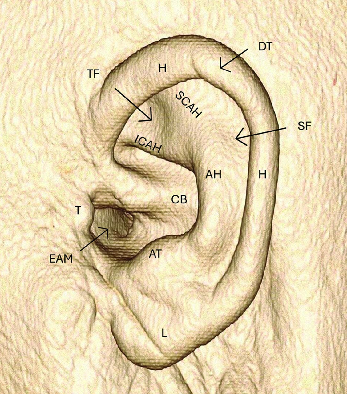
- FIG 1.
3D VRT demonstrating the external ear anatomy. AH indicates antihelix; AT, antitragus; CB, concha bowl; DT, Darwin’s tubercle; EAM, external auditory meatus; H, helix; ICAH, inferior crus antihelix; L, lobule; SCAH, superior crus antihelix; SF, scaphoid fossa; T, tragus; TF, triangular fossa; VRT, volume-rendered technique.
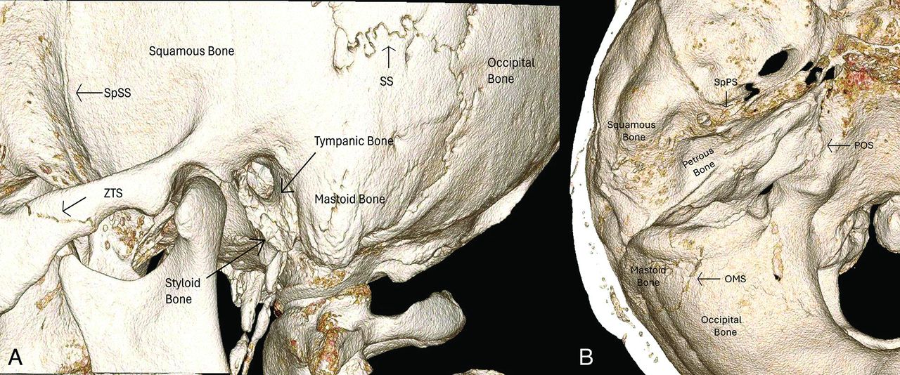
- FIG 2.
3D VRT image in lateral (A) and superior (B) projection. OMS indicates occipital mastoid suture; POS, petro-occipital suture; SpPS, sphenopetrosal suture; SpSS, sphenosquamosal suture; SS, squamosal suture; VRT, volume-rendered technique; ZTS, zygomaticotemporal suture.
- FIG 3.
Coronal (A) and sagittal (B) images of the EAC. CEAC indicates cartilaginous external auditory canal; GF, glenoid fossa; FT, foramen tympanicum; MC, mandibular condyle; OEAC, osseous external auditory canal; PF, pars flaccida; PT, pars tensa; TA, tympanic annulus; TS, tympanic sulcus.
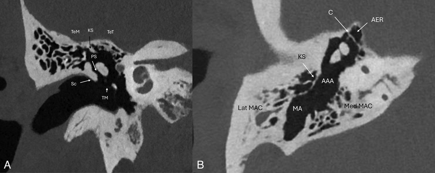
- FIG 4.
Coronal (A) and axial (B) PCT images demonstrating the anatomy of the epitympanum: AAA indicates aditus ad antrum; AER, anterior epitympanic recess; C, cog; KS, Koerner septum; Lat MAC, lateral mastoid air cells; MA, mastoid antrum; Med MAC, medial mastoid air cells; PS, Prussak space; Sc, scutum; TeM, tegmen mastoideum; TeT, tegmen tympani; TM, tympanic membrane.
- FIG 5.
Axial (A) and oblique (B) PCT images of the hypotympanum. ET indicates Eustachian tube; FS, foramen spinosum; ICA, internal carotid artery; MC, mandibular condyle; TTM, tensor tympani muscle.
- FIG 6.
Axial PCT images (A and B) of the mesotympanum and retrotympanic structures. BTC indicates basal turn cochlea; CP, cochlear promontory; FN, facial nerve mastoid segment; RWR, round window recess; SM, stapedius muscle; ST, sinus tympani; Sub, subiculum; PE, pyramidal eminence; Pont, ponticulus.
- FIG 7.
Coronal oblique (A and C) and axial oblique (B and D) images of the malleus. AML indicates anterior malleal ligament; CFP, cochleariform process; H, head of malleus; I, incus; LML, lateral malleal ligament; LP, lateral process; M, manubrium attaching to the tympanic membrane; Ma, manubrium; N, neck of malleus; SML, superior malleal ligament; TTM, tensor tympani muscle; TTT, tensor tympani tendon.
- FIG 8.
Axial oblique (A), sagittal oblique (B and E), axial (C), and coronal oblique (D and F) PCT images of the middle ear cavity with anatomic details of the ossicular chain. Sagittal oblique image (E) depicts the “molar tooth” configuration of the malleus manubrium and long process of the incus in parallel. Axial oblique and axial images of the incudomalleolar joint depicts the classic “ice cream cone” configuration. APM indicates anterior process of malleus; B, body of incus; FN, facial nerve tympanic segment; H, head of malleus; IF, incudal fossa; LeP, lenticular process of incus; LP, long process of incus; LPIL, lateral band of posterior incudal ligament; MPIL, medial band of posterior incudal ligament; OW, oval window; SP, short process of incus; St, stapes.
- FIG 9.
Axial and axial oblique PCT images of the stapes. A, LeP indicates lenticular process of incus; SHN, stapes head and neck; StT, stapedius tendon. B, AC, anterior crus of stapes; PC, posterior crus of stapes; SC, stapes capitulum (head); SN, stapes neck; StT, stapedius tendon.


Jump to section
Related Articles
Cited By...
- No citing articles found.