
Article Figures & Data
Figures
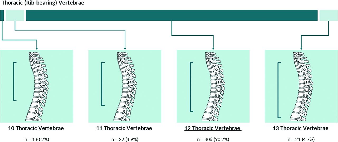
- FIG 1.
Allocation of spinal segment distribution variants in thoracic (rib-bearing) vertebrae.
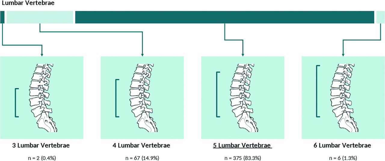
- FIG 2.
Allocation of spinal segment distribution variants in lumbar vertebrae.
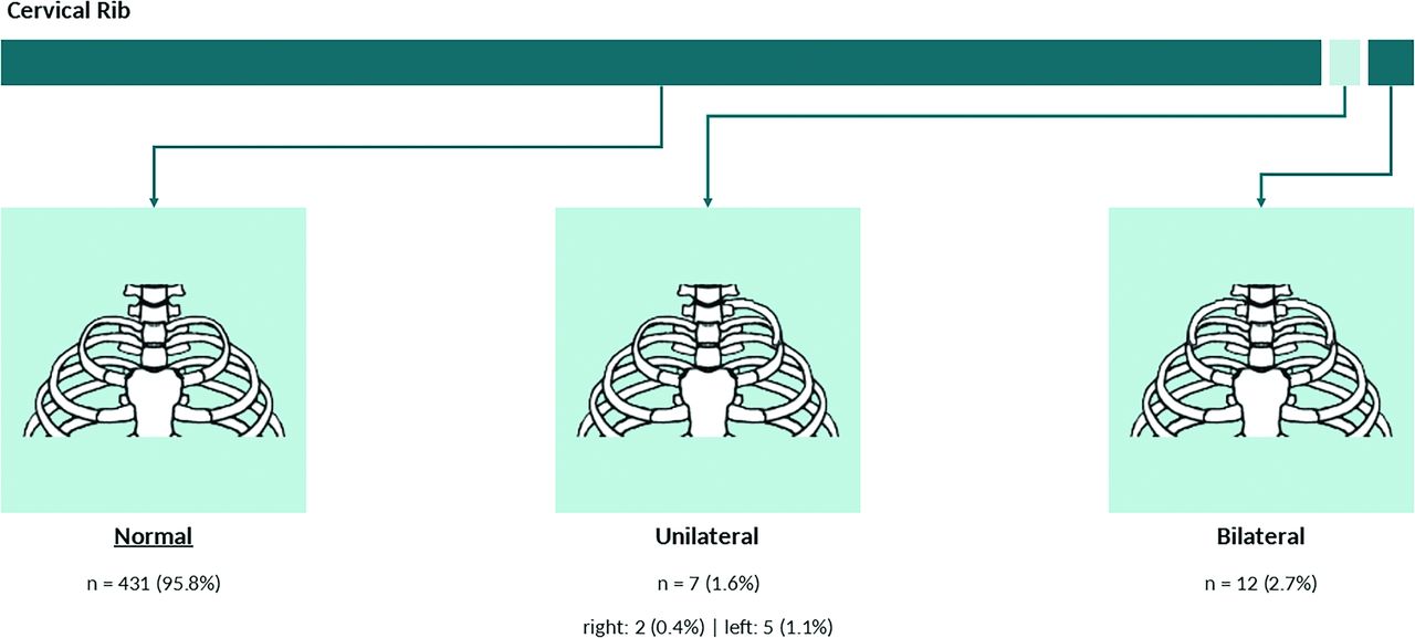
- FIG 3.
Frequency and distribution of the cervical rib.
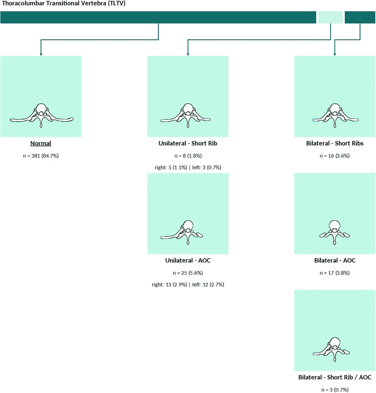
- FIG 4.
Frequency and distribution of the TLTV. AOC indicates accessory ossification center.
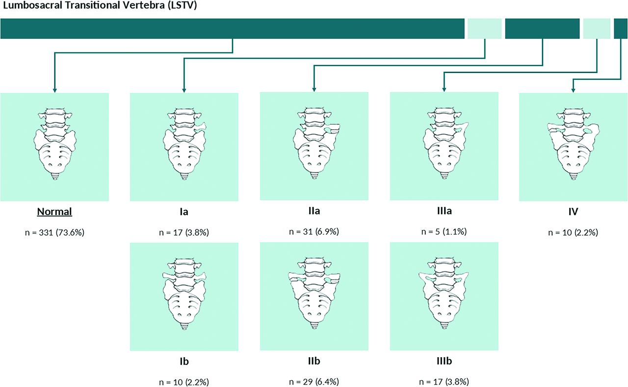
- FIG 5.
Frequency and distribution of the LSTV.
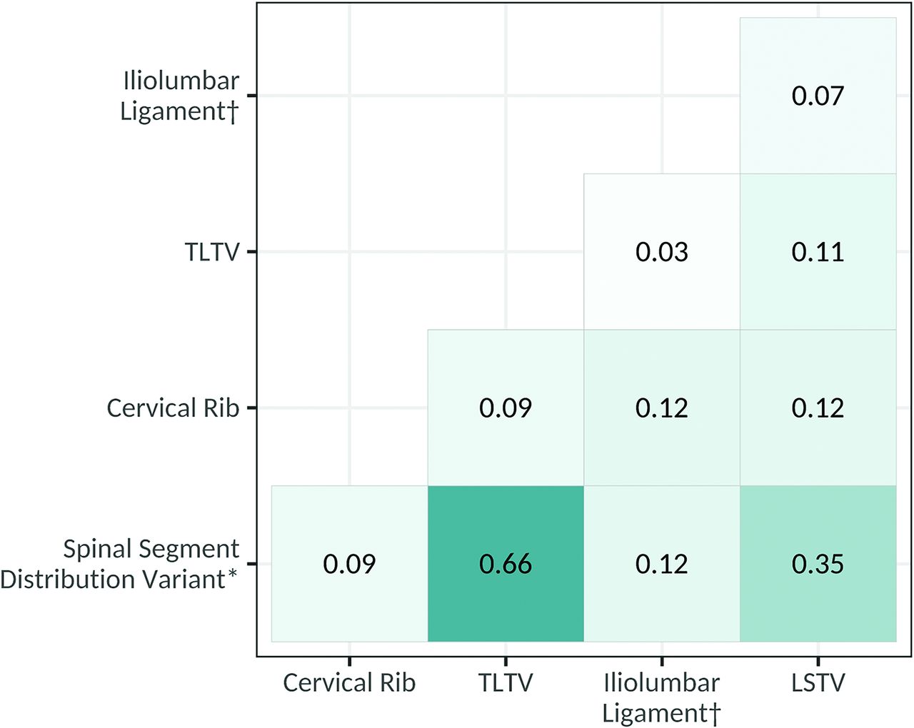
- FIG 6.
Correlation between spinal segment variants. The asterisk indicates an atypical number of presacral segments or an atypical distribution of thoracolumbar vertebrae; dagger, the inability to visualize the ILL.
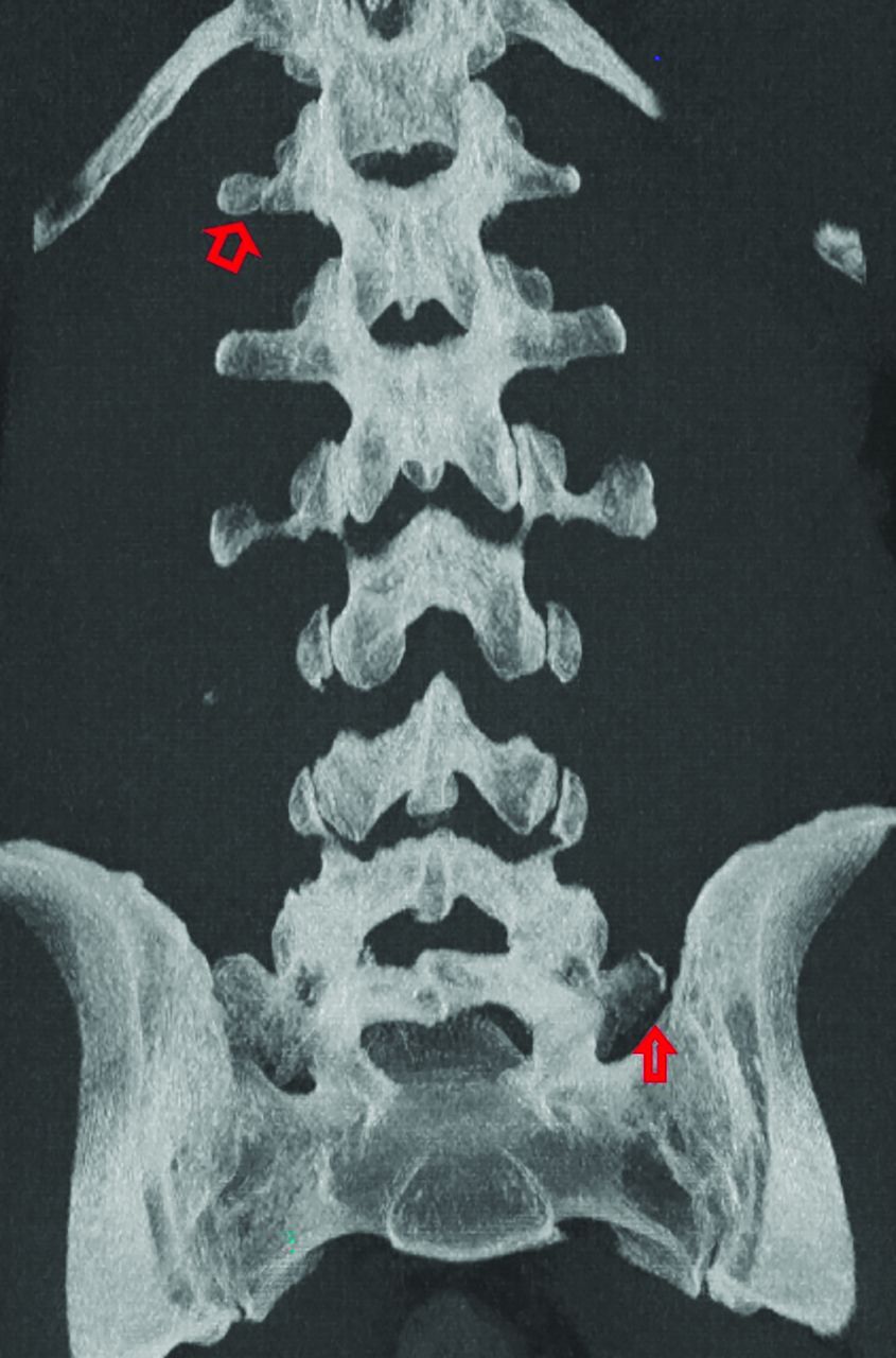
- FIG 7.
Example of thoracolumbar and lumbosacral transitional anatomy. Coronal MIP CT image of the lumbar spine demonstrates thoracolumbar and lumbosacral anatomy with an accessory ossification center on the right at T12 (open arrow) as well as a partially sacralized L5 vertebral body with an unfused left transverse process (closed arrow).


Tables
Classification Laterality Description I Unilateral (a) Bilateral (b) Dysplastic transverse process with a ≥19-mm width on the craniocaudal dimension II Lumbarization or sacralization with an enlarged transverse process that has a diarthrodial joint with the sacrum III Lumbarization or sacralization with complete osseous fusion of the transverse process to the sacrum IV – Unilateral IIa transition with a IIIa transition on the contralateral side Note:—The en dash indicates duplicate entries.
Cervical Rib TLTV ILLa LSTV OR (95% CI), P Value OR (95% CI), P Value OR (95% CI), P Value OR (95% CI), P Value ILLa – – – 2.10 (0.79–5.31), .121 TLTV – – 1.50 (0.42–4.29), .482 1.87 (1.08–3.20), .023 Cervical rib – 2.70 (0.92–7.09), .053 4.86 (1.06–16.54), .020 3.28 (1.29–8.47), .012 Spinal segment distribution variantb 2.44 (0.92–6.19), .062 66.13 (30.34–166.85), <.001 3.06 (1.18–7.80), .018 5.47 (3.43–8.80), <.001


