
This article requires a subscription to view the full text. If you have a subscription you may use the login form below to view the article. Access to this article can also be purchased.
Graphical Abstract

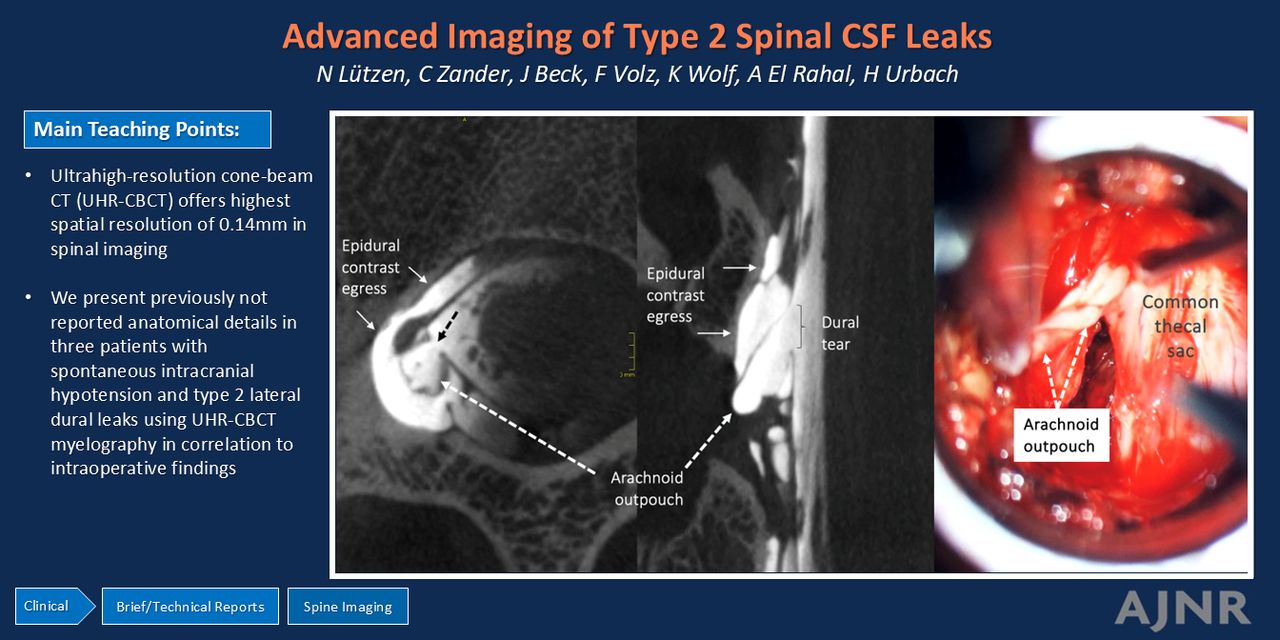
SUMMARY:
Type 2 CSF leaks are spinal lateral dural tears, causing spontaneous intracranial hypotension. They may be visualized with digital subtraction myelography, conebeam CT myelography, and energy-integrating detector or photon-counting CT myelography. A recently introduced ultra-high-resolution conebeam CT myelography has shown beneficial visualization of CSF-venous fistulas, another cause of spontaneous intracranial hypotension. However, the use of this technique has not yet been reported in imaging of type 2 leaks. In this technical report, we describe our first experiences and highlight the advantages of ultra-high-resolution conebeam CT for visualizing type 2 leaks in spontaneous intracranial hypotension.
ABBREVIATIONS:
- CBCT
- conebeam CT
- DSM
- digital subtraction myelography
- SIH
- spontaneous intracranial hypotension
- UHR-CBCT
- ultra-high-resolution conebeam CT
Footnotes
N. Lützen and C. Zander contributed equally to this work.
Disclosure forms provided by the authors are available with the full text and PDF of this article at www.ajnr.org.
- © 2025 by American Journal of Neuroradiology


