Advertisement

Main menu

  • Home
  • Content
    • Current Issue
    • Accepted Manuscripts
    • Article Preview
    • Past Issue Archive
    • Video Articles
    • AJNR Case Collection
    • Case of the Week Archive
    • Case of the Month Archive
    • Classic Case Archive
  • Special Collections
    • AJNR Awards
    • Low-Field MRI
    • Alzheimer Disease
    • ASNR Foundation Special Collection
    • Photon-Counting CT
    • View All
  • Multimedia
    • AJNR Podcasts
    • AJNR SCANtastic
    • Trainee Corner
    • MRI Safety Corner
    • Imaging Protocols
  • For Authors
    • Submit a Manuscript
    • Submit a Video Article
    • Submit an eLetter to the Editor/Response
    • Manuscript Submission Guidelines
    • Statistical Tips
    • Fast Publishing of Accepted Manuscripts
    • Graphical Abstract Preparation
    • Imaging Protocol Submission
    • Author Policies
  • About Us
    • About AJNR
    • Editorial Board
    • Editorial Board Alumni
  • More
    • Become a Reviewer/Academy of Reviewers
    • Subscribers
    • Permissions
    • Alerts
    • Feedback
    • Advertisers
    • ASNR Home

User menu

  • Alerts
  • Log in

Search

  • Advanced search
American Journal of Neuroradiology

American Journal of Neuroradiology

ASHNR American Society of Functional Neuroradiology ASHNR American Society of Pediatric Neuroradiology ASSR
  • Alerts
  • Log in

Advanced Search

  • Home
  • Content
    • Current Issue
    • Accepted Manuscripts
    • Article Preview
    • Past Issue Archive
    • Video Articles
    • AJNR Case Collection
    • Case of the Week Archive
    • Case of the Month Archive
    • Classic Case Archive
  • Special Collections
    • AJNR Awards
    • Low-Field MRI
    • Alzheimer Disease
    • ASNR Foundation Special Collection
    • Photon-Counting CT
    • View All
  • Multimedia
    • AJNR Podcasts
    • AJNR SCANtastic
    • Trainee Corner
    • MRI Safety Corner
    • Imaging Protocols
  • For Authors
    • Submit a Manuscript
    • Submit a Video Article
    • Submit an eLetter to the Editor/Response
    • Manuscript Submission Guidelines
    • Statistical Tips
    • Fast Publishing of Accepted Manuscripts
    • Graphical Abstract Preparation
    • Imaging Protocol Submission
    • Author Policies
  • About Us
    • About AJNR
    • Editorial Board
    • Editorial Board Alumni
  • More
    • Become a Reviewer/Academy of Reviewers
    • Subscribers
    • Permissions
    • Alerts
    • Feedback
    • Advertisers
    • ASNR Home
  • Follow AJNR on Twitter
  • Visit AJNR on Facebook
  • Follow AJNR on Instagram
  • Join AJNR on LinkedIn
  • RSS Feeds

AJNR Awards, New Junior Editors, and more. Read the latest AJNR updates

Research ArticleFunctional

Connectomic Basis for Tremor Control in Stereotactic Radiosurgical Thalamotomy

E.H. Middlebrooks, R.A. Popple, E. Greco, L. Okromelidze, H.C. Walker, D.A. Lakhani, A.R. Anderson, E.M. Thomas, H.D. Deshpande, B.A. McCullough, N.P. Stover, V.W. Sung, A.P. Nicholas, D.G. Standaert, T. Yacoubian, M.N. Dean, J.A. Roper, S.S. Grewal, M.T. Holland, J.N. Bentley, B.L. Guthrie and M. Bredel
American Journal of Neuroradiology February 2023, 44 (2) 157-164; DOI: https://doi.org/10.3174/ajnr.A7778
E.H. Middlebrooks
aFrom the Departments of Radiology (E.H.M., E.G., L.O., D.A.L.)
bNeurosurgery (E.H.M., S.S.G.), Mayo Clinic, Jacksonville, Florida
  • Find this author on Google Scholar
  • Find this author on PubMed
  • Search for this author on this site
  • ORCID record for E.H. Middlebrooks
R.A. Popple
cDepartments of Radiation Oncology (R.A.P., A.R.A., E.M.T., M.B.)
  • Find this author on Google Scholar
  • Find this author on PubMed
  • Search for this author on this site
  • ORCID record for R.A. Popple
E. Greco
aFrom the Departments of Radiology (E.H.M., E.G., L.O., D.A.L.)
  • Find this author on Google Scholar
  • Find this author on PubMed
  • Search for this author on this site
  • ORCID record for E. Greco
L. Okromelidze
aFrom the Departments of Radiology (E.H.M., E.G., L.O., D.A.L.)
  • Find this author on Google Scholar
  • Find this author on PubMed
  • Search for this author on this site
  • ORCID record for L. Okromelidze
H.C. Walker
dNeurology (H.C.W., B.A.M., N.P.S., V.W.S., A.P.N., D.G.S., T.Y., M.N.D.)
  • Find this author on Google Scholar
  • Find this author on PubMed
  • Search for this author on this site
  • ORCID record for H.C. Walker
D.A. Lakhani
aFrom the Departments of Radiology (E.H.M., E.G., L.O., D.A.L.)
gDepartment of Radiology (D.A.L.), West Virginia University, Morgantown, West Virginia
  • Find this author on Google Scholar
  • Find this author on PubMed
  • Search for this author on this site
  • ORCID record for D.A. Lakhani
A.R. Anderson
cDepartments of Radiation Oncology (R.A.P., A.R.A., E.M.T., M.B.)
  • Find this author on Google Scholar
  • Find this author on PubMed
  • Search for this author on this site
  • ORCID record for A.R. Anderson
E.M. Thomas
cDepartments of Radiation Oncology (R.A.P., A.R.A., E.M.T., M.B.)
hDepartment of Radiation Oncology (E.M.T.), Ohio State University, Columbus, Ohio
  • Find this author on Google Scholar
  • Find this author on PubMed
  • Search for this author on this site
H.D. Deshpande
eRadiology (H.D.D.)
  • Find this author on Google Scholar
  • Find this author on PubMed
  • Search for this author on this site
  • ORCID record for H.D. Deshpande
B.A. McCullough
dNeurology (H.C.W., B.A.M., N.P.S., V.W.S., A.P.N., D.G.S., T.Y., M.N.D.)
  • Find this author on Google Scholar
  • Find this author on PubMed
  • Search for this author on this site
N.P. Stover
dNeurology (H.C.W., B.A.M., N.P.S., V.W.S., A.P.N., D.G.S., T.Y., M.N.D.)
  • Find this author on Google Scholar
  • Find this author on PubMed
  • Search for this author on this site
  • ORCID record for N.P. Stover
V.W. Sung
dNeurology (H.C.W., B.A.M., N.P.S., V.W.S., A.P.N., D.G.S., T.Y., M.N.D.)
  • Find this author on Google Scholar
  • Find this author on PubMed
  • Search for this author on this site
  • ORCID record for V.W. Sung
A.P. Nicholas
dNeurology (H.C.W., B.A.M., N.P.S., V.W.S., A.P.N., D.G.S., T.Y., M.N.D.)
  • Find this author on Google Scholar
  • Find this author on PubMed
  • Search for this author on this site
D.G. Standaert
dNeurology (H.C.W., B.A.M., N.P.S., V.W.S., A.P.N., D.G.S., T.Y., M.N.D.)
  • Find this author on Google Scholar
  • Find this author on PubMed
  • Search for this author on this site
  • ORCID record for D.G. Standaert
T. Yacoubian
dNeurology (H.C.W., B.A.M., N.P.S., V.W.S., A.P.N., D.G.S., T.Y., M.N.D.)
  • Find this author on Google Scholar
  • Find this author on PubMed
  • Search for this author on this site
  • ORCID record for T. Yacoubian
M.N. Dean
dNeurology (H.C.W., B.A.M., N.P.S., V.W.S., A.P.N., D.G.S., T.Y., M.N.D.)
  • Find this author on Google Scholar
  • Find this author on PubMed
  • Search for this author on this site
  • ORCID record for M.N. Dean
J.A. Roper
iSchool of Kinesiology (J.A.R.), Auburn University, Auburn, Alabama
  • Find this author on Google Scholar
  • Find this author on PubMed
  • Search for this author on this site
  • ORCID record for J.A. Roper
S.S. Grewal
bNeurosurgery (E.H.M., S.S.G.), Mayo Clinic, Jacksonville, Florida
  • Find this author on Google Scholar
  • Find this author on PubMed
  • Search for this author on this site
  • ORCID record for S.S. Grewal
M.T. Holland
fNeurosurgery (M.T.H., J.N.B., B.L.G.), University of Alabama at Birmingham, Birmingham, Alabama
  • Find this author on Google Scholar
  • Find this author on PubMed
  • Search for this author on this site
  • ORCID record for M.T. Holland
J.N. Bentley
fNeurosurgery (M.T.H., J.N.B., B.L.G.), University of Alabama at Birmingham, Birmingham, Alabama
  • Find this author on Google Scholar
  • Find this author on PubMed
  • Search for this author on this site
  • ORCID record for J.N. Bentley
B.L. Guthrie
fNeurosurgery (M.T.H., J.N.B., B.L.G.), University of Alabama at Birmingham, Birmingham, Alabama
  • Find this author on Google Scholar
  • Find this author on PubMed
  • Search for this author on this site
  • ORCID record for B.L. Guthrie
M. Bredel
cDepartments of Radiation Oncology (R.A.P., A.R.A., E.M.T., M.B.)
  • Find this author on Google Scholar
  • Find this author on PubMed
  • Search for this author on this site
  • ORCID record for M. Bredel
  • Article
  • Figures & Data
  • Supplemental
  • Info & Metrics
  • Responses
  • References
  • PDF
Loading

Abstract

BACKGROUND AND PURPOSE: Given the increased use of stereotactic radiosurgical thalamotomy and other ablative therapies for tremor, new biomarkers are needed to improve outcomes. Using resting-state fMRI and MR tractography, we hypothesized that a “connectome fingerprint” can predict tremor outcomes and potentially serve as a targeting biomarker for stereotactic radiosurgical thalamotomy.

MATERIALS AND METHODS: We evaluated 27 patients who underwent unilateral stereotactic radiosurgical thalamotomy for essential tremor or tremor-predominant Parkinson disease. Percentage postoperative improvement in the contralateral limb Fahn‐Tolosa‐Marin Clinical Tremor Rating Scale (TRS) was the primary end point. Connectome-style resting-state fMRI and MR tractography were performed before stereotactic radiosurgery. Using the final lesion volume as a seed, “connectivity fingerprints” representing ideal connectivity maps were generated as whole-brain R-maps using a voxelwise nonparametric Spearman correlation. A leave-one-out cross-validation was performed using the generated R-maps.

RESULTS: The mean improvement in the contralateral tremor score was 55.1% (SD, 38.9%) at a mean follow-up of 10.0 (SD, 5.0) months. Structural connectivity correlated with contralateral TRS improvement (r = 0.52; P  = .006) and explained 27.0% of the variance in outcome. Functional connectivity correlated with contralateral TRS improvement (r = 0.50; P  = .008) and explained 25.0% of the variance in outcome. Nodes most correlated with tremor improvement corresponded to areas of known network dysfunction in tremor, including the cerebello-thalamo-cortical pathway and the primary and extrastriate visual cortices.

CONCLUSIONS: Stereotactic radiosurgical targets with a distinct connectivity profile predict improvement in tremor after treatment. Such connectomic fingerprints show promise for developing patient-specific biomarkers to guide therapy with stereotactic radiosurgical thalamotomy.

ABBREVIATIONS:

BOLD
blood oxygen level–dependent MRI
DBS
deep brain stimulation
DRTT
dentato-rubro-thalamic tract
SMS
simultaneous multislice
SRS
stereotactic radiosurgery
TRS
Fahn‐Tolosa‐Marin Clinical Tremor Rating Scale
VIM
ventral intermediate nucleus

Tremor is a debilitating neurologic condition that is seen with multiple disorders, most commonly in essential tremor and Parkinson’s disease.1 Patients who are refractory to pharmacologic therapies are potentially candidates for surgical intervention. Deep brain stimulation (DBS) is the current criterion standard surgical treatment; however, not all patients are candidates or wish to undergo DBS. Recently, there has been a resurgence in ablative therapies using incisionless ablative techniques such as stereotactic radiosurgery (SRS) and MR imaging–guided focused ultrasound. While ablative treatment mechanisms might overlap with DBS, little is known on the connectomics of SRS and tremor improvement.

In contrast to traditional “localizationist” models of the brain, a more recent shift to a network, or “connectomic,” model has greatly enhanced our understanding of brain function and pathology.2 Such a shift has also occurred in the realm of neuromodulation with recognition of many disorders as “circuitopathies”3 and the need for targeted network surgery, or “connectomic surgery.”4 The connectomic model, using MR tractography as a measure of structural connectivity and resting-state fMRI as a measure of functional connectivity, has been previously applied to better understand treatment effects in DBS surgery.5⇓⇓⇓⇓⇓⇓⇓⇓⇓⇓⇓⇓⇓⇓⇓⇓-22 In treatment of tremor, this has led to the consolidation of the role of the cerebello-thalamo-cortical sensorimotor network in explaining many previously reported “sweet spots” in the DBS literature.5⇓⇓⇓⇓⇓⇓⇓⇓-14

To date, there has been limited application of the connectomic model to SRS in tremor. Using a combination of resting-state functional MR imaging and MR tractography, we explored whether the therapeutic network for SRS in treatment of tremor displays a “connectome fingerprint” that correlates with tremor improvement. Identification of such a network may provide further insight into the mechanism of tremor improvement and guide targeting in SRS. We also hypothesized that the similarity of any patient's individual connectivity map to the connectome fingerprint of tremor improvement will predict his or her measured tremor improvement, which may establish feasibility for the potential of connectivity fingerprints to guide SRS targeting.

MATERIALS AND METHODS

This prospective clinical trial was approved by the University of Alabama at Birmingham institutional review board. All patients gave verbal and written consent. The trial was registered at https://www.clinicaltrials.gov/ under trial NCT03305588. Subjects were recruited in collaboration with the University of Alabama at Birmingham Neurology, Neurosurgery, Movement Disorders, and Radiation Oncology Departments. Patients with medically refractory essential tremor or tremor-dominant Parkinson disease older than 18 years of age with Eastern Cooperative Oncology Group Performance Status of ≤2 were included. Patients were not candidates for DBS on the basis of either medical/surgical comorbidities or by their own choice. Patients were ineligible if they had prior brain radiosurgery or therapeutic brain radiation therapy or if there was a contraindication to MR imaging.

Imaging Protocol

Before thalamotomy, patients were scanned on a 3T Magnetom Prisma (Siemens) scanner with a high-performance, 80 mT/m gradient system and a 64-channel head/neck coil. Patients’ heads were tightly packed in the head coil to minimize motion.

Diffusion imaging was obtained with a spin-echo EPI sequence using simultaneous multislice (SMS) excitation. A total of 64 diffusion directions were obtained with a b-value = 1500 s/mm2 plus 6 B0 volumes without diffusion-weighting. The diffusion scan was repeated with an opposing phase-encoding direction for distortion correction. Additional relevant parameters include TR = 3.3 seconds, TE = 90 ms, SMS factor = 4, in-plane acceleration = 2, phase partial Fourier = 6/8, receiver bandwidth = 1232 Hz/px, echo spacing = 0.94 ms, EPI factor = 140, in-plane resolution = 1.5 × 1.5 mm, section thickness = 1.5 mm, with scan time = 4 minutes and 21 seconds per acquisition.

A connectome-style resting-state fMRI was acquired with a multiecho blood oxygen level–dependent (BOLD) gradient-echo EPI sequence using SMS excitation. The temporal SNR of BOLD imaging is generally lower in subcortical regions due to many factors, including the distance from the receive coil and increased physiologic noise. To address this issue, we used a multiecho BOLD approach that significantly improves subcortical connectivity measures.23 Additionally, the use of a reduced TR with high spatial resolution and a long, connectome-style BOLD acquisition has all been shown to improve the statistical analysis of resting-state fMRI data. Relevant parameters include the following: TR = 1.5 seconds, echo times = 12.4, 34.3, and 56.2 ms, flip angle = 65°, SMS factor = 4, in-plane acceleration = 2, phase partial Fourier = 6/8, receiver bandwidth = 1850 Hz/px, echo spacing = 0.66 ms, EPI factor = 81, in-plane resolution = 2.0 × 2.0 mm, section thickness = 2.0 mm. The scan was repeated with opposing phase-encoding directions to account for directional bias. A total of 1180 measurements were obtained with a total scan time of 31 minutes and 28 seconds.

A T1-weighted MPRAGE sequence was used for structural registration and performed before SRS. At 3 months after SRS, 3D T1-weighted images were repeated with intravenous contrast. See the Online Supplemental Data for sequence details.

SRS Procedure

Frameless SRS was performed using a thermoplastic mask with an open face molded to the patient’s head. Plans were created using the Eclipse treatment planning system (Varian Medical Systems), and the dose was calculated using either the Analytical Anisotropic Algorithm or AcurosXB algorithm (Varian Eclipse Treatment Planning System) with grid spacing of 1 mm. Patients were treated using an Edge (Varian) linear accelerator equipped with a high-definition multileaf collimator and a 10 MV flattening-filter-free beam by using the virtual cone technique.24 Optical surface guidance was used to monitor the patient position during treatment.25 For full details of the procedure, see the Online Supplemental Data .

Clinical Testing

Before SRS, a baseline Fahn‐Tolosa‐Marin Clinical Tremor Rating Scale (TRS) was assessed. The TRS was repeated at each follow-up assessment after SRS, and the last documented TRS score was used for analysis. TRS was assessed by a single examiner who was blinded to the imaging results at the time of assessment. The primary outcome measure for the study was a percentage decrease in contralateral upper and lower body tremors, measured as the sum of the lateralized TRS scores from Part I on the body side opposite of the treatment hemisphere. Any disease-related medications were maintained at preoperative doses for follow-up.

Image Preprocessing

The postoperative postcontrast T1-weighted images were coregistered to the preoperative T1-weighted images using Advanced Normalization Tools (http://stnava.github.io/ANTs/). The final lesion location was manually segmented as the enhancing region on the 3-month postoperative postcontrast T1-weighted images.

The diffusion data underwent eddy current correction, section-to-volume movement correction, and outlier replacement in FSL “eddy_cuda” (https://git.fmrib.ox.ac.uk/fsl/conda/fsl-eddy-cuda) on a custom-built Linux workstation using 3 NVIDIA Quadro P5000 GPUs. Susceptibility-induced distortions were corrected with FSL “topup” (https://fsl.fmrib.ox.ac.uk/fsl/fslwiki/topup) using the repeat acquisitions in opposing phase-encoding directions.26 FSL “BEDPOSTX_GPU” (https://users.fmrib.ox.ac.uk/∼moisesf/Bedpostx_GPU/) was used to estimate the orientation distribution functions with 3 fiber orientations modeled per voxel. The diffusion data were coregistered to the T1 MPRAGE using a boundary-based registration implemented in FreeSurfer (http://surfer.nmr.mgh.harvard.edu). An exclusion mask was generated to include the ventricles and contralateral hemisphere white matter.

The multiecho BOLD data were initially preprocessed in AFNI, Version 22.0.06 (https://afni.nimh.nih.gov/). Preprocessing steps included realignment, spatial smoothing of 4-mm full width at half maximum, and coregistration to the T1 MPRAGE. Denoising of the data was performed with multiecho independent component analysis using TE-dependent analysis implemented in AFNI. Last, motion censoring (framewise displacement of >0.3 mm) and outlier detection were performed using default values in AFNI. Global mean signal regression and regression of motion parameters were also performed.

Connectome Generation

Probabilistic tractography was performed using “probtrackx2_gpu” (https://users.fmrib.ox.ac.uk/∼moisesf/Probtrackx_GPU/index.html) from the FMRIB Software Library, Version 6.0.3 (http://fsl.fmrib.ox.ac.uk) in each of the subjects, with 250,000 samples, a curvature threshold of 0.2, modified Euler streamlining (https://mycareerwise.com/programming/category/numerical-analysis/modified-euler-method), and a step length of 0.5 mm. A region-of-avoidance was added to exclude the internal capsule and deep white matter of the contralateral hemisphere. Because no plausible fibers or existing evidence implicates tracts connecting the thalamus with the contralateral hemisphere via the capsular fibers, such connections are most likely spurious fibers and their inclusion leads to an artificial increase in directions of freedom with resulting inflation of P values. Probabilistic tractography was performed using the segmented lesion as the seed region to generate “lesion tract maps” of all tracts passing through the lesion (Fig 1).

FIG 1.
  • Download figure
  • Open in new tab
  • Download powerpoint
FIG 1.

Pipeline for generation of a single-subject lesion connectivity map and lesion tract map (A and B). The lesion is segmented from the posttreatment MR imaging (C). The mean BOLD time-series for the lesion is extracted and correlated to all other brain voxels. D, The resulting t-score map is a patient-specific lesion connectivity map. E, The lesion is used as a seed region to generate a patient-specific lesion tract map representing the probability of all streamlines connected to the lesion.

The preprocessed BOLD resting-state fMRI data underwent a seed-based correlation analysis across the entire brain for each subject using the segmented lesion as the seed point to generate a whole-brain “lesion connectivity map” (Fig 1). The resulting t-maps were used for subsequent analysis.

The structural images, lesion connectivity maps, and lesion tract maps were then normalized into Montreal Neurological Institute template space, as described in the Online Supplemental Data. The right hemisphere lesion connectivity maps and lesion tract maps were nonlinearly flipped to the left hemisphere for comparison across the cohort.

Connectome Analysis

To determine the relationship between structural connectivity and contralateral tremor improvement, we generated an ideal tract fingerprint for the cohort. First, the individual lesion tract maps for each subject were correlated with percentage improvement in the contralateral tremor to generate a Spearman rank-correlation coefficient on a voxel-by-voxel basis. The resultant group R-map, or tract fingerprint, represents an optimal voxel-by-voxel tract map for contralateral tremor improvement.

To assess the significance of the tract fingerprint in predicting outcomes, we performed a leave-one-out cross-validation. An R-map was created by correlating tracts traversing the lesion with the contralateral TRS improvement using all patients except one, withheld for validation. The probabilistic tract map from the left-out patient was then used to calculate spatial similarity (measured via the Fisher z-transformed spatial correlation coefficient) with the tract fingerprint generated from the remainder of the cohort. The similarity index for each left-out subject was then correlated versus the measured tremor improvement using the Spearman correlation.

Next, the process was repeated using the lesion connectivity map generated from the resting-state fMRI for each individual subject to create a functional fingerprint.

Statistics

Demographic data were expressed as mean, range, and SD, as appropriate. The primary outcome measure was the percentage change in the contralateral TRS. Correlation of lesion volume and contralateral TRS improvement was assessed with a nonparametric Spearman correlation. Statistical significance was considered as P < .05.

RESULTS

Demographic and Clinical Results

Forty total patients were enrolled in the clinical trial. Three died of unrelated causes before follow-up, 1 declined treatment, 3 patients were not eligible for 3T MR imaging, and 6 patients were lost to follow-up for their imaging and/or clinical assessment, leaving 27 patients for analysis. Demographic and group outcome data are listed in the Table. Twenty-three (85.2%) patients underwent left thalamotomy, while 4 (14.8%) had right thalamotomy. Twenty patients (74.0%) had a ≥50% reduction in contralateral tremor, while 7 patients (26.0%) had  <50% reduction in contralateral tremor. The mean improvement in the contralateral tremor score was 55.1% (SD, 38.9%). All patients had at least 6 months of clinical follow-up with the mean duration of follow-up being 10.0 (SD, 5.0) months. Adverse events determined to be related to radiosurgery included 1 subject with grade I sensory deficits consisting of numbness in the left first and second digits and left-sided mouth and buccal mucosa dysesthesia occurring 10 months after radiosurgery. Two subjects experienced grade II radiation necrosis consistent with hyper-response. One of these subjects experienced right-foot drag, right-hand grip weakness, right-arm proprioceptive loss, mild expressive aphasia, bilateral lip paresthesia, and left-sided headache occurring 7 months after radiosurgery. One subject experienced falls, dysarthria, right-arm and right-leg weakness, and incoordination occurring 11 months after radiosurgery. Both subjects with grade II radionecrosis reported near-resolution of tremor at 4 months after radiosurgery. All 3 subjects with radiosurgery-related adverse events were treated with tapered corticosteroids and bevacizumab infusions. There were no grade III–V adverse events related to radiosurgery.

View this table:
  • View inline
  • View popup

Demographic and clinical outcome data

Structural Connectome Analysis

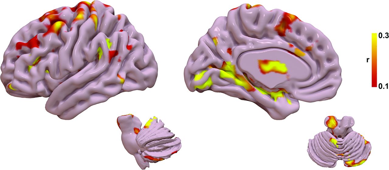
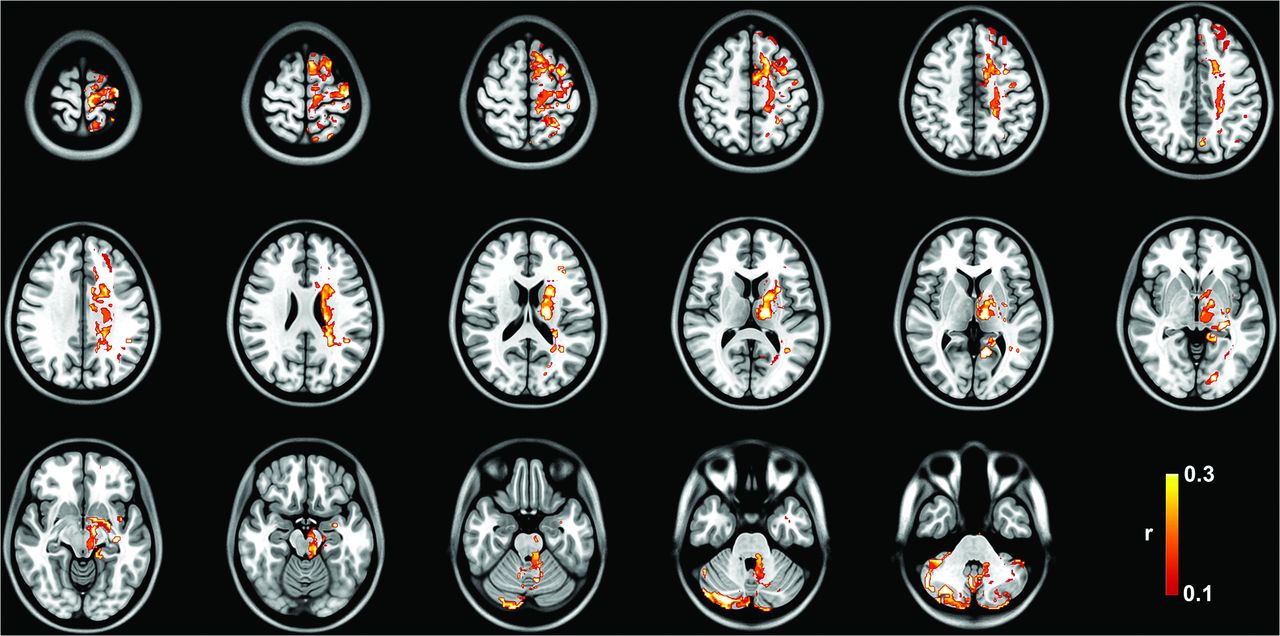
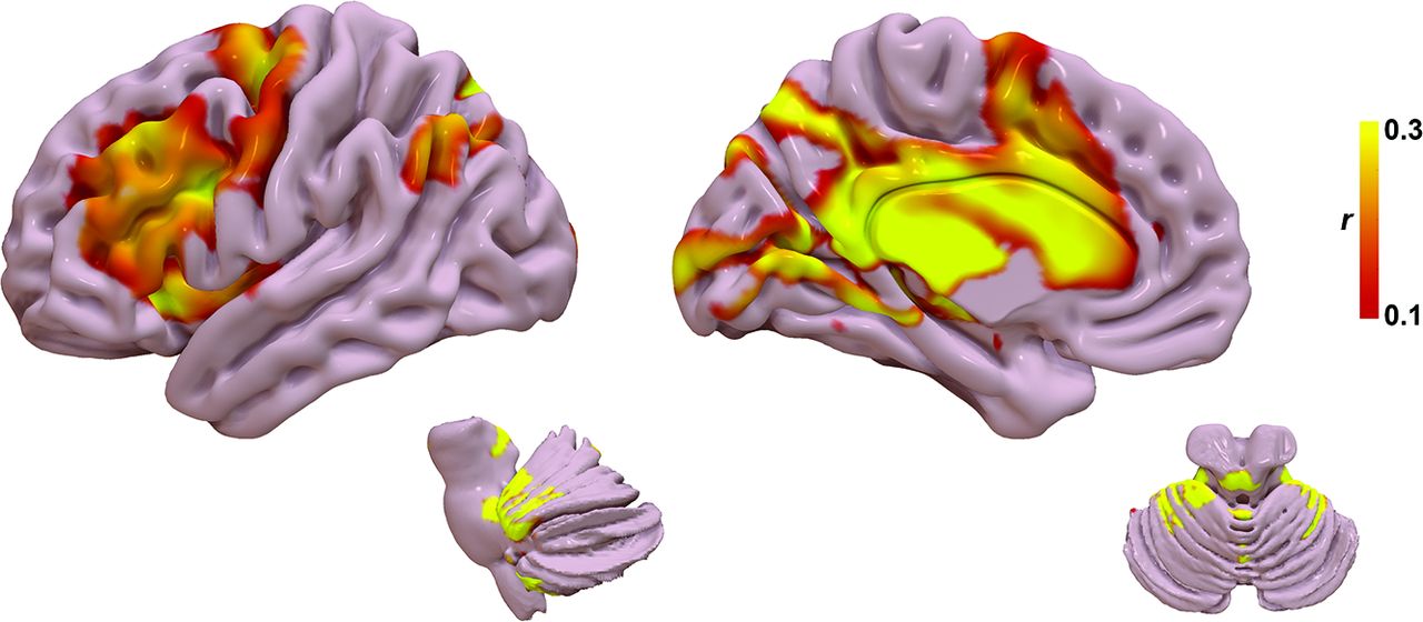
The tract R-map, or tract fingerprint, is shown in Fig 2. The cerebello-thalamo-cortical somatosensory network is a primary component of the affected network, including the primary somatosensory cortex, supplementary motor area, premotor cortex, ventral thalamic nuclei, and cerebellum—primarily cerebellar lobules IV, V, VI, and VIIIB, all areas implicated in the cerebellar somatosensory network.27 Additional connectivity to temporal, occipital, and prefrontal areas is also present. The tract pathways correlated to tremor improvement (Fig 3) show primary involvement of the dentato-rubro-thalamic tract (DRTT); thalamocortical tracts to the primary sensorimotor, supplementary motor area, and prefrontal cortex; as well as the pallidothalamic tracts.

FIG 2.
  • Download figure
  • Open in new tab
  • Download powerpoint
FIG 2.

Tract fingerprint representing the ideal tract connectivity pattern for tremor improvement. Commonly implicated areas of abnormality in tremor are identified, including the cerebello-thalamo-cortical motor network, as well as striate and extrastriate cortical regions.

FIG 3.
  • Download figure
  • Open in new tab
  • Download powerpoint
FIG 3.

Group tract R-map of pathways most correlated with tremor improvement.

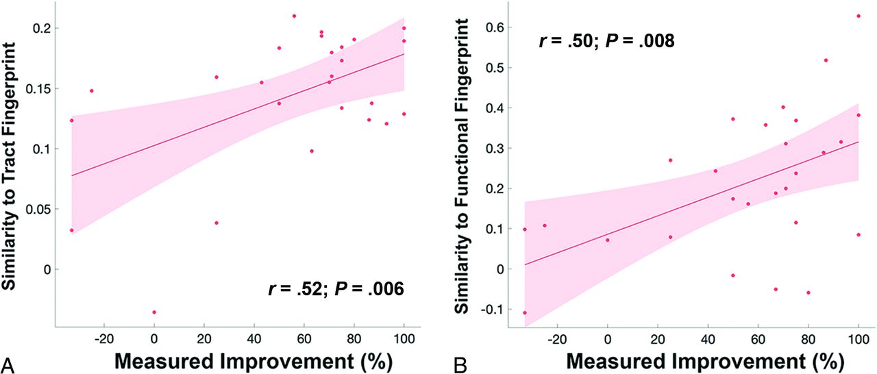
Results of the leave-one-out cross-validation of the tractography are shown in Fig 4A. The structural connectivity correlated with measured improvement in contralateral TRS (r = 0.52; P  = .006) and could explain 27.0% of the variance in outcome.

FIG 4.
  • Download figure
  • Open in new tab
  • Download powerpoint
FIG 4.

A, Leave-one-out cross-validation of the tract fingerprint shows greater similarity of the individual’s lesion tract map to the fingerprint map predicted measured improvement in tremor (r = 0.52; P  = .006). B, Leave-one-out cross-validation of the functional fingerprint also shows that greater similarity of the individual’s lesion functional map to the fingerprint map predicted measured improvement in tremor (r = 0.50; P  = .008).

Functional Connectome Analysis

The functional R-map, or functional fingerprint, is shown in Fig 5. The functional connectivity data further corroborate the structural connectivity, showing a correlation with cerebello-thalamo-cortical somatosensory network, in particular the primary somatosensory cortex, ventral thalamic nuclei, and cerebellar lobules IV, V, VI, and VIIIB. Additional connectivity to temporal, occipital, and prefrontal areas is also present. Results for the leave-one-out cross-validation of the functional connectivity are shown in Fig 4B. The functional connectivity correlated with measured improvement in contralateral TRS (r = 0.50; P  = .008) and could explain 25.0% of the variance in outcome.

FIG 5.
  • Download figure
  • Open in new tab
  • Download powerpoint
FIG 5.

Functional fingerprint representing the ideal tract connectivity pattern for tremor improvement. Similar to the tract fingerprint, commonly implicated areas of abnormality in tremor are identified, including the cerebello-thalamo-cortical motor network as well as the striate and extrastriate cortical regions.

DISCUSSION

In this study, we show that improvement in tremor after SRS thalamotomy is significantly correlated with a distinct pattern of structural and functional connectivity, primarily via the cerebello-thalamo-cortical pathway. This network has been previously demonstrated to be an important driver in tremor pathophysiology and a target for other forms of neuromodulation, such as DBS. Our findings indicate that a significant amount of variance in tremor outcomes after SRS are driven by an identifiable network topology, which may serve as a new therapeutic biomarker for SRS targeting. As opposed to other forms of neuromodulation, such as DBS, there are no intraprocedural biomarkers to validate target selection in SRS. By application of the concept of connectomic surgery,12 we have shown the potential of a lesion connectome fingerprint to predict tremor improvement from a given SRS target using both MR tractography and resting-state fMRI. Our approach may enhance network-specific targeting in SRS and provide patient-specific biomarkers.

The optimal target for neuromodulation of tremor has been debated for decades. Most commonly proposed targets have been the ventral intermediate nucleus (VIM) and the posterior subthalamic area, including the caudal zona incerta, prelemniscal radiations, and subthalamic nucleus.11 More recently, connectivity studies in DBS have suggested that these targets are unified by a common network and all targets overlap with the DRTT.7,14 The role of the DRTT in tremor improvement has been well-demonstrated in DBS,5⇓⇓⇓⇓⇓⇓⇓⇓-14,28⇓⇓⇓⇓⇓-34 but limited data exist on the use of tractography in surgical planning for SRS.35,36 Historically, the DRTT is defined by efferent cerebellar fibers originating in the dentate nucleus of the cerebellum that traverse superiorly through the ipsilateral superior cerebellar peduncle, decussate in the midbrain, pass the red nucleus (but without synapsing in the red nucleus), en route to the contralateral VIM and posterior vetralis oralis nucleus.37,38 Tracts ultimately reach the contralateral primary motor cortex and premotor/supplementary motor cortex from the VIM and vetralis oralis nucleus, respectively. More recently, a small number of DRTT fibers have been shown to follow a similar path but without decussation in the midbrain and extend to the ipsilateral thalamus; however, these tracts likely have less of a role in motor function.37,38 Our study adds further evidence to the role of the DRTT in the surgical treatment of tremor, particularly implicating a similar therapeutic target in SRS as seen in DBS.

From an anatomic perspective, nodes within the fingerprint maps correspond to areas known to exhibit network abnormalities in tremor. First, the fingerprint network distribution agrees with established network abnormalities attributed to the development of tremor via tremor-specific frequency synchronization within multiple nodes in the cerebello-thalamo-cortical pathway, including the cerebellum, thalamus, primary sensorimotor cortex, supplementary motor cortex, and premotor cortex.39,40 Similar to prior work in DBS for essential tremor, we found that a lesion network fingerprint that includes the motor and supplementary motor networks also correlates with tremor improvement in SRS.7,14 Additionally, there was correlation with cerebellar lobules IV, V, and VI, which are regions previously shown to be abnormal in essential tremor and part of the sensorimotor network.41 Taken together, our results suggest that the therapeutic network involved in SRS thalamotomy provides evidence for targeting biomarkers similar to those in the existing DBS literature.

Most interesting, we also found structural and functional connectivity to the primary and extrastriate visual cortex as part of the tremor connectome fingerprint. While not historically considered a major part of tremor pathogenesis, the primary and associative visual cortices have been the subject of more recent investigation of the role of the visuospatial network in tremor augmentation. Archer et al42 showed that visual feedback exacerbates tremor severity in essential tremor and functional changes in extrastriate areas correlate with the worsening of tremor. Likewise, postsurgical alterations in brain connectivity have been observed in the visual cortex that correlated with tremor improvement in MR imaging–guided focused ultrasound43 and SRS;44 however, direct activation of this region was not observed with active DBS.45 Pretreatment functional connectivity between the ventrolateral thalamus and visual association areas has also been shown as a predictor of tremor improvement after SRS.46 Further studies are needed to understand the true causal effect of visual network connectivity versus a coincident change in addition to the cerebello-thalamo-cortical motor network.

Several limitations to our study are noteworthy. Although tremor outcomes were gathered prospectively, the connectomics analyses were applied in retrospect. Thus, future studies would benefit from examining the effects of prospective targeting based on the proposed biomarkers. SRS thalamotomy was effective for tremor and well-tolerated overall. Larger cohorts, however, are needed to expand on these results and better assess the frequency of potential adverse effects. Additionally, intersubject variations in the temporal evolution of the lesion may result in fluctuation of the clinical response and are relatively unpredictable pre-SRS. We chose to use the 3-month post-SRS examination because this was thought to most accurately represent the intended target location with less variability among patients. Last, there are inherent limitations to MR tractography and resting-state fMRI that have been well-described.6,31,47 We used a robust probabilistic tracking approach and a lengthy fMRI acquisition to increase the robustness of our connectome modeling; however, these protocols may be challenging to implement in routine clinical care, and further studies will be needed to explore reproducibility with less robust data sets.

CONCLUSIONS

Using a connectomic approach, we have shown that SRS targets with a distinct connectivity profile predict posttreatment tremor improvement. Among these are networks implicated in tremor pathogenesis, including the cerebello-thalamo-cortical network and visual networks. The use of such connectomic fingerprints may provide a patient-specific biomarker for SRS thalamotomy; however, validation of this approach for use in targeting will require further studies to prospectively validate these results.

Footnotes

  • This prospective clinical trial was funded by Varian Medical Systems Inc under the study protocol RAD 1601: EDGE Radiosurgery for Intractable Essential Tremor and Tremor-Dominant Parkinson’s Disease (NCT03305588).

  • Disclosure forms provided by the authors are available with the full text and PDF of this article at www.ajnr.org.

References

  1. 1.↵
    1. Louis ED,
    2. Ferreira JJ
    . How common is the most common adult movement disorder? Update on the worldwide prevalence of essential tremor. Mov Disord 2010;25:534–41 doi:10.1002/mds.22838 pmid:20175185
    CrossRefPubMed
  2. 2.↵
    1. Sporns O,
    2. Tononi G,
    3. Kötter R
    . The human connectome: a structural description of the human brain. PLoS Comput Biol 2005;1:e42 doi:10.1371/journal.pcbi.0010042 pmid:16201007
    CrossRefPubMed
  3. 3.↵
    1. Lozano AM,
    2. Lipsman N
    . Probing and regulating dysfunctional circuits using deep brain stimulation. Neuron 2013;77:406–24 doi:10.1016/j.neuron.2013.01.020 pmid:23395370
    CrossRefPubMed
  4. 4.↵
    1. Henderson JM
    . “Connectomic surgery”: diffusion tensor imaging (DTI) tractography as a targeting modality for surgical modulation of neural networks. Front Integr Neurosci 2012;6:15 doi:10.3389/fnint.2012.00015 pmid:22536176
    CrossRefPubMed
  5. 5.↵
    1. Middlebrooks EH,
    2. Domingo RA,
    3. Vivas-Buitrago T, et al
    . Neuroimaging advances in deep brain stimulation: review of indications, anatomy, and brain connectomics. AJNR Am J Neuroradiol 2020;41:1558–68 doi:10.3174/ajnr.A6693 pmid:32816768
    Abstract/FREE Full Text
  6. 6.↵
    1. Middlebrooks EH,
    2. Okromelidze L,
    3. Carter RE, et al
    . Directed stimulation of the dentato-rubro-thalamic tract for deep brain stimulation in essential tremor: a blinded clinical trial. Neuroradiol J 2022;35:203–12 doi:10.1177/19714009211036689 pmid:34340623
    CrossRefPubMed
  7. 7.↵
    1. Middlebrooks EH,
    2. Okromelidze L,
    3. Wong JK, et al
    . Connectivity correlates to predict essential tremor deep brain stimulation outcome: evidence for a common treatment pathway. Neuroimage Clin 2021;32:102846 doi:10.1016/j.nicl.2021.102846 pmid:34624639
    CrossRefPubMed
  8. 8.↵
    1. Middlebrooks EH,
    2. Tuna IS,
    3. Almeida L, et al
    . Structural connectivity-based segmentation of the thalamus and prediction of tremor improvement following thalamic deep brain stimulation of the ventral intermediate nucleus. Neuroimage Clin 2018;20:1266–73 doi:10.1016/j.nicl.2018.10.009 pmid:30318403
    CrossRefPubMed
  9. 9.↵
    1. Neudorfer C,
    2. Kroneberg D,
    3. Al-Fatly B, et al
    . Personalizing deep brain stimulation using advanced imaging sequences. Ann Neurol 2022;91:613–28 doi:10.1002/ana.26326 pmid:35165921
    CrossRefPubMed
  10. 10.↵
    1. Tsuboi T,
    2. Wong JK,
    3. Eisinger RS, et al
    . Comparative connectivity correlates of dystonic and essential tremor deep brain stimulation. Brain 2021;144:1774–86 doi:10.1093/brain/awab074 pmid:33889943
    CrossRefPubMed
  11. 11.↵
    1. Wong JK,
    2. Hess CW,
    3. Almeida L, et al
    . Deep brain stimulation in essential tremor: targets, technology, and a comprehensive review of clinical outcomes. Expert Rev Neurother 2020;20:319–31 doi:10.1080/14737175.2020.1737017 pmid:32116065
    CrossRefPubMed
  12. 12.↵
    1. Wong JK,
    2. Middlebrooks EH,
    3. Grewal SS, et al
    . A comprehensive review of brain connectomics and imaging to improve deep brain stimulation outcomes. Mov Disord 2020;35:741–51 doi:10.1002/mds.28045 pmid:32281147
    CrossRefPubMed
  13. 13.↵
    1. Wong JK,
    2. Patel B,
    3. Middlebrooks EH, et al
    . Connectomic analysis of unilateral dual-lead thalamic deep brain stimulation for treatment of multiple sclerosis tremor. Brain Commun 2022;4:fcac063 doi:10.1093/braincomms/fcac063 pmid:35368612
    CrossRefPubMed
  14. 14.↵
    1. Al-Fatly B,
    2. Ewert S,
    3. Kubler D, et al
    . Connectivity profile of thalamic deep brain stimulation to effectively treat essential tremor. Brain 2019;142:3086–98 doi:10.1093/brain/awz236 pmid:31377766
    CrossRefPubMed
  15. 15.↵
    1. Middlebrooks EH,
    2. Grewal SS,
    3. Stead M, et al
    . Differences in functional connectivity profiles as a predictor of response to anterior thalamic nucleus deep brain stimulation for epilepsy: a hypothesis for the mechanism of action and a potential biomarker for outcomes. Neurosurg Focus 2018;45:E7 doi:10.3171/2018.5.FOCUS18151 pmid:30064322
    CrossRefPubMed
  16. 16.↵
    1. Middlebrooks EH,
    2. He X,
    3. Grewal SS, et al
    . Neuroimaging and thalamic connectomics in epilepsy neuromodulation. Epilepsy Res 2022;182:106916 doi:10.1016/j.eplepsyres.2022.106916 pmid:35367691
    CrossRefPubMed
  17. 17.↵
    1. Middlebrooks EH,
    2. Jain A,
    3. Okromelidze L, et al
    . Acute brain activation patterns of high- versus low-frequency stimulation of the anterior nucleus of the thalamus during deep brain stimulation for epilepsy. Neurosurgery 2021;89:901–08 doi:10.1093/neuros/nyab306 pmid:34460925
    CrossRefPubMed
  18. 18.↵
    1. Middlebrooks EH,
    2. Lin C,
    3. Okromelidze L, et al
    . Functional activation patterns of deep brain stimulation of the anterior nucleus of the thalamus. World Neurosurg 2020;136:357–63.e2 doi:10.1016/j.wneu.2020.01.108 pmid:32001414
    CrossRefPubMed
  19. 19.↵
    1. Tsuboi T,
    2. Charbel M,
    3. Peterside DT, et al
    . Pallidal connectivity profiling of stimulation-induced dyskinesia in Parkinson’s disease. Mov Disord 2021;36:380–88 doi:10.1016/j.wneu.2020.01.108 pmid:33002233
    CrossRefPubMed
  20. 20.↵
    1. Horn A,
    2. Reich M,
    3. Vorwerk J, et al
    . Connectivity predicts deep brain stimulation outcome in Parkinson disease. Ann Neurol 2017;82:67–78 doi:10.1002/ana.24974 pmid:28586141
    CrossRefPubMed
  21. 21.↵
    1. Li N,
    2. Baldermann JC,
    3. Kibleur A, et al
    . A unified connectomic target for deep brain stimulation in obsessive-compulsive disorder. Nat Commun 2020;11:3364 doi:10.1038/s41467-020-16734-3 pmid:32620886
    CrossRefPubMed
  22. 22.↵
    1. Reich MM,
    2. Horn A,
    3. Lange F, et al
    . Probabilistic mapping of the antidystonic effect of pallidal neurostimulation: a multicentre imaging study. Brain 2019;142:1386–98 doi:10.1093/brain/awz046 pmid:30851091
    CrossRefPubMed
  23. 23.↵
    1. Kundu P,
    2. Inati SJ,
    3. Evans JW, et al
    . Differentiating BOLD and non-BOLD signals in fMRI time series using multi-echo EPI. Neuroimage 2012;60:1759–70 doi:10.1016/j.neuroimage.2011.12.028 pmid:22209809
    CrossRefPubMed
  24. 24.↵
    1. Popple RA,
    2. Wu X,
    3. Brezovich IA, et al
    . The virtual cone: a novel technique to generate spherical dose distributions using a multileaf collimator and standardized control-point sequence for small target radiation surgery. Adv Radiat Oncol 2018;3:421–30 doi:10.1016/j.adro.2018.02.011 pmid:30197943
    CrossRefPubMed
  25. 25.↵
    1. Li G,
    2. Ballangrud A,
    3. Kuo LC, et al
    . Motion monitoring for cranial frameless stereotactic radiosurgery using video-based three-dimensional optical surface imaging. Med Phys 2011;38:3981–94 doi:10.1118/1.3596526 pmid:21858995
    CrossRefPubMed
  26. 26.↵
    1. Graham MS,
    2. Drobnjak I,
    3. Jenkinson M, et al
    . Quantitative assessment of the susceptibility artefact and its interaction with motion in diffusion MRI. PLoS One 2017;12:e0185647 doi:10.1371/journal.pone.0185647 pmid:28968429
    CrossRefPubMed
  27. 27.↵
    1. Buckner RL,
    2. Krienen FM,
    3. Castellanos A, et al
    . The organization of the human cerebellum estimated by intrinsic functional connectivity. J Neurophysiol 2011;106:2322–45 doi:10.1152/jn.00339.2011 pmid:21795627
    CrossRefPubMed
  28. 28.↵
    1. Middlebrooks EH,
    2. Grewal SS,
    3. Holanda VM
    . Complexities of connectivity-based DBS targeting: rebirth of the debate on thalamic and subthalamic treatment of tremor. Neuroimage Clin 2019;22:101761 doi:10.1016/j.nicl.2019.101761 pmid:30987840
    CrossRefPubMed
  29. 29.↵
    1. Middlebrooks EH,
    2. Tipton P,
    3. Okromelidze L, et al
    . Deep brain stimulation for tremor: direct targeting of a novel imaging biomarker. Ann Neurol 2022;92:341–42 doi:10.1002/ana.26422 pmid:35596936
    CrossRefPubMed
  30. 30.↵
    1. Neudorfer C,
    2. Kroneberg D,
    3. Al-Fatly B, et al
    . Reply to “Deep Brain Stimulation for Tremor: Direct Targeting of a Novel Imaging Biomarker.” Ann Neurol 2022;92:343–44 doi:10.1002/ana.26420 pmid:35608332
    CrossRefPubMed
  31. 31.↵
    1. Akram H,
    2. Dayal V,
    3. Mahlknecht P, et al
    . Connectivity derived thalamic segmentation in deep brain stimulation for tremor. Neuroimage Clin 2018;18:130–42 doi:10.1016/j.nicl.2018.01.008 pmid:29387530
    CrossRefPubMed
  32. 32.↵
    1. Coenen VA,
    2. Allert N,
    3. Paus S, et al
    . Modulation of the cerebello-thalamo-cortical network in thalamic deep brain stimulation for tremor: a diffusion tensor imaging study. Neurosurgery 2014;75:657–69; discussion 6697–70 doi:10.1227/NEU.0000000000000540 pmid:25161000
    CrossRefPubMed
  33. 33.↵
    1. Coenen VA,
    2. Sajonz B,
    3. Prokop T, et al
    . The dentato-rubro-thalamic tract as the potential common deep brain stimulation target for tremor of various origin: an observational case series. Acta Neurochir (Wien) 2020;162:1053–66 doi:10.1007/s00701-020-04248-2 pmid:31997069
    CrossRefPubMed
  34. 34.↵
    1. Coenen VA,
    2. Varkuti B,
    3. Parpaley Y, et al
    . Postoperative neuroimaging analysis of DRT deep brain stimulation revision surgery for complicated essential tremor. Acta Neurochir (Wien) 2017;159:779–87 doi:10.1007/s00701-017-3134-z pmid:28283867
    CrossRefPubMed
  35. 35.↵
    1. Kim W,
    2. Chivukula S,
    3. Hauptman J, et al
    . Diffusion tensor imaging-based thalamic segmentation in deep brain stimulation for chronic pain conditions. Stereotact Funct Neurosurg 2016;94:225–34 doi:10.1159/000448079 pmid:27537848
    CrossRefPubMed
  36. 36.↵
    1. Kim W,
    2. Sharim J,
    3. Tenn S, et al
    . Diffusion tractography imaging-guided frameless linear accelerator stereotactic radiosurgical thalamotomy for tremor: case report. J Neurosurg 2018;128:215–21 doi:10.3171/2016.10.JNS161603 pmid:28298033
    CrossRefPubMed
  37. 37.↵
    1. Gallay MN,
    2. Jeanmonod D,
    3. Liu J, et al
    . Human pallidothalamic and cerebellothalamic tracts: anatomical basis for functional stereotactic neurosurgery. Brain Struct Funct 2008;212:443–63 doi:10.1007/s00429-007-0170-0 pmid:18193279
    CrossRefPubMed
  38. 38.↵
    1. Petersen KJ,
    2. Reid JA,
    3. Chakravorti S, et al
    . Structural and functional connectivity of the nondecussating dentato-rubro-thalamic tract. Neuroimage 2018;176:364–71 doi:10.1016/j.neuroimage.2018.04.074 pmid:29733955
    CrossRefPubMed
  39. 39.↵
    1. Hellwig B,
    2. Haussler S,
    3. Schelter B, et al
    . Tremor-correlated cortical activity in essential tremor. Lancet 2001;357:519–23 doi:10.1016/S0140-6736(00)04044-7 pmid:11229671
    CrossRefPubMed
  40. 40.↵
    1. McAuley JH,
    2. Marsden CD
    . Physiological and pathological tremors and rhythmic central motor control. Brain 2000;123:1545–67 doi:10.1093/brain/123.8.1545
    CrossRefPubMed
  41. 41.↵
    1. Nicoletti V,
    2. Cecchi P,
    3. Pesaresi I, et al
    . Cerebello-thalamo-cortical network is intrinsically altered in essential tremor: evidence from a resting state functional MRI study. Sci Rep 2020;10:16661 doi:10.1038/s41598-020-73714-9 pmid:33028912
    CrossRefPubMed
  42. 42.↵
    1. Archer DB,
    2. Coombes SA,
    3. Chu WT, et al
    . A widespread visually-sensitive functional network relates to symptoms in essential tremor. Brain 2018;141:472–85 doi:10.1093/brain/awx338 pmid:29293948
    CrossRefPubMed
  43. 43.↵
    1. Xiong Y,
    2. Han D,
    3. He J, et al
    . Correlation of visual area with tremor improvement after MRgFUS thalamotomy in Parkinson’s disease. J Neurosurg 2022;136:681–88 doi:10.3171/2021.3.JNS204329 pmid:34479209
    CrossRefPubMed
  44. 44.↵
    1. Tuleasca C,
    2. Najdenovska E,
    3. Regis J, et al
    . Clinical response to VIM’s thalamic stereotactic radiosurgery for essential tremor is associated with distinctive functional connectivity patterns. Acta Neurochir (Wien) 2018;160:611–24 doi:10.1007/s00701-017-3456-x pmid:29335882
    CrossRefPubMed
  45. 45.↵
    1. Gibson WS,
    2. Jo HJ,
    3. Testini P, et al
    . Functional correlates of the therapeutic and adverse effects evoked by thalamic stimulation for essential tremor. Brain 2016;139:2198–210 doi:10.1093/brain/aww145 pmid:27329768
    CrossRefPubMed
  46. 46.↵
    1. Tuleasca C,
    2. Najdenovska E,
    3. Régis J, et al
    . Pretherapeutic motor thalamus resting-state functional connectivity with visual areas predicts tremor arrest after thalamotomy for essential tremor: tracing the cerebello-thalamo-visuo-motor network. World Neurosurg 2018;117:e438–49 doi:10.1016/j.wneu.2018.06.049 pmid:29920392
    CrossRefPubMed
  47. 47.↵
    1. Schlaier JR,
    2. Beer AL,
    3. Faltermeier R, et al
    . Probabilistic vs. deterministic fiber tracking and the influence of different seed regions to delineate cerebellar-thalamic fibers in deep brain stimulation. Eur J Neurosci 2017;45:1623–33 doi:10.1111/ejn.13575 pmid:28391647
    CrossRefPubMed
  • Received September 8, 2022.
  • Accepted after revision December 30, 2022.
  • © 2023 by American Journal of Neuroradiology
PreviousNext
Back to top

In this issue

American Journal of Neuroradiology: 44 (2)
American Journal of Neuroradiology
Vol. 44, Issue 2
1 Feb 2023
  • Table of Contents
  • Index by author
  • Complete Issue (PDF)
Advertisement
Print
Download PDF
Email Article

Thank you for your interest in spreading the word on American Journal of Neuroradiology.

NOTE: We only request your email address so that the person you are recommending the page to knows that you wanted them to see it, and that it is not junk mail. We do not capture any email address.

Enter multiple addresses on separate lines or separate them with commas.
Connectomic Basis for Tremor Control in Stereotactic Radiosurgical Thalamotomy
(Your Name) has sent you a message from American Journal of Neuroradiology
(Your Name) thought you would like to see the American Journal of Neuroradiology web site.
CAPTCHA
This question is for testing whether or not you are a human visitor and to prevent automated spam submissions.
Cite this article
E.H. Middlebrooks, R.A. Popple, E. Greco, L. Okromelidze, H.C. Walker, D.A. Lakhani, A.R. Anderson, E.M. Thomas, H.D. Deshpande, B.A. McCullough, N.P. Stover, V.W. Sung, A.P. Nicholas, D.G. Standaert, T. Yacoubian, M.N. Dean, J.A. Roper, S.S. Grewal, M.T. Holland, J.N. Bentley, B.L. Guthrie, M. Bredel
Connectomic Basis for Tremor Control in Stereotactic Radiosurgical Thalamotomy
American Journal of Neuroradiology Feb 2023, 44 (2) 157-164; DOI: 10.3174/ajnr.A7778

Citation Manager Formats

  • BibTeX
  • Bookends
  • EasyBib
  • EndNote (tagged)
  • EndNote 8 (xml)
  • Medlars
  • Mendeley
  • Papers
  • RefWorks Tagged
  • Ref Manager
  • RIS
  • Zotero
0 Responses
Respond to this article
Share
Bookmark this article
Connectomics for Tremor Control in Radiosurgery
E.H. Middlebrooks, R.A. Popple, E. Greco, L. Okromelidze, H.C. Walker, D.A. Lakhani, A.R. Anderson, E.M. Thomas, H.D. Deshpande, B.A. McCullough, N.P. Stover, V.W. Sung, A.P. Nicholas, D.G. Standaert, T. Yacoubian, M.N. Dean, J.A. Roper, S.S. Grewal, M.T. Holland, J.N. Bentley, B.L. Guthrie, M. Bredel
American Journal of Neuroradiology Feb 2023, 44 (2) 157-164; DOI: 10.3174/ajnr.A7778
del.icio.us logo Twitter logo Facebook logo Mendeley logo
  • Tweet Widget
  • Facebook Like
  • Google Plus One
Purchase

Jump to section

  • Article
    • Abstract
    • ABBREVIATIONS:
    • MATERIALS AND METHODS
    • RESULTS
    • DISCUSSION
    • CONCLUSIONS
    • Footnotes
    • References
  • Figures & Data
  • Supplemental
  • Info & Metrics
  • Responses
  • References
  • PDF

Related Articles

  • PubMed
  • Google Scholar

Cited By...

  • No citing articles found.
  • Crossref (3)
  • Google Scholar

This article has been cited by the following articles in journals that are participating in Crossref Cited-by Linking.

  • Stereotactic radiosurgery in the treatment of essential tremor – a systematic review
    Mateusz Bilski, Katarzyna Szklener, Sebastian Szklener, Anna Rudzińska, Natalia Kluz, Jakub Klas, Anna Rodzajewska, Weronika Kuryło, Mateusz Korga, Izabela Baranowska, Sławomir Mańdziuk
    Frontiers in Neurology 2024 15
  • Gamma knife versus linear accelerator thalamotomy for essential tremor and Parkinson’s disease: A systematic review and meta-analysis
    Renuka Chintapalli, Stephano Chang, Tania Kaprealian, Ricky Savjani, Stephen Tenn, Ausaf Bari
    Journal of Clinical Neuroscience 2025 133
  • The variation of functional connectivity and activity before and after thalamotomy surgery (review)
    Mohammad-Hossein H. K. Nili, Shahrzad M. Esfahan, Yamin Bagheri, Abdol-Hossein Vahabie, Mehdi Sanayei, Abolhassan Ertiaei, Mohammad Shirani, Mohammad-Reza A. Dehaqani, Ehsan Rezayat
    Frontiers in Human Neuroscience 2023 17

More in this TOC Section

  • Kurtosis and Epileptogenic Tubers: A Pilot Study
  • Glutaric Aciduria Type 1: DK vs. Conventional MRI
  • Multiparametric MRI in PEDS Pontine Glioma
Show more FUNCTIONAL

Similar Articles

Advertisement

Indexed Content

  • Current Issue
  • Accepted Manuscripts
  • Article Preview
  • Past Issues
  • Editorials
  • Editor's Choice
  • Fellows' Journal Club
  • Letters to the Editor
  • Video Articles

Cases

  • Case Collection
  • Archive - Case of the Week
  • Archive - Case of the Month
  • Archive - Classic Case

More from AJNR

  • Trainee Corner
  • Imaging Protocols
  • MRI Safety Corner

Multimedia

  • AJNR Podcasts
  • AJNR Scantastics

Resources

  • Turnaround Time
  • Submit a Manuscript
  • Submit a Video Article
  • Submit an eLetter to the Editor/Response
  • Manuscript Submission Guidelines
  • Statistical Tips
  • Fast Publishing of Accepted Manuscripts
  • Graphical Abstract Preparation
  • Imaging Protocol Submission
  • Evidence-Based Medicine Level Guide
  • Publishing Checklists
  • Author Policies
  • Become a Reviewer/Academy of Reviewers
  • News and Updates

About Us

  • About AJNR
  • Editorial Board
  • Editorial Board Alumni
  • Alerts
  • Permissions
  • Not an AJNR Subscriber? Join Now
  • Advertise with Us
  • Librarian Resources
  • Feedback
  • Terms and Conditions
  • AJNR Editorial Board Alumni

American Society of Neuroradiology

  • Not an ASNR Member? Join Now

© 2025 by the American Society of Neuroradiology All rights, including for text and data mining, AI training, and similar technologies, are reserved.
Print ISSN: 0195-6108 Online ISSN: 1936-959X

Powered by HighWire