Advertisement

Main menu

  • Home
  • Content
    • Current Issue
    • Accepted Manuscripts
    • Article Preview
    • Past Issue Archive
    • Video Articles
    • AJNR Case Collection
    • Case of the Week Archive
    • Case of the Month Archive
    • Classic Case Archive
  • Special Collections
    • AJNR Awards
    • Low-Field MRI
    • Alzheimer Disease
    • ASNR Foundation Special Collection
    • Photon-Counting CT
    • View All
  • Multimedia
    • AJNR Podcasts
    • AJNR SCANtastic
    • Trainee Corner
    • MRI Safety Corner
    • Imaging Protocols
  • For Authors
    • Submit a Manuscript
    • Submit a Video Article
    • Submit an eLetter to the Editor/Response
    • Manuscript Submission Guidelines
    • Statistical Tips
    • Fast Publishing of Accepted Manuscripts
    • Graphical Abstract Preparation
    • Imaging Protocol Submission
    • Author Policies
  • About Us
    • About AJNR
    • Editorial Board
    • Editorial Board Alumni
  • More
    • Become a Reviewer/Academy of Reviewers
    • Subscribers
    • Permissions
    • Alerts
    • Feedback
    • Advertisers
    • ASNR Home

User menu

  • Alerts
  • Log in

Search

  • Advanced search
American Journal of Neuroradiology

American Journal of Neuroradiology

ASHNR American Society of Functional Neuroradiology ASHNR American Society of Pediatric Neuroradiology ASSR
  • Alerts
  • Log in

Advanced Search

  • Home
  • Content
    • Current Issue
    • Accepted Manuscripts
    • Article Preview
    • Past Issue Archive
    • Video Articles
    • AJNR Case Collection
    • Case of the Week Archive
    • Case of the Month Archive
    • Classic Case Archive
  • Special Collections
    • AJNR Awards
    • Low-Field MRI
    • Alzheimer Disease
    • ASNR Foundation Special Collection
    • Photon-Counting CT
    • View All
  • Multimedia
    • AJNR Podcasts
    • AJNR SCANtastic
    • Trainee Corner
    • MRI Safety Corner
    • Imaging Protocols
  • For Authors
    • Submit a Manuscript
    • Submit a Video Article
    • Submit an eLetter to the Editor/Response
    • Manuscript Submission Guidelines
    • Statistical Tips
    • Fast Publishing of Accepted Manuscripts
    • Graphical Abstract Preparation
    • Imaging Protocol Submission
    • Author Policies
  • About Us
    • About AJNR
    • Editorial Board
    • Editorial Board Alumni
  • More
    • Become a Reviewer/Academy of Reviewers
    • Subscribers
    • Permissions
    • Alerts
    • Feedback
    • Advertisers
    • ASNR Home
  • Follow AJNR on Twitter
  • Visit AJNR on Facebook
  • Follow AJNR on Instagram
  • Join AJNR on LinkedIn
  • RSS Feeds

AJNR Awards, New Junior Editors, and more. Read the latest AJNR updates

Research ArticleSpine
Open Access

Evaluation of 2 Novel Ratio-Based Metrics for Lumbar Spinal Stenosis

U.U. Bharadwaj, A.R. Ben-Natan, J. Huang, V. Pedoia, D. Chou, S. Majumdar, T.M. Link and C.T. Chin
American Journal of Neuroradiology October 2022, 43 (10) 1530-1538; DOI: https://doi.org/10.3174/ajnr.A7638
U.U. Bharadwaj
aFrom the Departments of Radiology and Biomedical Imaging (U.U.B., V.P., S.M., T.M.L., C.T.C.)
  • Find this author on Google Scholar
  • Find this author on PubMed
  • Search for this author on this site
  • ORCID record for U.U. Bharadwaj
A.R. Ben-Natan
bNeurological Surgery (A.R.B.-N., J.H., D.C.), University of California San Francisco, San Francisco, California
  • Find this author on Google Scholar
  • Find this author on PubMed
  • Search for this author on this site
  • ORCID record for A.R. Ben-Natan
J. Huang
bNeurological Surgery (A.R.B.-N., J.H., D.C.), University of California San Francisco, San Francisco, California
  • Find this author on Google Scholar
  • Find this author on PubMed
  • Search for this author on this site
  • ORCID record for J. Huang
V. Pedoia
aFrom the Departments of Radiology and Biomedical Imaging (U.U.B., V.P., S.M., T.M.L., C.T.C.)
  • Find this author on Google Scholar
  • Find this author on PubMed
  • Search for this author on this site
D. Chou
bNeurological Surgery (A.R.B.-N., J.H., D.C.), University of California San Francisco, San Francisco, California
  • Find this author on Google Scholar
  • Find this author on PubMed
  • Search for this author on this site
  • ORCID record for D. Chou
S. Majumdar
aFrom the Departments of Radiology and Biomedical Imaging (U.U.B., V.P., S.M., T.M.L., C.T.C.)
  • Find this author on Google Scholar
  • Find this author on PubMed
  • Search for this author on this site
  • ORCID record for S. Majumdar
T.M. Link
aFrom the Departments of Radiology and Biomedical Imaging (U.U.B., V.P., S.M., T.M.L., C.T.C.)
  • Find this author on Google Scholar
  • Find this author on PubMed
  • Search for this author on this site
C.T. Chin
aFrom the Departments of Radiology and Biomedical Imaging (U.U.B., V.P., S.M., T.M.L., C.T.C.)
  • Find this author on Google Scholar
  • Find this author on PubMed
  • Search for this author on this site
  • ORCID record for C.T. Chin
  • Article
  • Figures & Data
  • Supplemental
  • Info & Metrics
  • Responses
  • References
  • PDF
Loading

Article Figures & Data

Figures

  • Tables
  • FIG 1.
    • Download figure
    • Open in new tab
    • Download powerpoint
    FIG 1.

    Sample T2-weighted axial section at L2/L3 graded normal with the following: A, Free-form annotation around the DSCA of 166 mm2. B, Line annotation with a DSDIA of 14 mm. C, Line annotations with a DDRDIA of 0.4. D, Free-form annotations with DDRCA of 0.41. The square root is used as a normalization step to account for the quadraticity of area measures.

  • FIG 2.
    • Download figure
    • Open in new tab
    • Download powerpoint
    FIG 2.

    Sample T2-weighted axial MR imaging slices of the lumbar spine for each stenosis grade, determined qualitatively by a neuroradiologist, with the metrics annotated. A, Grade: normal; level, L1/L2; DSCA, 167 mm2; DSDIA, 15 mm; DDRDIA, 0.42; DDRCA, 0.40. B, Grade: mild; level, L3/L4; DSCA, 104 mm2; DSDIA, 10 mm; DDRDIA, 0.24; DDRCA, 0.27. C, Grade: moderate; level L4/L5; DSCA, 115 mm2; DSDIA, 10 mm; DDRDIA, 0.22; DDRCA, 0.24. D, Grade: severe; level, L2/L3; DSCA, 64 mm2; DSDIA, 7 mm; DDRDIA, 0.14; DDRCA, 0.16.
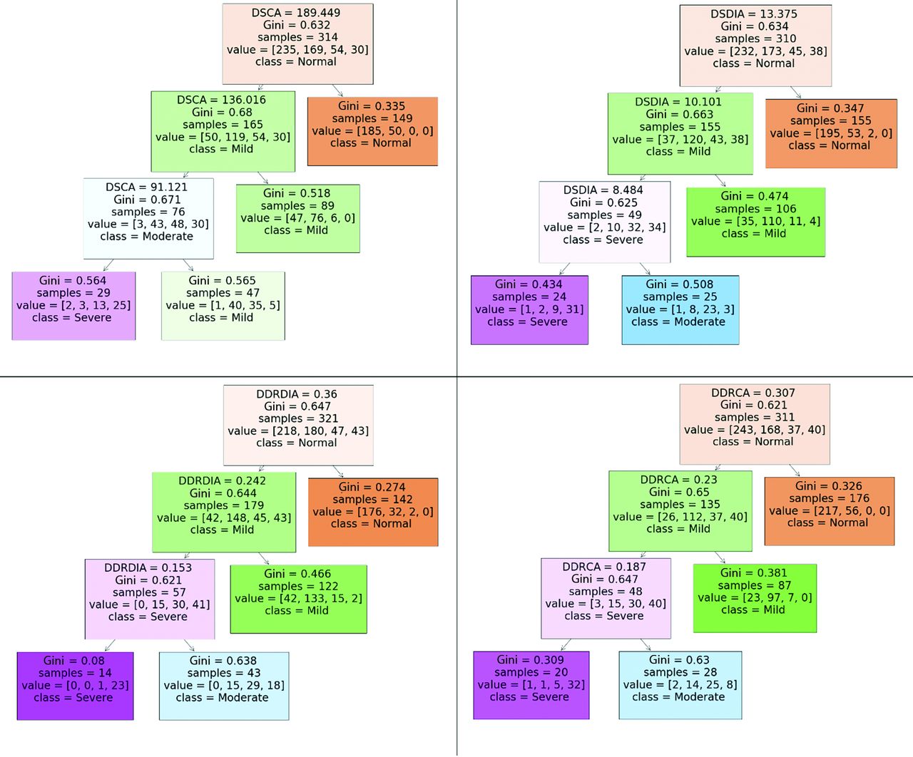
  • FIG 3.
    • Download figure
    • Open in new tab
    • Download powerpoint
    FIG 3.

    Decision rules and cutoff thresholds generated by a decision the tree classifier (maximum depth = 3, maximum leaves = 4, criterion = Gini impurity) for each quantitative metric.
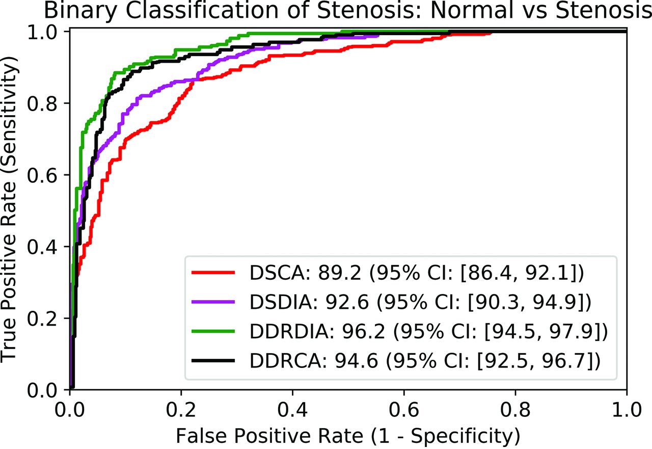
  • FIG 4.
    • Download figure
    • Open in new tab
    • Download powerpoint
    FIG 4.

    Receiver operator characteristic curve using each quantitative metric as a score for binary classification of stenosis. AUROC values are reported in the legend.

Tables

  • Figures
    • View popup
    Table 1:

    Classification of each spinal level as normal versus stenosed on the evaluation cohort (n = 130) using each quantitative metrica

    Metrics ComparedAUROC: Group 1AUROC: Group 2Z-StatisticSignificance
    DSCA vs DSDIA96.5 (95.0–97.7)96.6 (95.1–97.8)0.19P = .85
    DSCA vs DDRDIA96.5 (95.0–97.7)98.0 (96.7–98.9)b2.24bP = .02b
    DSCA vs DDRCA96.5 (95.0–97.7)98.6 (97.4–99.3)b3.54bP = .004b
    DSDIA vs DDRDIA96.6 (95.1–97.8)98.0 (96.7–98.9)b2.56bP = .01b
    DSDIA vs DDRCA96.6 (95.1–97.8)98.6 (97.4–99.3)b2.89bP = .004b
    DDRDIA vs DDRCA98.0 (96.7–98.9)98.6 (97.4–99.3)1.57P = .12
    • ↵a Reported are pair-wise comparisons between the quantitative metrics using the DeLong paired test for AUROCs. Data in parentheses represent 95% confidence intervals.

    • ↵b Difference in AUROC is statistically significant (P < .05).

    • View popup
    Table 2:

    Classification of each spinal level as normal, mild, moderate, and severe stenosis on the evaluation cohort (n = 130) using decision trees trained on the development cohort (n = 130)

    MetricAccuracyAUROCCohen κ
    Accuracy95% CIAUROC95% CIκ95% CI
    DSCA64.9(60.9–69.0)76.6(73.9–79.3)0.62(0.57–0.66)
    DSDIA71.4(67.1–75.7)80.9(78.0–83.8)0.69(0.64–0.75)
    DDRDIA76.5(72.6–80.4)a84.3a(81.7–86.9)a0.75a(0.71–0.79)a
    DDRCA78.9(75.0–82.9)a86.0a(83.3–88.5)a0.80a(0.75–0.83)a
    • ↵a Ratio-based metrics with higher κ scores (P < .001).

    • View popup
    Table 3:

    AUROC for binary classification based on each metric across symptomatic splits (VAS <7 versus VAS ≥7) of low back pain and radicular paina

    MetricSymptomatology Analysis of Low Back Pain
    VAS < 7VAS ≥ 7Significancea
    AUROC95% CIAUROC95% CIP Value
    Low Back Pain
     DSCA97.7(96.7–98.6)95.1(92.9–97.3)P < .001
     DSDIA96.6(95.4–97.8)96.5(94.9–98.1)P = .43
     DDRDIA96.8(95.6–97.9)97.5(96.1–98.8)P < .001
     DDRCA98.5(97.8–99.2)96.3(94.8–97.9)P < .001
    Radicular back pain
     DSCA98.4(97.4–99.4)96.3(94.5–98.1)P < .001
     DSDIA98.2(96.8–99.6)96.9(95.5–98.2)P < .001
     DDRDIA98.7(97.9–99.5)97.1(95.6–98.5)P < .001
     DDRCA99.0(98.4–99.6)97.1(95.5–98.7)P < .001
    • ↵a The P values represent a comparison of AUROCs among the symptomatic splits.

    • View popup
    Table 4:

    AUROC for predicting surgery using each quantitative metric on the prognostic cohort (n = 58)

    MetricPredicting Surgery at Each Spinal Level
    AUROC95% CISignificancea
    DSCA82.4(75.5–90.4)P = 1.0
    DSDIA81.3(73.2–89.4)P < .001
    DDRDIA80.8(73.0–89.5)P < .001
    DDRCA83.5b(76.6–90.1)bP < .001b
    • ↵a The P values represent comparison between each metric and AUROC obtained with the baseline metric DSCA.

    • ↵b Quantitative metric with the highest AUROC.

    • View popup
    Table 5:

    Classification of each spinal level as normal, mild, moderate, and severe stenosis on the prognostic cohort (n = 58) using decision trees trained on the development cohort (n = 130)

    κ Scores for Grading Stenosis across Surgical vs Nonsurgical Levels
    MetricNonsurgical LevelsSurgical LevelsSignificancea
    κ95% CIκ95% CIP Value
    DSCA0.65(0.57–0.75)0.74(0.63–0.85)P < .001
    DSDIA0.67(0.58–0.77)0.68(0.51–0.85)P = .23
    DDRDIA0.69(0.61–0.76)b0.77b(0.65–0.90)bP < .001b
    DDRCA0.71(0.62–0.79)0.73(0.58–0.87)P < .001
    • ↵a The P values represent a comparison of κ scores between nonsurgical and surgical levels.

    • ↵b The metric with the highest κ score for surgical levels.

PreviousNext
Back to top

In this issue

American Journal of Neuroradiology: 43 (10)
American Journal of Neuroradiology
Vol. 43, Issue 10
1 Oct 2022
  • Table of Contents
  • Index by author
  • Complete Issue (PDF)
Advertisement
Print
Download PDF
Email Article

Thank you for your interest in spreading the word on American Journal of Neuroradiology.

NOTE: We only request your email address so that the person you are recommending the page to knows that you wanted them to see it, and that it is not junk mail. We do not capture any email address.

Enter multiple addresses on separate lines or separate them with commas.
Evaluation of 2 Novel Ratio-Based Metrics for Lumbar Spinal Stenosis
(Your Name) has sent you a message from American Journal of Neuroradiology
(Your Name) thought you would like to see the American Journal of Neuroradiology web site.
CAPTCHA
This question is for testing whether or not you are a human visitor and to prevent automated spam submissions.
Cite this article
U.U. Bharadwaj, A.R. Ben-Natan, J. Huang, V. Pedoia, D. Chou, S. Majumdar, T.M. Link, C.T. Chin
Evaluation of 2 Novel Ratio-Based Metrics for Lumbar Spinal Stenosis
American Journal of Neuroradiology Oct 2022, 43 (10) 1530-1538; DOI: 10.3174/ajnr.A7638

Citation Manager Formats

  • BibTeX
  • Bookends
  • EasyBib
  • EndNote (tagged)
  • EndNote 8 (xml)
  • Medlars
  • Mendeley
  • Papers
  • RefWorks Tagged
  • Ref Manager
  • RIS
  • Zotero
0 Responses
Respond to this article
Share
Bookmark this article
Novel Metrics for Lumbar Spinal Stenosis
U.U. Bharadwaj, A.R. Ben-Natan, J. Huang, V. Pedoia, D. Chou, S. Majumdar, T.M. Link, C.T. Chin
American Journal of Neuroradiology Oct 2022, 43 (10) 1530-1538; DOI: 10.3174/ajnr.A7638
del.icio.us logo Twitter logo Facebook logo Mendeley logo
  • Tweet Widget
  • Facebook Like
  • Google Plus One
Purchase

Jump to section

  • Article
    • Abstract
    • ABBREVIATIONS:
    • MATERIALS AND METHODS
    • RESULTS
    • DISCUSSION
    • CONCLUSIONS
    • Acknowledgments
    • Footnotes
    • References
  • Figures & Data
  • Supplemental
  • Info & Metrics
  • Responses
  • References
  • PDF

Related Articles

  • PubMed
  • Google Scholar

Cited By...

  • An Optimized CT Protocol for Detecting Suspected Cauda Equina Syndrome: A Comparative Analysis with MRI
  • Crossref (6)
  • Google Scholar

This article has been cited by the following articles in journals that are participating in Crossref Cited-by Linking.

  • Comparison of Long‐term Follow‐Up of n‐HA PA66 Cage and PEEK Cage of Lumbar Interbody Fusion in Multi‐level Degenerative Lumbar Diseases: A Stepwise Propensity Score Matching Analysis
    Qiujiang Li, Qingyang Gao, Lei Wang, Limin Liu, Huiliang Yang, Yueming Song
    Orthopaedic Surgery 2024 16 1
  • Patients with achondroplasia have increased risk of 90-day adverse events following laminectomy: A matched comparison using a national database
    Anusha Patil, Peter Y. Joo, Jay Moran, Lancelot Benn, Addisu Mesfin
    Spine Deformity 2025 13 1
  • An Optimized CT Protocol for Detecting Suspected Cauda Equina Syndrome: A Comparative Analysis with MRI
    Philip J. Dempsey, David T. Ryan, Gerard Lambe, Jack W. Power, Andrew H. Yates, Grace Kenny, Peter J. MacMahon
    American Journal of Neuroradiology 2024 45 9
  • Magnetic resonance morphometry of the lumbar spinal canal in Zenica - Doboj Canton in Bosnia and Herzegovina
    Belma Jaganjac, Amina Džidić-Krivić, Hakija Bečulić, Armin Šljivo, Emir Begagić, Adis Šišić
    Medicinski Glasnik 2023 20 2
  • Posterior concave reconstruction with cage in the surgical treatment of complex lumbar deformity caused by lumbosacral hemivertebrae
    Qiujiang Li, Bowen Hu, Huiliang Yang, Ganjun Feng, Limin Liu, Xi Yang, Yueming Song
    European Spine Journal 2024 33 5
  • Neue Messparameter für die Einschätzung einer LWS-Spinalkanalstenose
    Neuroradiologie Scan 2023 13 02

More in this TOC Section

  • Bern Score Validity for SIH
  • MP2RAGE 7T in MS Lesions of the Cervical Spine
  • Resisted Inspiration for CSF-Venous Fistula
Show more Spine

Similar Articles

Advertisement

Indexed Content

  • Current Issue
  • Accepted Manuscripts
  • Article Preview
  • Past Issues
  • Editorials
  • Editor's Choice
  • Fellows' Journal Club
  • Letters to the Editor
  • Video Articles

Cases

  • Case Collection
  • Archive - Case of the Week
  • Archive - Case of the Month
  • Archive - Classic Case

More from AJNR

  • Trainee Corner
  • Imaging Protocols
  • MRI Safety Corner
  • Book Reviews

Multimedia

  • AJNR Podcasts
  • AJNR Scantastics

Resources

  • Turnaround Time
  • Submit a Manuscript
  • Submit a Video Article
  • Submit an eLetter to the Editor/Response
  • Manuscript Submission Guidelines
  • Statistical Tips
  • Fast Publishing of Accepted Manuscripts
  • Graphical Abstract Preparation
  • Imaging Protocol Submission
  • Evidence-Based Medicine Level Guide
  • Publishing Checklists
  • Author Policies
  • Become a Reviewer/Academy of Reviewers
  • News and Updates

About Us

  • About AJNR
  • Editorial Board
  • Editorial Board Alumni
  • Alerts
  • Permissions
  • Not an AJNR Subscriber? Join Now
  • Advertise with Us
  • Librarian Resources
  • Feedback
  • Terms and Conditions
  • AJNR Editorial Board Alumni

American Society of Neuroradiology

  • Not an ASNR Member? Join Now

© 2025 by the American Society of Neuroradiology All rights, including for text and data mining, AI training, and similar technologies, are reserved.
Print ISSN: 0195-6108 Online ISSN: 1936-959X

Powered by HighWire