
Article Figures & Data
Figures
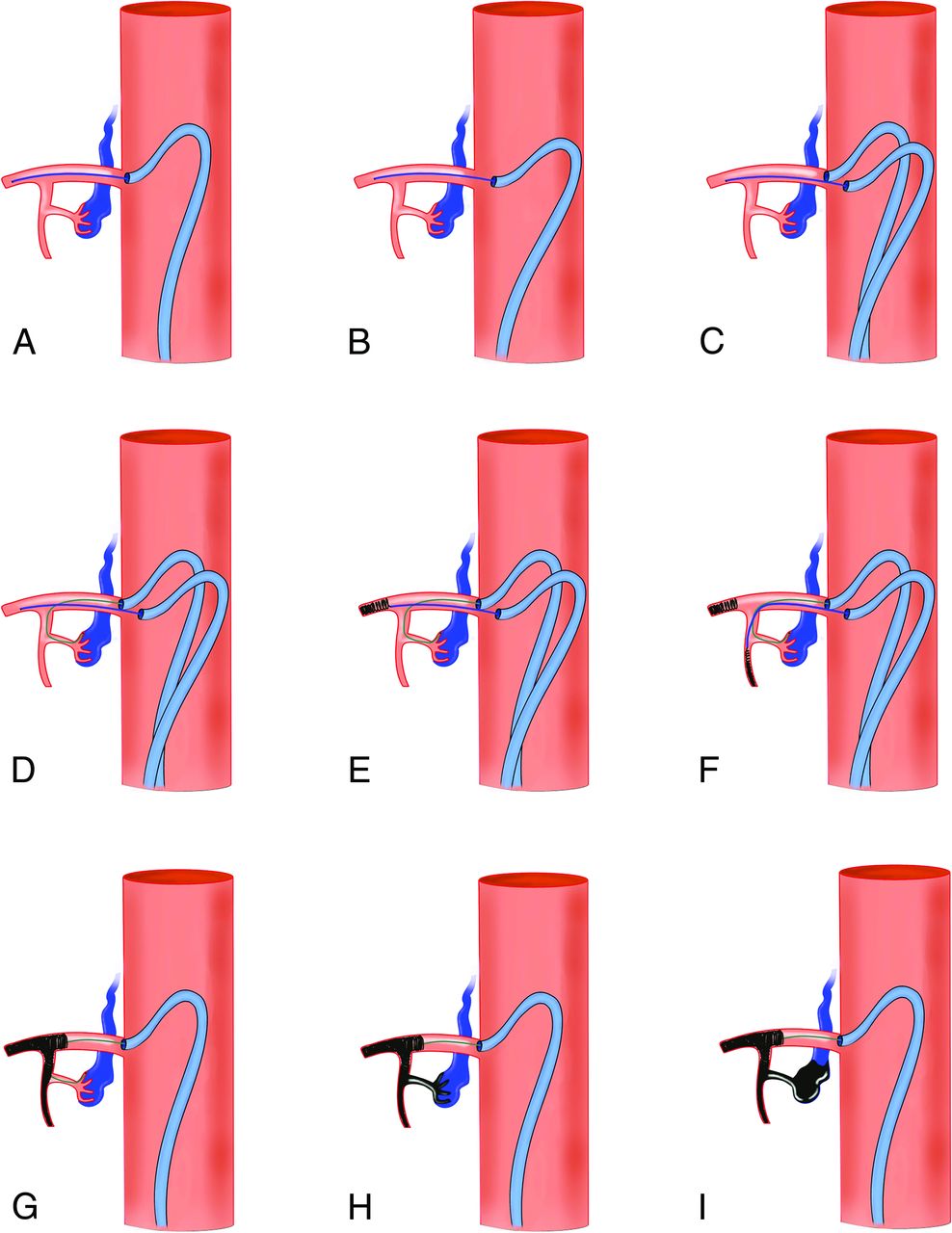
- FIG 1.
Drawing summarizing the different steps for the PCT in spinal vascular malformation embolization. A, Positioning the microcatheter No. 1 (for coiling) distally in the main trunk of the segmental artery, via the guiding catheter No. 1. B, The guiding catheter No. 1 is disengaged from the segmental artery ostium. C, The guiding catheter No. 2 is subsequently positioned at the ostium of the segmental artery. D, The microcatheter No. 2 (dedicated to the liquid embolic agent injection for the arteriovenous fistula treatment) is navigated via the guiding catheter No. 2 as close as possible to the shunt point. E, Coiling of the main trunk is performed via microcatheter No. 1 until occlusion. F and G, The dorsal branch and then the dorsospinal trunk are subsequently coiled until occlusion via the microcatheter No. 1 to obtain the proximal plug of the pressure cooker. H and I, Liquid embolic injection via the microcatheter No. 2 is performed until the filling of the origin of the draining vein.
- FIG 2.
Patient in his or her 40’s presenting with lower limb paresthesia, constipation, and bladder disturbance lasting for 36 months. A, Spinal cord MR imaging. T2WI, sagittal section, shows the hyperintense signal of the spinal cord extending up to T8 and multiple flow void signals in the enlarged spinal veins. B, Spinal DSA. Right T11 angiogram in a postero-anterior projection shows a spinal arteriovenous fistula with both ascending and descending venous drainage. C, Navigation of the microcatheter No. 1 (dedicated to the PCT plug) (black arrow). Then, the guiding catheter No. 1 is disengaged from the right T11 intercostal artery (white arrow). The guiding catheter No. 2 is subsequently positioned at the ostium of the segmental artery (white arrowhead). D, Navigation of the second microcatheter (No. 2) (black arrowhead) from the guiding catheter No. 2 (white arrowhead) as close as possible to the shunt point. Note the tip of microcatheter No. 1, located proximal to the tip of microcatheter No. 2. E, Ultraselective angiogram in a postero-anterior projection from microcatheter No. 2. The origin of the main draining vein (radicular vein) is clearly seen (double white arrow). F, PCT. Injection of 50% n-BCA through microcatheter No. 1 to create the proximal plug (black arrow). Then, injection of a 20% dilution of n-BCA is performed to embolize the fistula (white arrow). G, Plain x-ray in a postero-anterior projection shows the glue cast. Note the filling of the origin of the radicular vein (white arrows) and also progression of the liquid embolic agent through the retrocorporeal anastomosis (white arrowhead). H, Right T11 angiogram at the end of the procedure shows complete occlusion of the fistula. I, Spinal cord MR imaging 14 months after embolization. T2WI, sagittal section, shows complete resolution of the spinal cord edema.
- FIG 3.
Patient in his or her 60’s presenting with a 4-year history of paresthesia of the inferior limbs, bladder disturbance, and constipation. Spinal cord MR imaging. T2WI, sagittal (A) and axial (B) slices show a centromedullar hyperintense signal (white arrows) and lumbar spinal cord enlargement. Spinal DSA. Right L3 selective angiogram in a postero-anterior projection at an early phase (C) and intermediate phase (D). Numerous dural branches from the right L3 spinal trunk are depicted (C, white arrows), with abnormal connection with the right aspect of the epidural plexus (C, asterisk). Reflux from the anterior epidural plexus to a radicular vein is seen (C and D, black arrows), with an ascending drainage. E, Navigation of a 0.017-inch microcatheter via a 5F Simmons 1 catheter (white arrowhead). The microcatheter is stabilized by deploying a Catch Mini stent retriever (black arrows); then the guiding catheter is gently pulled away from the right L3 ostium. F, An Apollo (5-cm detachable tip) microcatheter is then navigated in the spinal trunk of the right L3 artery, as close as possible to the shunt point (black arrow). Selective DSA via the Apollo microcatheter in a postero-anterior projection. Note the guiding catheter (5F Simmons 1) at the ostium of the right L3 artery (black arrowhead), while the second one is stabilized in front of it (white arrowhead). Note the second 0.017-inch microcatheter that has been left in the main trunk of right L3 artery (white arrows). Coiling, via the 0.017-inch microcatheter, of the main L3 trunk (G, white arrow), then the dorsospinal trunk (G, black arrow) and the arterial segment before arising from the dorsospinal trunk (H, white arrow) to obtain the proximal plug of the pressure cooker. Finally, a 50% dilution n-BCA injection was performed at the proximal aspect of the cast of the coils through the 0.017-inch microcatheter (I, white arrow) to ensure that the proximal plug would be occlusive. Then, Squid 12 (Balt) was injected under a blank roadmap through the Apollo catheter until occlusion of the shunt point along the anterior epidural venous plexus (J and K, white arrows) and filling of the origin of the radicular vein (J, black arrow). L, Plain x-ray in a postero-anterior projection at the end of the Squid 12 injection shows the EVOH cast. M, Final control DSA in a postero-anterior projection via the common trunk feeding both the right and left L3 arteries, showing complete occlusion of the fistula. N, Postprocedural unenhanced CT scan, bone windowing. Sagittal reconstruction shows the EVOH cast in the anterior epidural venous plexus (white arrows).


Jump to section
Related Articles
Cited By...
- Magnetic resonance imaging changes in spinal arteriovenous fistulae treated by endovascular means: are they reliable to predict complete cure of the fistula?
- 'Pressure cooker and 'balloon pressure techniques significantly increase 3-month complete occlusion rate after spinal arteriovenous fistula embolization as compared to glue: single center evaluation on 38 consecutive patients
- 'Balloon pressure technique' for endovascular treatment of spinal cord arteriovenous fistulas: preliminary results in 10 cases