
Article Figures & Data
Figures
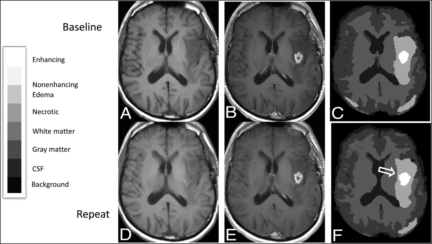
- FIG 1.
Registered baseline and repeat MR images from a 53-year-old man with multifocal left-hemispheric recurrent glioblastoma, with corresponding segmentations showing enlargement of enhancement segmentation on the repeat study compared with the baseline study. Registered T1-weighted axial slices obtained at baseline before (A) and after (B) administration of Gd-DTPA are shown along with segmentations obtained using BraTumIA 2.0 (C), as well as corresponding registered slices (D and E) and segmentation (F) from the repeat image set, aligned to match the baseline image set. The segmentation from the repeat image set appears larger along its anterior margin (open arrow). The overall enhancement volume increased by 2.2 mL on the repeat image set across all slices in the imaging volume. The legend for categorization of segmentation components within the segmentation is provided on the left.
- FIG 2.
Registered baseline and repeat MR images from a 62-year-old man with recurrent glioblastoma in the left temporal lobe, with corresponding segmentations showing a relatively high Dice coefficient for segmentation of enhancing tumor. Registered axial slices from the baseline image set, including T1-weighted images before (A) and after (B) administration of contrast agent, as well as FLAIR (C) and T2-weighted images (D) produced segmentation (E) using BraTumIA 1.2. Corresponding registered slices from the repeat imaging set (F – I) produced segmentation (K) using BraTumIA 1.2. The legend for categorization of segmentation components within the segmentation is provided on the left. The overlap of enhancing tumor segmentations from baseline (white) and repeat (black outline) time points at this slice is shown (J). For this case, the overall Dice coefficient is 0.94 and the 95% Hausdorff distance was 4.3 mm for the enhancing tumor segmentations. The legend for categorization of segmentation components within the segmentation is provided on the left.
- FIG 3.
Contrast-enhanced T1-weighted image from an MR imaging study of a 47-year-old man with recurrent glioblastoma in the right parietal lobe with corresponding segmentations obtained using BraTumIA 1.2 and BraTumIA 2.0. Registered axial contrast-enhanced T1-weighted image from the baseline image set (B) is compared to segmentations obtained by BraTumIA 1.2 (A) and BraTumIA 2.0 (C). For this case, the overall volume of the enhancing tumor segmentations was 23.4 mL for BraTumIA 1.2 and 20.0 mL for BraTumIA 2.0. The legend for categorization of segmentation components within the segmentation is the same as for Figs. 1 and 2.


Tables
- Table 1:
Differences between segmentation volumes obtained at baseline and repeat imaging (in mL)
Segmented Region Mean Segmentation Volumes, BraTumIA 1.2 [SD] Mean Segmentation Volumes, BraTumIA 2.0 [SD] Mean Difference in Volume between Baseline and Repeat Scans [SD] Baseline Repeat Baseline Repeat BraTumIA 1.2 BraTumIA2.0 Enhancing 24.2 [17.1] 26.3 [18.5] 22.9 [16.6] 24.9 [18.1] 2.1a 2.0b Edema 96.5 [34.3] 94.8 [39.2] 93.3 [33.2] 91.0 [37.7] –1.8 –2.2 Nonenhancing 3.0 [3.2] 3.0 [3.0] 2.8 [2.5] 2.6 [2.4] 0.03 –0.2 Necrotic 5.9 [13.0] 5.6 [12.4] 6.6 [12.6] 7.0 [12.9] –0.3 0.4 Total tumor-related abnormality 130 [51.3] 130 [56.7] 126 [48.4] 126 [52.8] 0.05 –0.1 Total nonenhancing tumor-related abnormality 105 [38.1] 103 [43.7] 103 [35.7] 101 [40.5] –2.0 –2.1 Nonenhancing non-necrotic tumor-related abnormality 99.5 [36.2] 97.8 [41.2] 96.1 [34.3] 93.6 [38.4] –1.7 –2.5 WM 545 [51.8] 543 [50.4] 549 [55.3] 546 [53.0] –2.7 –2.6 GM 594 [56.8] 597 [54.2] 608 [59.0] 611 [54.6] 2.7 2.8 BraTumIA Version RC (95% CI), mL %RC (95% CI) 1.2 2.0 1.2 2.0 Enhancing 6.9 (4.9–10) 5.2 (3.7–7.5) 46% (33%–67%) 39% (28%–57%) Edema 24 (17–35) 30 (21–43) 31% (22%–45%) 36% (26%–52%) Non-enhancing 2.1 (1.5–3.1) 2.0 (1.4–2.8) 95%a (68%–140%) 116%a (84%–174%) Necrotic 3.1 (2.2–4.5) 2.0 (1.4–2.9) 87% (62%–130%) 81% (58%–117%) Total tumor-related abnormality 25 (18–36) 31 (22–44) 26% (18%–37%) 32% (22%–47%) Total non-enhancing tumor-related abnormality 24 (17–34) 30 (21–43) 30% (21%–44%) 35% (25%–51%) Non-enhancing non-necrotic tumor-related abnormality 24 (17–35) 29 (21–42) 31% (22%–44%) 36% (25%–51%) White matter 40 (28–58) 47 (33–67) 7.1% (5.1%–10%) 8.3% (5.9%–12%) Gray matter 46 (33–67) 45 (32–65) 7.6% (5.3%–11%) 7.3% (5.1%–10%) ↵a N = 19 because of average non-enhancing volume of 0 for 1 patient.
BraTumIA Version Dice Coefficient (95% CI) 95%ile HD, mm (95% CI)b Average HD, mm (95% CI)b 1.2 2.0 1.2 2.0 1.2 2.0 Enhancing 0.81 (0.75–0.86) 0.75 (0.70–0.81) 19 (17–21) 14 (12–16) 0.88 (0.72–1.1) 0.72 (0.60–0.84) Edema 0.79 (0.75–0.84) 0.77 (0.73–0.81) 39 (34–45) 28 (24–31) 0.89 (0.72–1.1) 0.79 (0.66–0.94) Nonenhancing 0.27 (0.19–0.36) 0.24 (0.16–0.31)a 22 (20–25) 17 (16–19)a 3.9 (3.2–4.8) 3.1 (2.7–3.6)a Necrotic 0.51 (0.41–0.61) 0.45 (0.34–0.56) 18 (16–21) 16 (15–19) 2.3 (1.8–3.0) 2.3 (1.8–2.9) Total tumor-related abnormality 0.85 (0.81–0.88) 0.83 (0.80–0.87) 39 (33–46) 27 (24–31) 0.59 (0.47–0.73) 0.59 (0.48–0.73) Total nonenhancing tumor-related abnormality 0.80 (0.75–0.84) 0.77 (0.74–0.81) 40 (34–47) 27 (24–31) 0.70 (0.58–0.85) 0.76 (0.63–0.90) Nonenhancing non-necrotic tumor-related abnormality 0.80 (0.75–0.84) 0.77 (0.73–0.81) 39 (33–48) 28 (24–31) 0.71 (0.59–0.86) 0.78 (0.65–0.93) White matter 0.84 (0.83–0.85) 0.80 (0.79–0.82) 19 (18–20) 17 (17–18) 0.28 (0.26–0.30) 0.36 (0.34–0.38) Gray matter 0.76 (0.75–0.78) 0.72 (0.70–0.74) 13 (12–13) 14 (13–14) 0.37 (0.36–0.39) 0.48 (0.46–0.50) - Table 4:
Paired t test results for volumes reported by BraTumIA 1.2 and BraTumIA 2.0 (in mL)
Segmented Region Average Volume BraTumIA 1.2 Average Volume BraTumIA 2.0 Average Difference in Volumes (BraTumIA 2.0–BraTumIA 1.2) Mean SD Mean SD Mean Enhancing 25.2 17.6 23.9 17.1 –1.3a Edema 95.6 36.4 92.2 35.1 –3.4b Nonenhancing 3.02 3.04 2.69 2.43 –0.33 Necrotic 5.79 12.5 6.82 12.6 1.0b Total tumor-related abnormality 130 53.4 126 50.0 –4b Total nonenhancing tumor-related components 104 40.5 102 37.7 –2c Nonenhancing non-necrotic tumor-related components 98.7 38.3 94.8 36.0 –3.9b WM 544 50.5 547 53.5 3c GM 596 54.8 610 56.1 14a


Jump to section
Related Articles
Cited By...
- No citing articles found.