Advertisement

Main menu

  • Home
  • Content
    • Current Issue
    • Accepted Manuscripts
    • Article Preview
    • Past Issue Archive
    • Video Articles
    • AJNR Case Collection
    • Case of the Week Archive
    • Case of the Month Archive
    • Classic Case Archive
  • Special Collections
    • AJNR Awards
    • Low-Field MRI
    • Alzheimer Disease
    • ASNR Foundation Special Collection
    • Photon-Counting CT
    • View All
  • Multimedia
    • AJNR Podcasts
    • AJNR SCANtastic
    • Trainee Corner
    • MRI Safety Corner
    • Imaging Protocols
  • For Authors
    • Submit a Manuscript
    • Submit a Video Article
    • Submit an eLetter to the Editor/Response
    • Manuscript Submission Guidelines
    • Statistical Tips
    • Fast Publishing of Accepted Manuscripts
    • Graphical Abstract Preparation
    • Imaging Protocol Submission
    • Author Policies
  • About Us
    • About AJNR
    • Editorial Board
    • Editorial Board Alumni
  • More
    • Become a Reviewer/Academy of Reviewers
    • Subscribers
    • Permissions
    • Alerts
    • Feedback
    • Advertisers
    • ASNR Home

User menu

  • Alerts
  • Log in

Search

  • Advanced search
American Journal of Neuroradiology

American Journal of Neuroradiology

ASHNR American Society of Functional Neuroradiology ASHNR American Society of Pediatric Neuroradiology ASSR
  • Alerts
  • Log in

Advanced Search

  • Home
  • Content
    • Current Issue
    • Accepted Manuscripts
    • Article Preview
    • Past Issue Archive
    • Video Articles
    • AJNR Case Collection
    • Case of the Week Archive
    • Case of the Month Archive
    • Classic Case Archive
  • Special Collections
    • AJNR Awards
    • Low-Field MRI
    • Alzheimer Disease
    • ASNR Foundation Special Collection
    • Photon-Counting CT
    • View All
  • Multimedia
    • AJNR Podcasts
    • AJNR SCANtastic
    • Trainee Corner
    • MRI Safety Corner
    • Imaging Protocols
  • For Authors
    • Submit a Manuscript
    • Submit a Video Article
    • Submit an eLetter to the Editor/Response
    • Manuscript Submission Guidelines
    • Statistical Tips
    • Fast Publishing of Accepted Manuscripts
    • Graphical Abstract Preparation
    • Imaging Protocol Submission
    • Author Policies
  • About Us
    • About AJNR
    • Editorial Board
    • Editorial Board Alumni
  • More
    • Become a Reviewer/Academy of Reviewers
    • Subscribers
    • Permissions
    • Alerts
    • Feedback
    • Advertisers
    • ASNR Home
  • Follow AJNR on Twitter
  • Visit AJNR on Facebook
  • Follow AJNR on Instagram
  • Join AJNR on LinkedIn
  • RSS Feeds

AJNR Awards, New Junior Editors, and more. Read the latest AJNR updates

Research ArticleAdult Brain
Open Access

Manganese-Enhanced MRI in Patients with Multiple Sclerosis

D.J. Suto, G. Nair, D.M. Sudarshana, S.U. Steele, J. Dwyer, E.S. Beck, J. Ohayon, H. McFarland, A.P. Koretsky, I.C.M. Cortese and D.S. Reich
American Journal of Neuroradiology September 2020, 41 (9) 1569-1576; DOI: https://doi.org/10.3174/ajnr.A6665
D.J. Suto
aFrom the National Institute of Neurological Disorders and Stroke, National Institutes of Health, Bethesda, Maryland.
  • Find this author on Google Scholar
  • Find this author on PubMed
  • Search for this author on this site
  • ORCID record for D.J. Suto
G. Nair
aFrom the National Institute of Neurological Disorders and Stroke, National Institutes of Health, Bethesda, Maryland.
  • Find this author on Google Scholar
  • Find this author on PubMed
  • Search for this author on this site
  • ORCID record for G. Nair
D.M. Sudarshana
aFrom the National Institute of Neurological Disorders and Stroke, National Institutes of Health, Bethesda, Maryland.
  • Find this author on Google Scholar
  • Find this author on PubMed
  • Search for this author on this site
  • ORCID record for D.M. Sudarshana
S.U. Steele
aFrom the National Institute of Neurological Disorders and Stroke, National Institutes of Health, Bethesda, Maryland.
  • Find this author on Google Scholar
  • Find this author on PubMed
  • Search for this author on this site
  • ORCID record for S.U. Steele
J. Dwyer
aFrom the National Institute of Neurological Disorders and Stroke, National Institutes of Health, Bethesda, Maryland.
  • Find this author on Google Scholar
  • Find this author on PubMed
  • Search for this author on this site
  • ORCID record for J. Dwyer
E.S. Beck
aFrom the National Institute of Neurological Disorders and Stroke, National Institutes of Health, Bethesda, Maryland.
  • Find this author on Google Scholar
  • Find this author on PubMed
  • Search for this author on this site
  • ORCID record for E.S. Beck
J. Ohayon
aFrom the National Institute of Neurological Disorders and Stroke, National Institutes of Health, Bethesda, Maryland.
  • Find this author on Google Scholar
  • Find this author on PubMed
  • Search for this author on this site
  • ORCID record for J. Ohayon
H. McFarland
aFrom the National Institute of Neurological Disorders and Stroke, National Institutes of Health, Bethesda, Maryland.
  • Find this author on Google Scholar
  • Find this author on PubMed
  • Search for this author on this site
  • ORCID record for H. McFarland
A.P. Koretsky
aFrom the National Institute of Neurological Disorders and Stroke, National Institutes of Health, Bethesda, Maryland.
  • Find this author on Google Scholar
  • Find this author on PubMed
  • Search for this author on this site
  • ORCID record for A.P. Koretsky
I.C.M. Cortese
aFrom the National Institute of Neurological Disorders and Stroke, National Institutes of Health, Bethesda, Maryland.
  • Find this author on Google Scholar
  • Find this author on PubMed
  • Search for this author on this site
  • ORCID record for I.C.M. Cortese
D.S. Reich
aFrom the National Institute of Neurological Disorders and Stroke, National Institutes of Health, Bethesda, Maryland.
  • Find this author on Google Scholar
  • Find this author on PubMed
  • Search for this author on this site
  • ORCID record for D.S. Reich
  • Article
  • Figures & Data
  • Supplemental
  • Info & Metrics
  • Responses
  • References
  • PDF
Loading

Article Figures & Data

Figures

  • Tables
  • FIG 1.
    • Download figure
    • Open in new tab
    • Download powerpoint
    FIG 1.

    Summary of MR imaging time points and contrast enhancement. Participants were imaged at baseline, shortly after injection, and in the days to months after injection. Red vertical lines indicate MR imaging scans; blue bars, enhancement of MS lesions due to mangafodipir injection; yellow bars, gadolinium enhancement of MS lesions; the green bar, enhancement of a meningioma due to mangafodipir injection; and the orange bar, gadolinium enhancement of a meningioma. Gradients suggest decay of signal between time points but are not intended to be quantitative because temporal granularity is limited by infrequent imaging time points. D indicates days; Gd2+, gadolinium ion.

  • FIG 2.
    • Download figure
    • Open in new tab
    • Download powerpoint
    FIG 2.

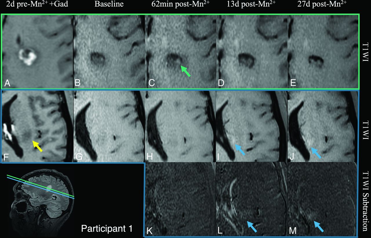
    Mangafodipir enhancement of an active gadolinium-enhancing lesion (participant 1). At screening, this lesion enhanced with gadolinium on T1-SPACE (A) with caudal edema visible on T1 (F, yellow arrow). At the center of the lesion (green plane, A–E), faint manganese enhancement is noted at 62 minutes (C) and resolved 13 days after injection (D). Caudal to the lesion (blue plane, F–M), manganese enhancement was noted outside the lesion border at 13 days (I, blue arrow) and on subtraction images (L). This enhancement persisted at 27 days (J and M, blue arrow). Additionally, ventricular narrowing, which commonly fluctuates,16 was noted on day 13 postmangafodipir in reference to the baseline (L); this appeared to resolve by day 27 postmangafodipir.

  • FIG 3.
    • Download figure
    • Open in new tab
    • Download powerpoint
    FIG 3.

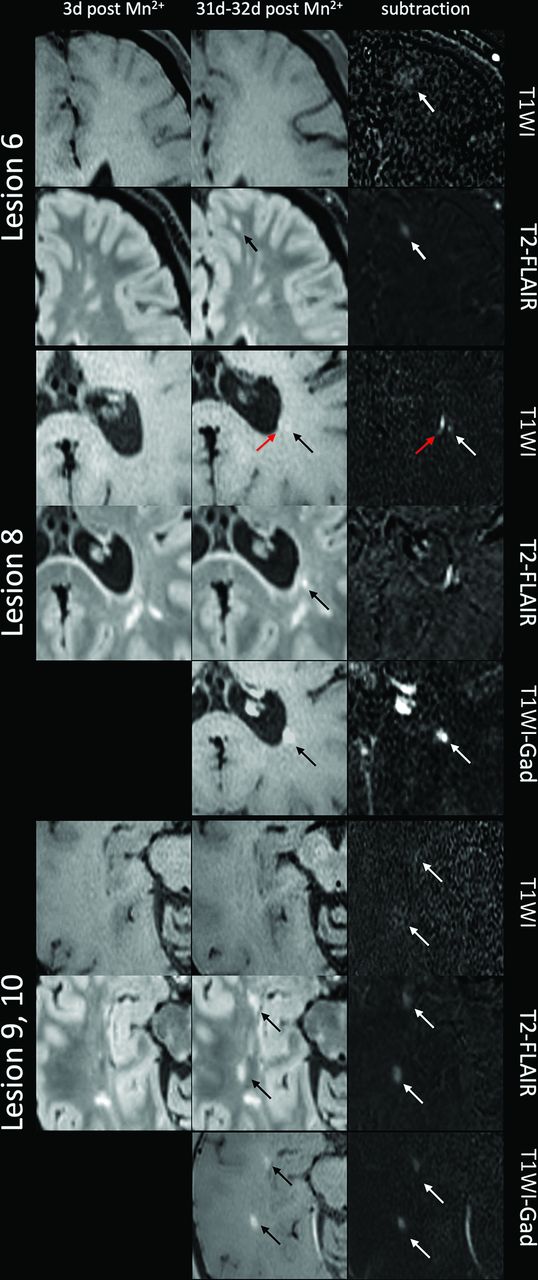
    Mangafodipir enhancement of MS lesions (participant 5). Upper part, Lesion 6 showed blush-like mangafodipir enhancement in a T2 lesion that formed between days 3 and 31 post-mangafodipir injection. There is subtle T1-hyperintense signal visible only on subtraction, suggesting manganese enhancement, in an area larger than the T2-FLAIR lesion. This lesion did not enhance with gadolinium on day 32 post-mangafodipir injection. Middle, lower part, Several lesions that formed between day 3 and day 31 post-mangafodipir injection enhanced with both manganese and gadolinium. Lesion 8 (middle) demonstrates nodular (black arrow) and ependymal (red arrow) enhancement with manganese but only nodular enhancement with gadolinium. Lesions 9 and 10 (lower part) demonstrate blush-like enhancement with manganese, visible on subtraction images, and nodular enhancement with gadolinium. Gad indicates gadolinium.

  • FIG 4.
    • Download figure
    • Open in new tab
    • Download powerpoint
    FIG 4.

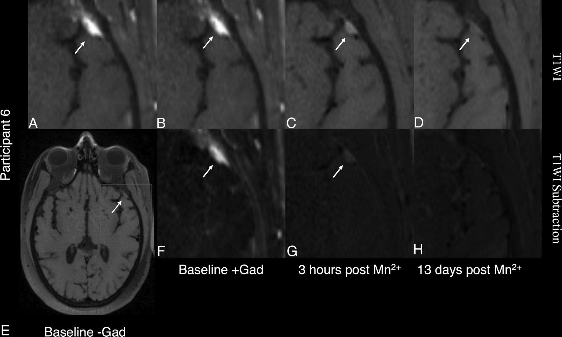
    Manganese enhancement of a presumed meningioma in participant 6, a 39-year-old man with secondary-progressive MS. The mass (arrow) is visible on precontrast T1-weighted images (A and E) with vivid gadolinium enhancement (gadolinium [Gad]) visible on both T1-weighted (B) and T1 gadolinium subtraction (F) images. Mangafodipir enhancement is visible within the mass 3 hours after mangafodipir (C and G) and had resolved by 13 days postinjection (D and H).

Tables

  • Figures
  • Clinical and demographic data

    ParticipantAge (yr)SexMS PhenotypeCurrent Disease-Modifying TherapyEDSSYears Since Symptom Onset
    138MRRMSNone1<1
    242FRRMSDimethyl fumarate1.55
    332FRRMSNone0<1
    440FRRMSInterferon β-1a36
    533FRRMSDaclizumab215
    639MSPMSAutologous stem cell transplant, conditioning with cyclophosphamide and rituximab613
    • Note:—SPMS indicates secondary-progressive MS; EDSS, Expanded Disability Status Scale.

PreviousNext
Back to top

In this issue

American Journal of Neuroradiology: 41 (9)
American Journal of Neuroradiology
Vol. 41, Issue 9
1 Sep 2020
  • Table of Contents
  • Index by author
  • Complete Issue (PDF)
Advertisement
Print
Download PDF
Email Article

Thank you for your interest in spreading the word on American Journal of Neuroradiology.

NOTE: We only request your email address so that the person you are recommending the page to knows that you wanted them to see it, and that it is not junk mail. We do not capture any email address.

Enter multiple addresses on separate lines or separate them with commas.
Manganese-Enhanced MRI in Patients with Multiple Sclerosis
(Your Name) has sent you a message from American Journal of Neuroradiology
(Your Name) thought you would like to see the American Journal of Neuroradiology web site.
CAPTCHA
This question is for testing whether or not you are a human visitor and to prevent automated spam submissions.
Cite this article
D.J. Suto, G. Nair, D.M. Sudarshana, S.U. Steele, J. Dwyer, E.S. Beck, J. Ohayon, H. McFarland, A.P. Koretsky, I.C.M. Cortese, D.S. Reich
Manganese-Enhanced MRI in Patients with Multiple Sclerosis
American Journal of Neuroradiology Sep 2020, 41 (9) 1569-1576; DOI: 10.3174/ajnr.A6665

Citation Manager Formats

  • BibTeX
  • Bookends
  • EasyBib
  • EndNote (tagged)
  • EndNote 8 (xml)
  • Medlars
  • Mendeley
  • Papers
  • RefWorks Tagged
  • Ref Manager
  • RIS
  • Zotero
0 Responses
Respond to this article
Share
Bookmark this article
Manganese-Enhanced MRI in Patients with Multiple Sclerosis
D.J. Suto, G. Nair, D.M. Sudarshana, S.U. Steele, J. Dwyer, E.S. Beck, J. Ohayon, H. McFarland, A.P. Koretsky, I.C.M. Cortese, D.S. Reich
American Journal of Neuroradiology Sep 2020, 41 (9) 1569-1576; DOI: 10.3174/ajnr.A6665
del.icio.us logo Twitter logo Facebook logo Mendeley logo
  • Tweet Widget
  • Facebook Like
  • Google Plus One
Purchase

Jump to section

  • Article
    • Abstract
    • ABBREVIATION:
    • MATERIALS AND METHODS
    • RESULTS
    • DISCUSSION
    • CONCLUSIONS
    • ACKNOWLEDGMENTS
    • Footnotes
    • References
  • Figures & Data
  • Supplemental
  • Info & Metrics
  • Responses
  • References
  • PDF

Related Articles

  • PubMed
  • Google Scholar

Cited By...

  • No citing articles found.
  • Crossref
  • Google Scholar

This article has not yet been cited by articles in journals that are participating in Crossref Cited-by Linking.

More in this TOC Section

Adult Brain

  • Diagnostic Neuroradiology of Monoclonal Antibodies
  • Clinical Outcomes After Chiari I Decompression
  • Segmentation of Brain Metastases with BLAST
Show more Adult Brain

Functional

  • Kurtosis and Epileptogenic Tubers: A Pilot Study
  • Glutaric Aciduria Type 1: DK vs. Conventional MRI
  • Multiparametric MRI in PEDS Pontine Glioma
Show more Functional

Similar Articles

Advertisement

Indexed Content

  • Current Issue
  • Accepted Manuscripts
  • Article Preview
  • Past Issues
  • Editorials
  • Editor's Choice
  • Fellows' Journal Club
  • Letters to the Editor
  • Video Articles

Cases

  • Case Collection
  • Archive - Case of the Week
  • Archive - Case of the Month
  • Archive - Classic Case

More from AJNR

  • Trainee Corner
  • Imaging Protocols
  • MRI Safety Corner
  • Book Reviews

Multimedia

  • AJNR Podcasts
  • AJNR Scantastics

Resources

  • Turnaround Time
  • Submit a Manuscript
  • Submit a Video Article
  • Submit an eLetter to the Editor/Response
  • Manuscript Submission Guidelines
  • Statistical Tips
  • Fast Publishing of Accepted Manuscripts
  • Graphical Abstract Preparation
  • Imaging Protocol Submission
  • Evidence-Based Medicine Level Guide
  • Publishing Checklists
  • Author Policies
  • Become a Reviewer/Academy of Reviewers
  • News and Updates

About Us

  • About AJNR
  • Editorial Board
  • Editorial Board Alumni
  • Alerts
  • Permissions
  • Not an AJNR Subscriber? Join Now
  • Advertise with Us
  • Librarian Resources
  • Feedback
  • Terms and Conditions
  • AJNR Editorial Board Alumni

American Society of Neuroradiology

  • Not an ASNR Member? Join Now

© 2025 by the American Society of Neuroradiology All rights, including for text and data mining, AI training, and similar technologies, are reserved.
Print ISSN: 0195-6108 Online ISSN: 1936-959X

Powered by HighWire