Advertisement

Main menu

  • Home
  • Content
    • Current Issue
    • Accepted Manuscripts
    • Article Preview
    • Past Issue Archive
    • Video Articles
    • AJNR Case Collection
    • Case of the Week Archive
    • Case of the Month Archive
    • Classic Case Archive
  • Special Collections
    • AJNR Awards
    • Low-Field MRI
    • Alzheimer Disease
    • ASNR Foundation Special Collection
    • Photon-Counting CT
    • View All
  • Multimedia
    • AJNR Podcasts
    • AJNR SCANtastic
    • Trainee Corner
    • MRI Safety Corner
    • Imaging Protocols
  • For Authors
    • Submit a Manuscript
    • Submit a Video Article
    • Submit an eLetter to the Editor/Response
    • Manuscript Submission Guidelines
    • Statistical Tips
    • Fast Publishing of Accepted Manuscripts
    • Graphical Abstract Preparation
    • Imaging Protocol Submission
    • Author Policies
  • About Us
    • About AJNR
    • Editorial Board
    • Editorial Board Alumni
  • More
    • Become a Reviewer/Academy of Reviewers
    • Subscribers
    • Permissions
    • Alerts
    • Feedback
    • Advertisers
    • ASNR Home

User menu

  • Alerts
  • Log in

Search

  • Advanced search
American Journal of Neuroradiology

American Journal of Neuroradiology

ASHNR American Society of Functional Neuroradiology ASHNR American Society of Pediatric Neuroradiology ASSR
  • Alerts
  • Log in

Advanced Search

  • Home
  • Content
    • Current Issue
    • Accepted Manuscripts
    • Article Preview
    • Past Issue Archive
    • Video Articles
    • AJNR Case Collection
    • Case of the Week Archive
    • Case of the Month Archive
    • Classic Case Archive
  • Special Collections
    • AJNR Awards
    • Low-Field MRI
    • Alzheimer Disease
    • ASNR Foundation Special Collection
    • Photon-Counting CT
    • View All
  • Multimedia
    • AJNR Podcasts
    • AJNR SCANtastic
    • Trainee Corner
    • MRI Safety Corner
    • Imaging Protocols
  • For Authors
    • Submit a Manuscript
    • Submit a Video Article
    • Submit an eLetter to the Editor/Response
    • Manuscript Submission Guidelines
    • Statistical Tips
    • Fast Publishing of Accepted Manuscripts
    • Graphical Abstract Preparation
    • Imaging Protocol Submission
    • Author Policies
  • About Us
    • About AJNR
    • Editorial Board
    • Editorial Board Alumni
  • More
    • Become a Reviewer/Academy of Reviewers
    • Subscribers
    • Permissions
    • Alerts
    • Feedback
    • Advertisers
    • ASNR Home
  • Follow AJNR on Twitter
  • Visit AJNR on Facebook
  • Follow AJNR on Instagram
  • Join AJNR on LinkedIn
  • RSS Feeds

AJNR Awards, New Junior Editors, and more. Read the latest AJNR updates

Research ArticleAdult Brain
Open Access

Discrimination between Glioblastoma and Solitary Brain Metastasis: Comparison of Inflow-Based Vascular-Space-Occupancy and Dynamic Susceptibility Contrast MR Imaging

X. Li, D. Wang, S. Liao, L. Guo, X. Xiao, X. Liu, Y. Xu, J. Hua, J.J. Pillai and Y. Wu
American Journal of Neuroradiology April 2020, 41 (4) 583-590; DOI: https://doi.org/10.3174/ajnr.A6466
X. Li
aFrom the Department of Medical Imaging (X. Li, S.L., L.G., X.X., X. Liu, Y.X., Y.W.), Nanfang Hospital, Southern Medical University, Guangzhou, P.R. China
  • Find this author on Google Scholar
  • Find this author on PubMed
  • Search for this author on this site
  • ORCID record for X. Li
D. Wang
bSchool of Biomedical Engineering (D.W.), Shanghai Jiao Tong University, Shanghai, P.R. China
  • Find this author on Google Scholar
  • Find this author on PubMed
  • Search for this author on this site
  • ORCID record for D. Wang
S. Liao
aFrom the Department of Medical Imaging (X. Li, S.L., L.G., X.X., X. Liu, Y.X., Y.W.), Nanfang Hospital, Southern Medical University, Guangzhou, P.R. China
cDivision of CT and MR, Radiology Department (S.L.), First Affiliated Hospital of Gannan Medical University, Ganzhou, P.R. China
  • Find this author on Google Scholar
  • Find this author on PubMed
  • Search for this author on this site
  • ORCID record for S. Liao
L. Guo
aFrom the Department of Medical Imaging (X. Li, S.L., L.G., X.X., X. Liu, Y.X., Y.W.), Nanfang Hospital, Southern Medical University, Guangzhou, P.R. China
  • Find this author on Google Scholar
  • Find this author on PubMed
  • Search for this author on this site
  • ORCID record for L. Guo
X. Xiao
aFrom the Department of Medical Imaging (X. Li, S.L., L.G., X.X., X. Liu, Y.X., Y.W.), Nanfang Hospital, Southern Medical University, Guangzhou, P.R. China
  • Find this author on Google Scholar
  • Find this author on PubMed
  • Search for this author on this site
  • ORCID record for X. Xiao
X. Liu
aFrom the Department of Medical Imaging (X. Li, S.L., L.G., X.X., X. Liu, Y.X., Y.W.), Nanfang Hospital, Southern Medical University, Guangzhou, P.R. China
  • Find this author on Google Scholar
  • Find this author on PubMed
  • Search for this author on this site
  • ORCID record for X. Liu
Y. Xu
aFrom the Department of Medical Imaging (X. Li, S.L., L.G., X.X., X. Liu, Y.X., Y.W.), Nanfang Hospital, Southern Medical University, Guangzhou, P.R. China
  • Find this author on Google Scholar
  • Find this author on PubMed
  • Search for this author on this site
  • ORCID record for Y. Xu
J. Hua
dNeurosection, Division of MR Research (J.H.)
gF.M. Kirby Research Center for Functional Brain Imaging (J.H.), Kennedy Krieger Institute, Baltimore, Maryland.
  • Find this author on Google Scholar
  • Find this author on PubMed
  • Search for this author on this site
  • ORCID record for J. Hua
J.J. Pillai
eDivision of Neuroradiology (J.P.); Russell H. Morgan Department of Radiology and Radiological Science and
fDepartment of Neurosurgery (J.P.), Johns Hopkins University School of Medicine, Baltimore, Maryland
  • Find this author on Google Scholar
  • Find this author on PubMed
  • Search for this author on this site
  • ORCID record for J.J. Pillai
Y. Wu
aFrom the Department of Medical Imaging (X. Li, S.L., L.G., X.X., X. Liu, Y.X., Y.W.), Nanfang Hospital, Southern Medical University, Guangzhou, P.R. China
  • Find this author on Google Scholar
  • Find this author on PubMed
  • Search for this author on this site
  • ORCID record for Y. Wu
  • Article
  • Figures & Data
  • Supplemental
  • Info & Metrics
  • Responses
  • References
  • PDF
Loading

Article Figures & Data

Figures

  • Tables
  • Fig 1.
    • Download figure
    • Open in new tab
    • Download powerpoint
    Fig 1.

    ROI placements. Four to six ROIs were drawn in both intratumoral (circle with solid line) and peritumoral (circle with dotted line) regions, and the maximum value was recorded. Also, an ROI in the contralateral white matter (circle with dotted line) was chosen as a reference. The insert is the magnification of lesion area.

  • Fig 2.
    • Download figure
    • Open in new tab
    • Download powerpoint
    Fig 2.

    iVASO-CBVa, rCBVa, and DSC-rCBV in the intratumoral region (A) and in peritumoral T2-hyperintense region (B) of glioblastoma and single brain metastasis. Data are presented as mean value ± SD. Ns indicates not significant.
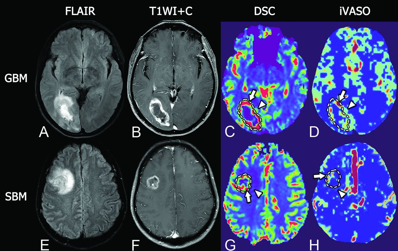
  • Fig 3.
    • Download figure
    • Open in new tab
    • Download powerpoint
    Fig 3.

    Representative MR images of glioblastoma and single brain metastasis. Upper row, A GBM in a 51-year-old woman. Lower row, SBM in a 46-year-old man. Both GBM and SBM present as hyperintense masses on T2 FLAIR with extensive peritumoral edema (A and E) and show a ring-enhancement pattern on fat-suppressed postcontrast T1WI with prominent necrosis in the tumor center (B and F). In intratumoral regions, GBM shows maximum DSC-rCBV similar to that of SBM (5.29 versus 4.98, arrows in C and G), but higher maximum iVASO-CBVa than SBM (5.90/100 mL versus 1.30/100 mL, arrows in D and H). In the peritumoral region, GBM shows prominently higher DSC-rCBV and iVASO-CBVa than SBM (DSC-rCBV, 3.11 versus 1.25, arrowheads in C and G; iVASO-CBVa, 1.20/100 mL versus 0.55/100 mL, arrowheads in D and H).

  • Fig 4.
    • Download figure
    • Open in new tab
    • Download powerpoint
    Fig 4.

    Receiver operating characteristic curves of parameters of iVASO and DSC MR imaging in differentiating glioblastoma and solitary brain metastasis. In the intratumoral region (A), both iVASO-CBVa (AUC  = 0.91) and rCBVa (AUC = 0.90) show higher AUCs than DSC-rCBV (AUC = 0.51). In the PTH (B), the AUC of DSC-rCBV (0.94) is higher than that of iVASO-CBVa (0.83) or rCBVa (0.72).

Tables

  • Figures
  • Results of receiver operating characteristic analysis of each parameter

    Technique/ParameterAUCP ValueCutoffSe (%)Sp (%)
    Intratumoral region
     iVASO
      CBVa0.91<.0013.2580.0100.0
      rCBVa0.90<.0015.2870.0100.0
     DSC
      rCBV0.51.9203.11100.018.2
    Peritumoral region
     iVASO
      CBVa0.83<.0011.0380.077.3
      rCBVa0.72.0142.0455.095.5
     DSC
      rCBV0.94<.0012.2680.0100.0
    • Note: Se indicates sensitivity; Sp, specificity.

PreviousNext
Back to top

In this issue

American Journal of Neuroradiology: 41 (4)
American Journal of Neuroradiology
Vol. 41, Issue 4
1 Apr 2020
  • Table of Contents
  • Index by author
  • Complete Issue (PDF)
Advertisement
Print
Download PDF
Email Article

Thank you for your interest in spreading the word on American Journal of Neuroradiology.

NOTE: We only request your email address so that the person you are recommending the page to knows that you wanted them to see it, and that it is not junk mail. We do not capture any email address.

Enter multiple addresses on separate lines or separate them with commas.
Discrimination between Glioblastoma and Solitary Brain Metastasis: Comparison of Inflow-Based Vascular-Space-Occupancy and Dynamic Susceptibility Contrast MR Imaging
(Your Name) has sent you a message from American Journal of Neuroradiology
(Your Name) thought you would like to see the American Journal of Neuroradiology web site.
CAPTCHA
This question is for testing whether or not you are a human visitor and to prevent automated spam submissions.
Cite this article
X. Li, D. Wang, S. Liao, L. Guo, X. Xiao, X. Liu, Y. Xu, J. Hua, J.J. Pillai, Y. Wu
Discrimination between Glioblastoma and Solitary Brain Metastasis: Comparison of Inflow-Based Vascular-Space-Occupancy and Dynamic Susceptibility Contrast MR Imaging
American Journal of Neuroradiology Apr 2020, 41 (4) 583-590; DOI: 10.3174/ajnr.A6466

Citation Manager Formats

  • BibTeX
  • Bookends
  • EasyBib
  • EndNote (tagged)
  • EndNote 8 (xml)
  • Medlars
  • Mendeley
  • Papers
  • RefWorks Tagged
  • Ref Manager
  • RIS
  • Zotero
0 Responses
Respond to this article
Share
Bookmark this article
Discrimination between Glioblastoma and Solitary Brain Metastasis: Comparison of Inflow-Based Vascular-Space-Occupancy and Dynamic Susceptibility Contrast MR Imaging
X. Li, D. Wang, S. Liao, L. Guo, X. Xiao, X. Liu, Y. Xu, J. Hua, J.J. Pillai, Y. Wu
American Journal of Neuroradiology Apr 2020, 41 (4) 583-590; DOI: 10.3174/ajnr.A6466
del.icio.us logo Twitter logo Facebook logo Mendeley logo
  • Tweet Widget
  • Facebook Like
  • Google Plus One
Purchase

Jump to section

  • Article
    • Abstract
    • ABBREVIATIONS:
    • MATERIALS AND METHODS
    • RESULTS
    • DISCUSSION
    • CONCLUSIONS
    • Footnotes
    • References
  • Figures & Data
  • Supplemental
  • Info & Metrics
  • Responses
  • References
  • PDF

Related Articles

  • PubMed
  • Google Scholar

Cited By...

  • No citing articles found.
  • Crossref (17)
  • Google Scholar

This article has been cited by the following articles in journals that are participating in Crossref Cited-by Linking.

  • Machine learning based differentiation of glioblastoma from brain metastasis using MRI derived radiomics
    Sarv Priya, Yanan Liu, Caitlin Ward, Nam H. Le, Neetu Soni, Ravishankar Pillenahalli Maheshwarappa, Varun Monga, Honghai Zhang, Milan Sonka, Girish Bathla
    Scientific Reports 2021 11 1
  • Single brain metastasis versus glioblastoma multiforme: a VOI-based multiparametric analysis for differential diagnosis
    Andrea Romano, Giulia Moltoni, Alessia Guarnera, Luca Pasquini, Alberto Di Napoli, Antonio Napolitano, Maria Camilla Rossi Espagnet, Alessandro Bozzao
    La radiologia medica 2022 127 5
  • Differentiating Glioblastoma Multiforme from Brain Metastases Using Multidimensional Radiomics Features Derived from MRI and Multiple Machine Learning Models
    Salar Bijari, Amin Jahanbakhshi, Parham Hajishafiezahramini, Parviz Abdolmaleki, Jiang Du
    BioMed Research International 2022 2022 1
  • The Role of 3D-pCASL MRI in the Differential Diagnosis of Glioblastoma and Brain Metastases
    Kristina Solozhentseva, Artem Batalov, Natalia Zakharova, Sergey Goryaynov, Eduard Pogosbekyan, Igor Pronin
    Frontiers in Oncology 2022 12
  • Artificial Neural Network-Derived Cerebral Metabolic Rate of Oxygen for Differentiating Glioblastoma and Brain Metastasis in MRI: A Feasibility Study
    Hakim Baazaoui, Simon Hubertus, Máté E. Maros, Sherif A. Mohamed, Alex Förster, Lothar R. Schad, Holger Wenz
    Applied Sciences 2021 11 21
  • Clinical application of brain perfusion imaging in detecting stroke mimics: A review
    Neda Khalili, Richard Wang, Tushar Garg, Amara Ahmed, Meisam Hoseinyazdi, Haris I. Sair, Licia P. Luna, Jarunee Intrapiromkul, Francis Deng, Vivek Yedavalli
    Journal of Neuroimaging 2023 33 1
  • Noninvasive Assessment of O(6)‐Methylguanine‐DNA Methyltransferase Promoter Methylation Status in World Health Organization Grade II–IV Glioma Using Histogram Analysis of Inflow‐Based Vascular‐Space‐Occupancy Combined with Structural Magnetic Resonance Imaging
    Wenle He, Xiaodan Li, Jun Hua, Shukun Liao, Liuji Guo, Xiang Xiao, Xiaomin Liu, Jun Zhou, Wensheng Wang, Yikai Xu, Yuankui Wu
    Journal of Magnetic Resonance Imaging 2021 54 1
  • Differential detection of metastatic and inflammatory lymph nodes using inflow-based vascular-space-occupancy (iVASO) MR imaging
    Liuji Guo, Xiaomin Liu, Jun Hua, Lin Dai, Ying Tao, Haimei Cao, Jie Qin, Xiaodan Li, Yuankui Wu
    Magnetic Resonance Imaging 2022 85
  • Texture analysis: A potential tool to differentiate primary brain tumors and solitary brain metastasis
    S. A. S. Souza, R. A. C. Guassu, A. F. F. Alves, M. Alvarez, L. C. C. Pitanga, F. Reis, A. Vacavant, J. R. A. Miranda, J. C. S. Trindade Filho, D. R. Pina
    Multimedia Tools and Applications 2023 83 13
  • Advanced magnetic resonance imaging findings of cerebellar hemangioblastomas: A report of three cases and a literature review
    Eiji Matsusue, Chie Inoue, Sadaharu Tabuchi, Hiroki Yoshioka, Yuichiro Nagao, Kensuke Matsumoto, Kazuhiko Nakamura, Shinya Fujii
    Acta Radiologica Open 2022 11 2

More in this TOC Section

Adult Brain

  • Diagnostic Neuroradiology of Monoclonal Antibodies
  • Clinical Outcomes After Chiari I Decompression
  • Segmentation of Brain Metastases with BLAST
Show more Adult Brain

Functional

  • Kurtosis and Epileptogenic Tubers: A Pilot Study
  • Glutaric Aciduria Type 1: DK vs. Conventional MRI
  • Brain Iron in Niemann-Pick Type C: 7T Study
Show more Functional

Similar Articles

Advertisement

Indexed Content

  • Current Issue
  • Accepted Manuscripts
  • Article Preview
  • Past Issues
  • Editorials
  • Editor's Choice
  • Fellows' Journal Club
  • Letters to the Editor
  • Video Articles

Cases

  • Case Collection
  • Archive - Case of the Week
  • Archive - Case of the Month
  • Archive - Classic Case

More from AJNR

  • Trainee Corner
  • Imaging Protocols
  • MRI Safety Corner
  • Book Reviews

Multimedia

  • AJNR Podcasts
  • AJNR Scantastics

Resources

  • Turnaround Time
  • Submit a Manuscript
  • Submit a Video Article
  • Submit an eLetter to the Editor/Response
  • Manuscript Submission Guidelines
  • Statistical Tips
  • Fast Publishing of Accepted Manuscripts
  • Graphical Abstract Preparation
  • Imaging Protocol Submission
  • Evidence-Based Medicine Level Guide
  • Publishing Checklists
  • Author Policies
  • Become a Reviewer/Academy of Reviewers
  • News and Updates

About Us

  • About AJNR
  • Editorial Board
  • Editorial Board Alumni
  • Alerts
  • Permissions
  • Not an AJNR Subscriber? Join Now
  • Advertise with Us
  • Librarian Resources
  • Feedback
  • Terms and Conditions
  • AJNR Editorial Board Alumni

American Society of Neuroradiology

  • Not an ASNR Member? Join Now

© 2025 by the American Society of Neuroradiology All rights, including for text and data mining, AI training, and similar technologies, are reserved.
Print ISSN: 0195-6108 Online ISSN: 1936-959X

Powered by HighWire