
Article Figures & Data
Figures
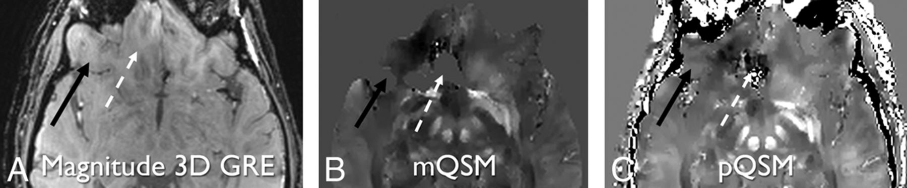
- Fig 1.
Paramagnetic right temporal hemorrhage is hypointense on magnitude 3D GRE (TE = 17.3 ms) (A) and hyperintense on mQSM (B) and pQSM (C) images (white dashed arrows). The diamagnetic calcification in bone (white solid arrows) is mostly hypointense on 3D GRE (A) and pQSM (C) and not present on mQSM (B) secondary to masking. Some areas of hyperintensity within the bone may represent diamagnetic structures, such as veins or artifacts. Left temporal lobe parenchyma next to hemorrhage is eroded on the mQSM (B), but preserved on magnitude and pQSM (C) images (white circles).
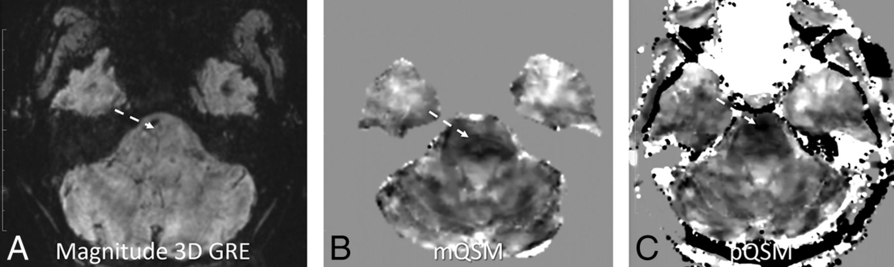
- Fig 2.
Right cerebellar hemorrhage is hypointense on magnitude 3D GRE (TE = 14.3 ms) (A). This part of the cerebellar parenchyma has been eroded on mQSM (B) and so is not visible due to masking. The same anatomy is preserved on pQSM (C), and hemorrhage is demonstrated (white solid arrows and circles). Additionally, an absent section of the right temporal brain parenchyma on mQSM (B) is partially preserved on the pQSM (C) relative to the 3D GRE image (A) (white dashed arrows and dashed circles). Note areas of black pixels within the white dashed circles on the mQSM and pQSM images (B and C) that correspond to brain tissue seen on 3D GRE image (A) indicate areas of nonvisualized brain.
- Fig 3.
Magnitude 3D GRE (TE = 14.3 ms) (A), mQSM (B), and pQSM (C) images demonstrating partial pQSM image depiction relative to mQSM. An area of the right inferior frontal lobe present on the 3D GRE image is present on the pQSM image and absent on mQSM image (black solid arrows). An adjacent inferior frontal lobe is present on the 3D GRE image, distorted on the pQSM image, and absent on the mQSM (white dashed arrows).
- Fig 4.
pQSM (C) demonstrates worse nonspecific diffuse low signal in the pons compared with mQSM (B), with no corresponding abnormality on the magnitude 3D GRE (TE = 41.8 ms) image (A) (white dashed arrows). Note that areas of hypointensity are in different positions within the pons.


Tables
Ohio TBI Score Sex Age (yr) Blood on GRE Nonvisualized Brain Male Female Mean Age SD Age Range on mQSM on pQSM All 34 22 42.1 14.42778 20–74 10 31 5 1 (none) 13 9 41.7 14.6878 20–74 1 13 3 2 (mild) 2 2 44.0 15.71623 27–58 1 3 0 3 (mild) 8 5 47.0 14.94434 30–71 4 8 1 4 (moderate) 2 1 45.7 13.50309 32–59 0 1 0 5 (severe) 9 5 37.9 13.96621 23–64 4 5 1 Correlation across TBI severity χ2 = 0.17, P = .98a ANOVA F(3,52) = 0.74, P = .53 P = .11b χ2 = 2.20, P = .53a P = .88b Correlation with blood products on 3D GRE χ2 = 0.04, P = .86a P = .49c – χ2 = 2.99, P = .08a P = .10b Feature Count % Blood products 3D GRE 10 18 mQSM 4 7 pQSM 10 18 Nonvisualized brain tissue on mQSM, pQSM visible 31 55 mQSM and pQSM, less on pQSM 5 9 pQSM, mQSM visible 0 0 Pons probable artifacts, more visible on pQSM 6 11 mQSM 0 0


Jump to section
Related Articles
Cited By...
- No citing articles found.