Advertisement

Main menu

  • Home
  • Content
    • Current Issue
    • Accepted Manuscripts
    • Article Preview
    • Past Issue Archive
    • Video Articles
    • AJNR Case Collection
    • Case of the Week Archive
    • Case of the Month Archive
    • Classic Case Archive
  • Special Collections
    • AJNR Awards
    • Low-Field MRI
    • Alzheimer Disease
    • ASNR Foundation Special Collection
    • Photon-Counting CT
    • View All
  • Multimedia
    • AJNR Podcasts
    • AJNR SCANtastic
    • Trainee Corner
    • MRI Safety Corner
    • Imaging Protocols
  • For Authors
    • Submit a Manuscript
    • Submit a Video Article
    • Submit an eLetter to the Editor/Response
    • Manuscript Submission Guidelines
    • Statistical Tips
    • Fast Publishing of Accepted Manuscripts
    • Graphical Abstract Preparation
    • Imaging Protocol Submission
    • Author Policies
  • About Us
    • About AJNR
    • Editorial Board
    • Editorial Board Alumni
  • More
    • Become a Reviewer/Academy of Reviewers
    • Subscribers
    • Permissions
    • Alerts
    • Feedback
    • Advertisers
    • ASNR Home

User menu

  • Alerts
  • Log in

Search

  • Advanced search
American Journal of Neuroradiology

American Journal of Neuroradiology

ASHNR American Society of Functional Neuroradiology ASHNR American Society of Pediatric Neuroradiology ASSR
  • Alerts
  • Log in

Advanced Search

  • Home
  • Content
    • Current Issue
    • Accepted Manuscripts
    • Article Preview
    • Past Issue Archive
    • Video Articles
    • AJNR Case Collection
    • Case of the Week Archive
    • Case of the Month Archive
    • Classic Case Archive
  • Special Collections
    • AJNR Awards
    • Low-Field MRI
    • Alzheimer Disease
    • ASNR Foundation Special Collection
    • Photon-Counting CT
    • View All
  • Multimedia
    • AJNR Podcasts
    • AJNR SCANtastic
    • Trainee Corner
    • MRI Safety Corner
    • Imaging Protocols
  • For Authors
    • Submit a Manuscript
    • Submit a Video Article
    • Submit an eLetter to the Editor/Response
    • Manuscript Submission Guidelines
    • Statistical Tips
    • Fast Publishing of Accepted Manuscripts
    • Graphical Abstract Preparation
    • Imaging Protocol Submission
    • Author Policies
  • About Us
    • About AJNR
    • Editorial Board
    • Editorial Board Alumni
  • More
    • Become a Reviewer/Academy of Reviewers
    • Subscribers
    • Permissions
    • Alerts
    • Feedback
    • Advertisers
    • ASNR Home
  • Follow AJNR on Twitter
  • Visit AJNR on Facebook
  • Follow AJNR on Instagram
  • Join AJNR on LinkedIn
  • RSS Feeds

AJNR Awards, New Junior Editors, and more. Read the latest AJNR updates

Research ArticleADULT BRAIN
Open Access

Influence of T1-Weighted Signal Intensity on FSL Voxel-Based Morphometry and FreeSurfer Cortical Thickness

S. Chung, X. Wang and Y.W. Lui
American Journal of Neuroradiology April 2017, 38 (4) 726-728; DOI: https://doi.org/10.3174/ajnr.A5053
S. Chung
aFrom the Center for Advanced Imaging Innovation and Research (CAI2R), Department of Radiology, and Bernard and Irene Schwartz Center for Biomedical Imaging, Department of Radiology, New York University School of Medicine, New York, New York.
  • Find this author on Google Scholar
  • Find this author on PubMed
  • Search for this author on this site
  • ORCID record for S. Chung
X. Wang
aFrom the Center for Advanced Imaging Innovation and Research (CAI2R), Department of Radiology, and Bernard and Irene Schwartz Center for Biomedical Imaging, Department of Radiology, New York University School of Medicine, New York, New York.
  • Find this author on Google Scholar
  • Find this author on PubMed
  • Search for this author on this site
  • ORCID record for X. Wang
Y.W. Lui
aFrom the Center for Advanced Imaging Innovation and Research (CAI2R), Department of Radiology, and Bernard and Irene Schwartz Center for Biomedical Imaging, Department of Radiology, New York University School of Medicine, New York, New York.
  • Find this author on Google Scholar
  • Find this author on PubMed
  • Search for this author on this site
  • ORCID record for Y.W. Lui
  • Article
  • Figures & Data
  • Info & Metrics
  • Responses
  • References
  • PDF
Loading

Abstract

SUMMARY: The effect of T1 signal on FSL voxel-based morphometry modulated GM density and FreeSurfer cortical thickness is explored. The techniques rely on different analyses, but both are commonly used to detect spatial changes in GM. Standard pipelines show FSL voxel-based morphometry is sensitive to T1 signal alterations within a physiologic range, and results can appear discordant between FSL voxel-based morphometry and FreeSurfer cortical thickness. Care should be taken in extrapolating results to the effect on brain volume.

ABBREVIATIONS:

CT
cortical thickness
mGM
modulated GM density
mTBI
mild traumatic brain injury
VBM
voxel-based morphometry

Cortical segmentation methodologies vary and are used throughout neuroimaging research as well as, increasingly, in clinical care. Two commonly used methods to study cortical GM are FMRIB Software Library voxel-based morphometry (FSL-VBM [http://fsl.fmrib.ox.ac.uk/fsl/fsl-4.1.9/fslvbm/index.html])1 and FreeSurfer (http://surfer.nmr.mgh.harvard.edu) cortical thickness (CT).2 FSL-VBM outputs a 3D map of modulated GM density (mGM), and FreeSurfer-CT analysis outputs 1D measurements around the cortical ribbon. Both are commonly interpreted as informing cortical volume, though there are instances where volume was increased based on one technique and decreased based on the other,3,4 suggesting that factors beyond brain volume contribute to results.

GM and WM contrast naturally contributes to successful segmentation; however, the extent to which changes in cortical T1 signal affect mGM and CT is not known. Understanding this relationship is critical for an appropriate interpretation. This study explored the effect of subtle T1 signal alterations within a physiologic range on FSL-VBM1 and FreeSurfer-CT2 by using standard processing pipelines. We also illustrate discordance between the techniques through individual clinical examples.

Materials and Methods

This study is in compliance with our Department of Radiology institutional review board. MPRAGE was performed at 3T (Skyra [Siemens, Erlangen, Germany]; FOV, 256 × 256 mm2; resolution, 1 × 1 × 1 mm3; matrix, 256 × 256; sections, 192; TR, 2100 ms; TE, 3.19 ms; TI, 900 ms; bandwidth, 260 Hz/pixel; flip angle, 8°).

Signal Intensity Simulation

MPRAGE images from a healthy 25-year-old man were used to generate simulated signal changes within the frontal operculum (Fig 1). Forty percent of voxels within the ROI were randomly selected for signal intensity alteration of up to ±20% (5% increments), covering a physiologic T1 range of GM. Pearson correlation coefficient was measured (5% significance level).

Fig 1.
  • Download figure
  • Open in new tab
  • Download powerpoint
Fig 1.

Simulated cortical signal change. A, First row (left to right), Representative MPRAGE in a healthy control patient (25-year-old man). Forty percent of voxels through 7 slices within the ROI were randomly selected and used to simulate changes in T1 signal intensity via decreasing and increasing voxel intensity to 20% in increments of 5%. Selected ROI from the frontal operculum for simulating signal change is shown in the original image (white solid line). Second row, Corresponding segmented GM maps in the native space. Third row, mGM maps in the template space and, Fourth row, corresponding cortical boundaries from FreeSurfer. B, High correlation between signal intensity and mGM is observed (black dots [R = 0.964; P < .001]), but no relationship is seen between signal intensity and CT (gray triangles).

Clinical Examples

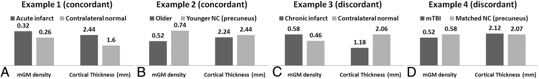
Several clinical cases were selected to illustrate concordant and discordant results: 1) acute and chronic infarcts; 2) healthy 19-year-old and 50-year-old patients; and, 3) a 28-year-old man with mild traumatic brain injury (mTBI) and an age- and sex-matched control patient.

Image Analysis

All images underwent denoising.5 Standard FSL-VBM (v1.1) processing steps included brain extraction, manual editing, automated tissue-type segmentation, nonlinear registration to GM template, modulation, and smoothing with isotropic Gaussian kernel (σ, 3 mm). FreeSurfer (v5.3.0) analysis was performed to estimate regional CT.

ROIs were drawn around areas of acute and chronic infarct in the native space and compared with contralateral normal-appearing analogous brain. For the control group and patients with TBI, a precuneal ROI from the Harvard-Oxford atlas was interrogated because previous studies report age-6 and TBI-associated7 morphometry changes in this region. ROIs drawn in the native space were warped to the target space by using transform matrices created by FSL-VBM and FreeSurfer-CT.

Results

Simulation results show a strong correlation between signal intensity and mGM (R = 0.964; P < .001). No correlation is present between signal intensity and CT (Fig 1B).

Concordant and discordant results are illustrated in Figs 2 and 3. The acute infarct showed 23% higher mGM and 52% higher CT compared with the contralateral analogous brain. The older healthy patient showed 30% lower mGM and 8% lower CT in the precuneus compared with the younger control patient. Discordant results included area of chronic infarct, demonstrating 26% higher mGM and 43% lower CT compared with the contralateral normal-appearing analogous brain. In the patient with mTBI, mGM was 12% lower and CT was 2% higher in a precuneal ROI compared with a matched control patient.

Fig 2.
  • Download figure
  • Open in new tab
  • Download powerpoint
Fig 2.

A 71-year-old man with acute onset weakness of the left upper extremity presented with an area of acute infarction (blue arrow) along the deep posterior cortex of the right precentral gyrus involving the hand motor region, with A, relative hypointensity on MPRAGE, B, hyperintensity on FLAIR, and C, restricted diffusion on the ADC map. The patient also has an area of chronic infarction more anteriorly (red arrow), showing A, relative hypointensity on MPRAGE, B, hyperintensity on FLAIR, and C, T2 shine through on the ADC map. ROIs in the affected areas (D) show higher and lower CT, respectively, compared with contralateral analogous brain (E); however, mGM as a marker of cortical volume was higher in the acute infarct, as expected, but also higher in the chronic infarct.

Fig 3.
  • Download figure
  • Open in new tab
  • Download powerpoint
Fig 3.

In vivo examples illustrate concordant results between FSL-VBM and FreeSurfer-CT in the ROIs of acute infarct (A) as well as when comparing younger and older healthy control patients (B), and discordant cortical morphometry results in ROIs of chronic infarct (C) and in the precuneus (D) in a patient with mTBI compared with a matched control patient. Of note, prior work reports morphometric changes to the precuneus in aging and traumatic brain injury. The mean values within the ROIs were reported.

Discussion

FSL-VBM is sensitive to T1 signal variations within a clinically relevant range, which was not found to be true for FreeSurfer-CT. Although mGM and CT are completely different measures, both are commonly used to assess GM volume. Discordant results between FSL-VBM and FreeSurfer-CT analyses may result from T1 effects on mGM. This observation is of clinical and research importance because there are myriad conditions that affect T1 signal. Careful interpretation of FSL-VBM is warranted, particularly in the setting of discordant findings.

Several FSL-VBM methodologic steps are worth comment: 1) bias correction may alter T1 signal, and 2) tissue-type segmentation can affect output mGM. All images underwent identical bias correction before both analyses, and we demonstrated the effect of T1 signal change on mGM and CT by applying standard pipelines to recreate commonly used approaches that have widespread availability. Future optimization is warranted to achieve accurate detection of pathology.

Other methods of cortical segmentation are not specifically addressed here. Whether a similar dependence on T1 signal is present in VBM approaches such as SPM12 (http://www.fil.ion.ucl.ac.uk/spm/software/spm12) is not known. The cases provided as part of this report are not meant as generalizable results regarding the specific pathologies described, but instead serve as in vivo examples of the phenomenon of discordance in terms of interpreting FSL-VBM and FreeSurfer-CT. Future studies with larger cohorts would be useful to study specific conditions; these results from single patients demonstrate that discordance may be present not only at a group-wise statistical level, but on an individual basis.

Conclusions

In summary, we demonstrated the dependence of mGM on T1 signal. Care should be taken in interpreting mGM results as volume change alone. Used in concert, FSL-VBM and FreeSurfer-CT analyses may be complementary.

Footnotes

  • Disclosures: Yvonne Lui—RELATED: Grant: NIH R01 NS039135-11, NIH R21 NS090349, NIH P41 EB017183.* *Money paid to the institution.

  • This work was supported by grant R01 NS039135-11 from the National Institute for Neurological Disorders and Stroke, a component of the National Institutes of Health. This work was also performed under the rubric of the Center for Advanced Imaging Innovation and Research (CAI2R, www.cai2r.net), a National Institute of Biomedical Imaging and Bioengineering Biomedical Technology Resource Center (NIH P41 EB017183).

Indicates open access to non-subscribers at www.ajnr.org

REFERENCES

  1. 1.↵
    1. Good CD,
    2. Johnsrude IS,
    3. Ashburner J, et al
    . A voxel-based morphometric study of ageing in 465 normal adult human brains. Neuroimage 2001;14:21–36 doi:10.1006/nimg.2001.0786 pmid:11525331
    CrossRefPubMed
  2. 2.↵
    1. Dale AM,
    2. Fischl B,
    3. Sereno MI
    . Cortical surface-based analysis. I. Segmentation and surface reconstruction. Neuroimage 1999;9:179–94 doi:10.1006/nimg.1998.0395 pmid:9931268
    CrossRefPubMed
  3. 3.↵
    1. Hutton C,
    2. Draganski B,
    3. Ashburner J, et al
    . A comparison between voxel-based cortical thickness and voxel-based morphometry in normal aging. Neuroimage 2009;48:371–80 doi:10.1016/j.neuroimage.2009.06.043 pmid:19559801
    CrossRefPubMed
  4. 4.↵
    1. Winkler AM,
    2. Kochunov P,
    3. Blangero J, et al
    . Cortical thickness or grey matter volume? The importance of selecting the phenotype for imaging genetics studies. Neuroimage 2010;53:1135–46 doi:10.1016/j.neuroimage.2009.12.028 pmid:20006715
    CrossRefPubMed
  5. 5.↵
    1. Smith SM,
    2. Brady JM
    . SUSAN—A new approach to low level image processing. Int J Comput Vis 1997;23:45–78 doi:10.1023/A:1007963824710
    CrossRefPubMed
  6. 6.↵
    1. Gaetz W,
    2. Roberts TP,
    3. Singh KD, et al
    . Functional and structural correlates of the aging brain: relating visual cortex (V1) gamma band responses to age-related structural change. Hum Brain Mapp 2012;33:2035–46 doi:10.1002/hbm.21339 pmid:21769992
    CrossRefPubMed
  7. 7.↵
    1. Zhou Y,
    2. Kierans A,
    3. Kenul D, et al
    . Mild traumatic brain injury: longitudinal regional brain volume changes. Radiology 2013;267:880–90 doi:10.1148/radiol.13122542 pmid:23481161
    CrossRefPubMed
  • Received June 24, 2016.
  • Accepted after revision October 26, 2016.
  • © 2017 by American Journal of Neuroradiology
View Abstract
PreviousNext
Back to top

In this issue

American Journal of Neuroradiology: 38 (4)
American Journal of Neuroradiology
Vol. 38, Issue 4
1 Apr 2017
  • Table of Contents
  • Index by author
  • Complete Issue (PDF)
Advertisement
Print
Download PDF
Email Article

Thank you for your interest in spreading the word on American Journal of Neuroradiology.

NOTE: We only request your email address so that the person you are recommending the page to knows that you wanted them to see it, and that it is not junk mail. We do not capture any email address.

Enter multiple addresses on separate lines or separate them with commas.
Influence of T1-Weighted Signal Intensity on FSL Voxel-Based Morphometry and FreeSurfer Cortical Thickness
(Your Name) has sent you a message from American Journal of Neuroradiology
(Your Name) thought you would like to see the American Journal of Neuroradiology web site.
CAPTCHA
This question is for testing whether or not you are a human visitor and to prevent automated spam submissions.
Cite this article
S. Chung, X. Wang, Y.W. Lui
Influence of T1-Weighted Signal Intensity on FSL Voxel-Based Morphometry and FreeSurfer Cortical Thickness
American Journal of Neuroradiology Apr 2017, 38 (4) 726-728; DOI: 10.3174/ajnr.A5053

Citation Manager Formats

  • BibTeX
  • Bookends
  • EasyBib
  • EndNote (tagged)
  • EndNote 8 (xml)
  • Medlars
  • Mendeley
  • Papers
  • RefWorks Tagged
  • Ref Manager
  • RIS
  • Zotero
0 Responses
Respond to this article
Share
Bookmark this article
Influence of T1-Weighted Signal Intensity on FSL Voxel-Based Morphometry and FreeSurfer Cortical Thickness
S. Chung, X. Wang, Y.W. Lui
American Journal of Neuroradiology Apr 2017, 38 (4) 726-728; DOI: 10.3174/ajnr.A5053
del.icio.us logo Twitter logo Facebook logo Mendeley logo
  • Tweet Widget
  • Facebook Like
  • Google Plus One
Purchase

Jump to section

  • Article
    • Abstract
    • ABBREVIATIONS:
    • Materials and Methods
    • Results
    • Discussion
    • Conclusions
    • Footnotes
    • REFERENCES
  • Figures & Data
  • Info & Metrics
  • Responses
  • References
  • PDF

Related Articles

  • No related articles found.
  • PubMed
  • Google Scholar

Cited By...

  • Fast and functionally specific cortical thickness changes induced by visual stimulation
  • Learning-related contraction of grey matter in rodent sensorimotor cortex is associated with adaptive myelination
  • Crossref (9)
  • Google Scholar

This article has been cited by the following articles in journals that are participating in Crossref Cited-by Linking.

  • Expansion and Renormalization of Human Brain Structure During Skill Acquisition
    Elisabeth Wenger, Claudio Brozzoli, Ulman Lindenberger, Martin Lövdén
    Trends in Cognitive Sciences 2017 21 12
  • Accelerating research on biological aging and mental health: Current challenges and future directions
    Laura K.M. Han, Josine E. Verhoeven, Audrey R. Tyrka, Brenda W.J.H. Penninx, Owen M. Wolkowitz, Kristoffer N.T. Månsson, Daniel Lindqvist, Marco P. Boks, Dóra Révész, Synthia H. Mellon, Martin Picard
    Psychoneuroendocrinology 2019 106
  • Volumetric gray matter measures of amygdala and accumbens in childhood overweight/obesity
    Gabor Perlaki, Denes Molnar, Paul A. M. Smeets, Wolfgang Ahrens, Maike Wolters, Gabriele Eiben, Lauren Lissner, Peter Erhard, Floor van Meer, Manfred Herrmann, Jozsef Janszky, Gergely Orsi, Francisco J. Esteban
    PLOS ONE 2018 13 10
  • Menstrual Cycle Variations in Gray Matter Volume, White Matter Volume and Functional Connectivity: Critical Impact on Parietal Lobe
    Timothy J. Meeker, Dieuwke S. Veldhuijzen, Michael L. Keaser, Rao P. Gullapalli, Joel D. Greenspan
    Frontiers in Neuroscience 2020 14
  • Learning-related contraction of gray matter in rodent sensorimotor cortex is associated with adaptive myelination
    Tomas Mediavilla, Özgün Özalay, Héctor M Estévez-Silva, Bárbara Frias, Greger Orädd, Fahad R Sultan, Claudio Brozzoli, Benjamín Garzón, Martin Lövdén, Daniel J Marcellino
    eLife 2022 11
  • Influence of hemispheric white matter lesions and migraine characteristics on cortical thickness and volume
    Hedvig Komáromy, Mingchen He, Gábor Perlaki, Gergely Orsi, Szilvia Anett Nagy, Edit Bosnyák, David Kamson Olayinka, Flóra John, Anita Trauninger, Zoltán Pfund
    The Journal of Headache and Pain 2019 20 1
  • Longitudinal changes in brain parenchyma due to mild traumatic brain injury during the first year after injury
    Angela M. Muller, William J. Panenka, Rael T. Lange, Grant L. Iverson, Jeffrey R. Brubacher, Naznin Virji‐Babul
    Brain and Behavior 2021 11 12
  • Cerebral changes and disrupted gray matter cortical networks in asymptomatic older adults at risk for Alzheimer&#039;s disease
    Jose L. Cantero, Mercedes Atienza, Pascual Sanchez-Juan, Eloy Rodriguez-Rodriguez, Jose Luis Vazquez-Higuera, Ana Pozueta, Andrea Gonzalez-Suarez, Eduard Vilaplana, Jordi Pegueroles, Victor Montal, Rafael Blesa, Daniel Alcolea, Alberto Lleo, Juan Fortea
    Neurobiology of Aging 2018 64
  • The Neurobiology and Treatment of OCD: Accelerating Progress
    D. J. Veltman
    2021 49

More in this TOC Section

  • Diagnostic Neuroradiology of Monoclonal Antibodies
  • Clinical Outcomes After Chiari I Decompression
  • Segmentation of Brain Metastases with BLAST
Show more ADULT BRAIN

Similar Articles

Advertisement

Indexed Content

  • Current Issue
  • Accepted Manuscripts
  • Article Preview
  • Past Issues
  • Editorials
  • Editor's Choice
  • Fellows' Journal Club
  • Letters to the Editor
  • Video Articles

Cases

  • Case Collection
  • Archive - Case of the Week
  • Archive - Case of the Month
  • Archive - Classic Case

More from AJNR

  • Trainee Corner
  • Imaging Protocols
  • MRI Safety Corner
  • Book Reviews

Multimedia

  • AJNR Podcasts
  • AJNR Scantastics

Resources

  • Turnaround Time
  • Submit a Manuscript
  • Submit a Video Article
  • Submit an eLetter to the Editor/Response
  • Manuscript Submission Guidelines
  • Statistical Tips
  • Fast Publishing of Accepted Manuscripts
  • Graphical Abstract Preparation
  • Imaging Protocol Submission
  • Evidence-Based Medicine Level Guide
  • Publishing Checklists
  • Author Policies
  • Become a Reviewer/Academy of Reviewers
  • News and Updates

About Us

  • About AJNR
  • Editorial Board
  • Editorial Board Alumni
  • Alerts
  • Permissions
  • Not an AJNR Subscriber? Join Now
  • Advertise with Us
  • Librarian Resources
  • Feedback
  • Terms and Conditions
  • AJNR Editorial Board Alumni

American Society of Neuroradiology

  • Not an ASNR Member? Join Now

© 2025 by the American Society of Neuroradiology All rights, including for text and data mining, AI training, and similar technologies, are reserved.
Print ISSN: 0195-6108 Online ISSN: 1936-959X

Powered by HighWire