Advertisement

Main menu

  • Home
  • Content
    • Current Issue
    • Accepted Manuscripts
    • Article Preview
    • Past Issue Archive
    • Video Articles
    • AJNR Case Collection
    • Case of the Week Archive
    • Case of the Month Archive
    • Classic Case Archive
  • Special Collections
    • AJNR Awards
    • Low-Field MRI
    • Alzheimer Disease
    • ASNR Foundation Special Collection
    • Photon-Counting CT
    • View All
  • Multimedia
    • AJNR Podcasts
    • AJNR SCANtastic
    • Trainee Corner
    • MRI Safety Corner
    • Imaging Protocols
  • For Authors
    • Submit a Manuscript
    • Submit a Video Article
    • Submit an eLetter to the Editor/Response
    • Manuscript Submission Guidelines
    • Statistical Tips
    • Fast Publishing of Accepted Manuscripts
    • Graphical Abstract Preparation
    • Imaging Protocol Submission
    • Author Policies
  • About Us
    • About AJNR
    • Editorial Board
    • Editorial Board Alumni
  • More
    • Become a Reviewer/Academy of Reviewers
    • Subscribers
    • Permissions
    • Alerts
    • Feedback
    • Advertisers
    • ASNR Home

User menu

  • Alerts
  • Log in

Search

  • Advanced search
American Journal of Neuroradiology

American Journal of Neuroradiology

ASHNR American Society of Functional Neuroradiology ASHNR American Society of Pediatric Neuroradiology ASSR
  • Alerts
  • Log in

Advanced Search

  • Home
  • Content
    • Current Issue
    • Accepted Manuscripts
    • Article Preview
    • Past Issue Archive
    • Video Articles
    • AJNR Case Collection
    • Case of the Week Archive
    • Case of the Month Archive
    • Classic Case Archive
  • Special Collections
    • AJNR Awards
    • Low-Field MRI
    • Alzheimer Disease
    • ASNR Foundation Special Collection
    • Photon-Counting CT
    • View All
  • Multimedia
    • AJNR Podcasts
    • AJNR SCANtastic
    • Trainee Corner
    • MRI Safety Corner
    • Imaging Protocols
  • For Authors
    • Submit a Manuscript
    • Submit a Video Article
    • Submit an eLetter to the Editor/Response
    • Manuscript Submission Guidelines
    • Statistical Tips
    • Fast Publishing of Accepted Manuscripts
    • Graphical Abstract Preparation
    • Imaging Protocol Submission
    • Author Policies
  • About Us
    • About AJNR
    • Editorial Board
    • Editorial Board Alumni
  • More
    • Become a Reviewer/Academy of Reviewers
    • Subscribers
    • Permissions
    • Alerts
    • Feedback
    • Advertisers
    • ASNR Home
  • Follow AJNR on Twitter
  • Visit AJNR on Facebook
  • Follow AJNR on Instagram
  • Join AJNR on LinkedIn
  • RSS Feeds

AJNR Awards, New Junior Editors, and more. Read the latest AJNR updates

Research ArticleADULT BRAIN
Open Access

Impact of Pial Collaterals on Infarct Growth Rate in Experimental Acute Ischemic Stroke

G.A. Christoforidis, P. Vakil, S.A. Ansari, F.H. Dehkordi and T.J. Carroll
American Journal of Neuroradiology February 2017, 38 (2) 270-275; DOI: https://doi.org/10.3174/ajnr.A5003
G.A. Christoforidis
aFrom the Department of Radiology (G.A.C., S.A.A., T.J.C.), University of Chicago, Chicago, Illinois
  • Find this author on Google Scholar
  • Find this author on PubMed
  • Search for this author on this site
  • ORCID record for G.A. Christoforidis
P. Vakil
bCollege of Medicine (P.V.), University of Illinois, Chicago, Illinois
  • Find this author on Google Scholar
  • Find this author on PubMed
  • Search for this author on this site
  • ORCID record for P. Vakil
S.A. Ansari
aFrom the Department of Radiology (G.A.C., S.A.A., T.J.C.), University of Chicago, Chicago, Illinois
cDepartments of Radiology, Neurology, and Neurological Surgery (S.A.A.), Northwestern University, Chicago, Illinois
  • Find this author on Google Scholar
  • Find this author on PubMed
  • Search for this author on this site
  • ORCID record for S.A. Ansari
F.H. Dehkordi
dDepartment of Economics and Decision Sciences (F.H.D.), Western Illinois University, Macomb, Illinois.
  • Find this author on Google Scholar
  • Find this author on PubMed
  • Search for this author on this site
  • ORCID record for F.H. Dehkordi
T.J. Carroll
aFrom the Department of Radiology (G.A.C., S.A.A., T.J.C.), University of Chicago, Chicago, Illinois
  • Find this author on Google Scholar
  • Find this author on PubMed
  • Search for this author on this site
  • ORCID record for T.J. Carroll
  • Article
  • Figures & Data
  • Info & Metrics
  • Responses
  • References
  • PDF
Loading

Article Figures & Data

Figures

  • Tables
  • Fig 1.
    • Download figure
    • Open in new tab
    • Download powerpoint
    Fig 1.

    Arterial arrival times measured from angiographic time-density curves. ROIs within the normal MCA proximal M1 segment (white arrow, A) and from collateralized MCA branches (double arrows, A) are identified on composite angiographic images. ROIs were used to calculate time-density curves (B). “Average arterial time” (in seconds) was defined as the time interval between contrast arrival at the normal M1 segment (interrupted curve, B) and the average of 3 ROIs at the M3/4 junction of the MCA corresponding to the occluded MCA (continuous curve, B). AAT is graphically depicted by the horizontal double arrow line.

  • Fig 2.
    • Download figure
    • Open in new tab
    • Download powerpoint
    Fig 2.

    Final infarct volumes by T2 FLAIR images acquired 24 hours post-MCA occlusion are compared with baseline (ie, 15 minutes postocclusion) angiographic measures of pial collateral recruitment. A, Angiographic scoring of pial collateral score and arterial arrival time (B) strongly correlates with final infarct volume. Both pial collateral score (C) and AAT (D) are predictive the infarct growth rate index (derived from the fits shown in Fig 3B) on the basis of a linear function.
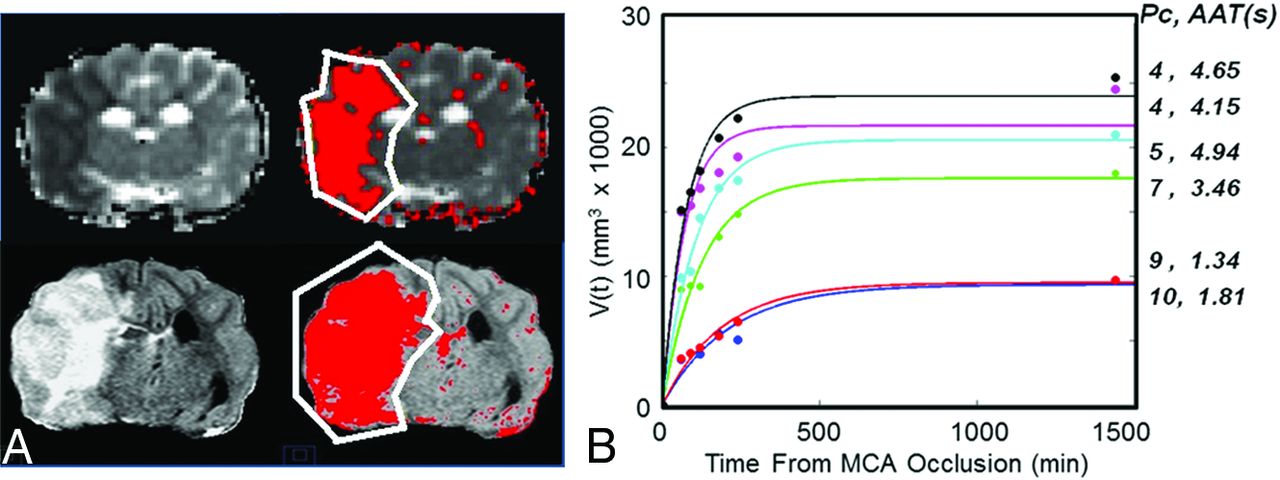
  • Fig 3.
    • Download figure
    • Open in new tab
    • Download powerpoint
    Fig 3.

    Single-section 120-minute mean diffusivity (upper part) and 24-hour FLAIR (lower part) images (A) were used to estimate and plot the growth of the infarct volume with time (B). A semiautomated algorithm was used to estimate the volume of the infarct on the basis of signal intensity within the affected hemisphere (red area), varying 1.5 times the SD from the mean of the signal in the contralateral normal-appearing hemisphere exclusive of spinal fluid. B, Infarct volume growth with time follows a predicable trend. Each curve corresponds to 1 experiment. The pial collateral scores and AAT measured immediately after occlusion (ie, at t = 15 minutes) for each curve are listed on the right.
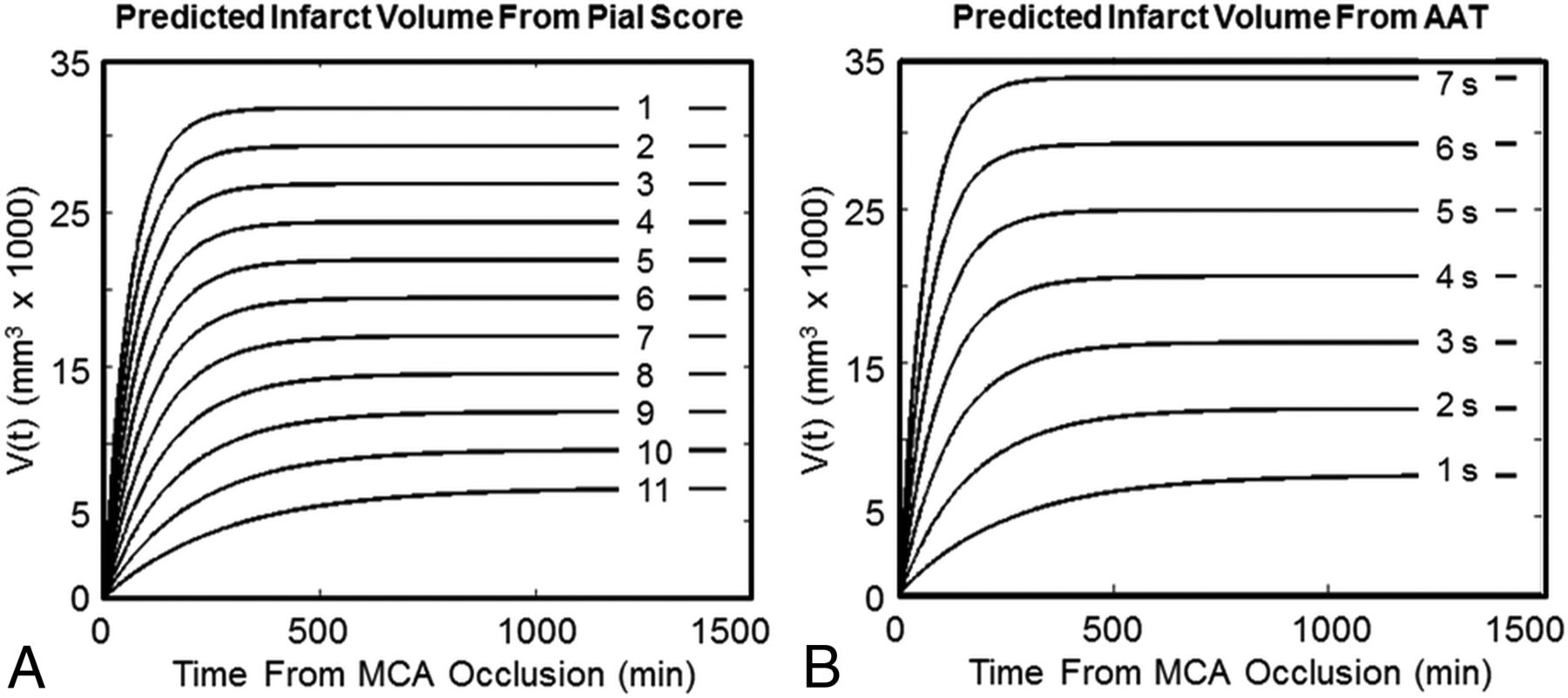
  • Fig 4.
    • Download figure
    • Open in new tab
    • Download powerpoint
    Fig 4.

    Families of infarct volume growth curves over 24 hours predicted by the pial collateral score and arterial arrival time.

Tables

  • Figures
    • View popup
    Table 1:

    Raw data

    Experiment No.
    123456
    Pc9.007.0010.505.504.003.50
    AAT (sec)1.3443.4691.8124.9384.6564.156
    V (60 min) (mm3)353389603575984214,94715,074
    V (90 min) (mm3)39769221400910,31015,42816,464
    V (120 min) (mm3)39229174443014,49316,80518,134
    V (180 min) (mm3)549213,043531216,81118,03420,694
    V (240 min) (mm3)505814,800646117,41919,25722,197
    V (24 hr) (mm3)961217,987966820,94624,47925,419
    • Note:—V (time) indicates infarct volume at “time” evaluated with diffusion-weighted MRI; V (24 hour), final infarct volume from FLAIR MRI.

    • View popup
    Table 2:

    Growth rate index and final infarct volumes

    ExperimentG (95% CI) ×(10−3)1/minVfit (95% CI) ×1000 mm3r2
    14.765 (2.37–7.16)9.31 (7.08–11.55)0.92
    27.951 (4.65–11.25)17.58 (14.52–20.63)0.96
    35.337 (3.629–7.05)9.467 (8.04–10.89)0.97
    49.23 (6.79–11.67)20.54 (18.41–22.67)0.98
    514.20 (5.64–22.75)21.65 (17.64–25.66)0.94
    613.22 (8.79–17.67)23.95 (21.36–26.54)0.98
    • Note:—G indicates growth rate index from fit; Vfit, final infarct volume from fit; r2 = coefficient of determination of fit.

PreviousNext
Back to top

In this issue

American Journal of Neuroradiology: 38 (2)
American Journal of Neuroradiology
Vol. 38, Issue 2
1 Feb 2017
  • Table of Contents
  • Index by author
  • Complete Issue (PDF)
Advertisement
Print
Download PDF
Email Article

Thank you for your interest in spreading the word on American Journal of Neuroradiology.

NOTE: We only request your email address so that the person you are recommending the page to knows that you wanted them to see it, and that it is not junk mail. We do not capture any email address.

Enter multiple addresses on separate lines or separate them with commas.
Impact of Pial Collaterals on Infarct Growth Rate in Experimental Acute Ischemic Stroke
(Your Name) has sent you a message from American Journal of Neuroradiology
(Your Name) thought you would like to see the American Journal of Neuroradiology web site.
CAPTCHA
This question is for testing whether or not you are a human visitor and to prevent automated spam submissions.
Cite this article
G.A. Christoforidis, P. Vakil, S.A. Ansari, F.H. Dehkordi, T.J. Carroll
Impact of Pial Collaterals on Infarct Growth Rate in Experimental Acute Ischemic Stroke
American Journal of Neuroradiology Feb 2017, 38 (2) 270-275; DOI: 10.3174/ajnr.A5003

Citation Manager Formats

  • BibTeX
  • Bookends
  • EasyBib
  • EndNote (tagged)
  • EndNote 8 (xml)
  • Medlars
  • Mendeley
  • Papers
  • RefWorks Tagged
  • Ref Manager
  • RIS
  • Zotero
0 Responses
Respond to this article
Share
Bookmark this article
Impact of Pial Collaterals on Infarct Growth Rate in Experimental Acute Ischemic Stroke
G.A. Christoforidis, P. Vakil, S.A. Ansari, F.H. Dehkordi, T.J. Carroll
American Journal of Neuroradiology Feb 2017, 38 (2) 270-275; DOI: 10.3174/ajnr.A5003
del.icio.us logo Twitter logo Facebook logo Mendeley logo
  • Tweet Widget
  • Facebook Like
  • Google Plus One
Purchase

Jump to section

  • Article
    • Abstract
    • ABBREVIATIONS:
    • Materials and Methods
    • Results
    • Discussion
    • Conclusions
    • Footnotes
    • References
  • Figures & Data
  • Info & Metrics
  • Responses
  • References
  • PDF

Related Articles

  • No related articles found.
  • PubMed
  • Google Scholar

Cited By...

  • A Method for Imaging the Ischemic Penumbra with MRI Using Intravoxel Incoherent Motion
  • Quantification of Collateral Supply with Local-AIF Dynamic Susceptibility Contrast MRI Predicts Infarct Growth
  • Association between intraprocedural drops in blood pressure and infarct growth rate patterns after acute large-vessel occlusions
  • Dynamic evolution of infarct volumes at MRI in ischemic stroke due to large vessel occlusion
  • Factors Associated With Fast Early Infarct Growth in Patients With Acute Ischemic Stroke With a Large Vessel Occlusion
  • Effect of early Sanguinate (PEGylated carboxyhemoglobin bovine) infusion on cerebral blood flow to the ischemic core in experimental middle cerebral artery occlusion
  • Infarct Progression in the Early and Late Phases of Acute Ischemic Stroke
  • Influence of simultaneous pressor and vasodilatory agents on the evolution of infarct growth in experimental acute middle cerebral artery occlusion
  • Association of Collateral Status and Ischemic Core Growth in Patients With Acute Ischemic Stroke
  • Focal cooling of brain parenchyma in a transient large vessel occlusion model: proof-of-concept
  • A canine model of mechanical thrombectomy in stroke
  • Crossref (43)
  • Google Scholar

This article has been cited by the following articles in journals that are participating in Crossref Cited-by Linking.

  • Association of Collateral Status and Ischemic Core Growth in Patients With Acute Ischemic Stroke
    Longting Lin, Jianhong Yang, Chushuang Chen, Huiqiao Tian, Andrew Bivard, Neil J. Spratt, Christopher R. Levi, Mark W. Parsons
    Neurology 2021 96 2
  • High Variability in Neuronal Loss
    Shashvat M. Desai, Marcelo Rocha, Tudor G. Jovin, Ashutosh P. Jadhav
    Stroke 2019 50 1
  • EphA4/Tie2 crosstalk regulates leptomeningeal collateral remodeling following ischemic stroke
    Benjamin Okyere, William A. Mills, Xia Wang, Michael Chen, Jiang Chen, Amanda Hazy, Yun Qian, John B. Matson, Michelle H. Theus
    Journal of Clinical Investigation 2020 130 2
  • Time Is Brain: The Stroke Theory of Relativity
    Camilo R. Gomez
    Journal of Stroke and Cerebrovascular Diseases 2018 27 8
  • Clinical and Imaging Parameters Associated With Hyperacute Infarction Growth in Large Vessel Occlusion Stroke
    Daniel Puhr-Westerheide, Steffen Tiedt, Lukas T. Rotkopf, Moriz Herzberg, Paul Reidler, Matthias P. Fabritius, Philipp M. Kazmierczak, Lars Kellert, Katharina Feil, Kolja M. Thierfelder, Franziska Dorn, Thomas Liebig, Frank A. Wollenweber, Wolfgang G. Kunz
    Stroke 2019 50 10
  • Early Collateral Recruitment After Stroke in Infants and Adults
    Philippe Bonnin, Mikaël Mazighi, Christiane Charriaut-Marlangue, Nathalie Kubis
    Stroke 2019 50 9
  • Factors influencing infarct growth including collateral status assessed using computed tomography in acute stroke patients with large artery occlusion
    Bin Jiang, Robyn L Ball, Patrik Michel, Ying Li, Guangming Zhu, Victoria Ding, Bochao Su, Zack Naqvi, Ashraf Eskandari, Manisha Desai, Max Wintermark
    International Journal of Stroke 2019 14 6
  • Focal cooling of brain parenchyma in a transient large vessel occlusion model: proof-of-concept
    Jildaz Caroff, Robert M King, Jennifer E Mitchell, Miklos Marosfoi, Joseph R Licwinko, Heather L Gray-Edwards, Ajit S Puri, Thomas L Merrill, Matthew J Gounis
    Journal of NeuroInterventional Surgery 2020 12 2
  • Systematic evaluation of computed tomography angiography collateral scores for estimation of long-term outcome after mechanical thrombectomy in acute ischaemic stroke
    Daniel Weiss, Bastian Kraus, Christian Rubbert, Marius Kaschner, Sebastian Jander, Michael Gliem, John-Ih Lee, Carl-Albrecht Haensch, Bernd Turowski, Julian Caspers
    The Neuroradiology Journal 2019 32 4
  • Factors Associated With Fast Early Infarct Growth in Patients With Acute Ischemic Stroke With a Large Vessel Occlusion
    Pierre Seners, Nicole Yuen, Jean-Marc Olivot, Michael Mlynash, Jeremy J. Heit, Soren Christensen, José Bernardo Escribano-Paredes, Emmanuel Carrera, Davide Strambo, Patrik Michel, Alexander Salerno, Max Wintermark, Hui Chen, Jean-François Albucher, Christophe Cognard, Igor Sibon, Michael Obadia, Julien Savatovsky, Maarten G. Lansberg, Gregory W. Albers
    Neurology 2023 101 21

More in this TOC Section

  • Diagnostic Neuroradiology of Monoclonal Antibodies
  • Clinical Outcomes After Chiari I Decompression
  • Segmentation of Brain Metastases with BLAST
Show more ADULT BRAIN

Similar Articles

Advertisement

Indexed Content

  • Current Issue
  • Accepted Manuscripts
  • Article Preview
  • Past Issues
  • Editorials
  • Editor's Choice
  • Fellows' Journal Club
  • Letters to the Editor
  • Video Articles

Cases

  • Case Collection
  • Archive - Case of the Week
  • Archive - Case of the Month
  • Archive - Classic Case

More from AJNR

  • Trainee Corner
  • Imaging Protocols
  • MRI Safety Corner
  • Book Reviews

Multimedia

  • AJNR Podcasts
  • AJNR Scantastics

Resources

  • Turnaround Time
  • Submit a Manuscript
  • Submit a Video Article
  • Submit an eLetter to the Editor/Response
  • Manuscript Submission Guidelines
  • Statistical Tips
  • Fast Publishing of Accepted Manuscripts
  • Graphical Abstract Preparation
  • Imaging Protocol Submission
  • Evidence-Based Medicine Level Guide
  • Publishing Checklists
  • Author Policies
  • Become a Reviewer/Academy of Reviewers
  • News and Updates

About Us

  • About AJNR
  • Editorial Board
  • Editorial Board Alumni
  • Alerts
  • Permissions
  • Not an AJNR Subscriber? Join Now
  • Advertise with Us
  • Librarian Resources
  • Feedback
  • Terms and Conditions
  • AJNR Editorial Board Alumni

American Society of Neuroradiology

  • Not an ASNR Member? Join Now

© 2025 by the American Society of Neuroradiology All rights, including for text and data mining, AI training, and similar technologies, are reserved.
Print ISSN: 0195-6108 Online ISSN: 1936-959X

Powered by HighWire