Advertisement

Main menu

  • Home
  • Content
    • Current Issue
    • Accepted Manuscripts
    • Article Preview
    • Past Issue Archive
    • Video Articles
    • AJNR Case Collection
    • Case of the Week Archive
    • Case of the Month Archive
    • Classic Case Archive
  • Special Collections
    • AJNR Awards
    • Low-Field MRI
    • Alzheimer Disease
    • ASNR Foundation Special Collection
    • Photon-Counting CT
    • View All
  • Multimedia
    • AJNR Podcasts
    • AJNR SCANtastic
    • Trainee Corner
    • MRI Safety Corner
    • Imaging Protocols
  • For Authors
    • Submit a Manuscript
    • Submit a Video Article
    • Submit an eLetter to the Editor/Response
    • Manuscript Submission Guidelines
    • Statistical Tips
    • Fast Publishing of Accepted Manuscripts
    • Graphical Abstract Preparation
    • Imaging Protocol Submission
    • Author Policies
  • About Us
    • About AJNR
    • Editorial Board
    • Editorial Board Alumni
  • More
    • Become a Reviewer/Academy of Reviewers
    • Subscribers
    • Permissions
    • Alerts
    • Feedback
    • Advertisers
    • ASNR Home

User menu

  • Alerts
  • Log in

Search

  • Advanced search
American Journal of Neuroradiology

American Journal of Neuroradiology

ASHNR American Society of Functional Neuroradiology ASHNR American Society of Pediatric Neuroradiology ASSR
  • Alerts
  • Log in

Advanced Search

  • Home
  • Content
    • Current Issue
    • Accepted Manuscripts
    • Article Preview
    • Past Issue Archive
    • Video Articles
    • AJNR Case Collection
    • Case of the Week Archive
    • Case of the Month Archive
    • Classic Case Archive
  • Special Collections
    • AJNR Awards
    • Low-Field MRI
    • Alzheimer Disease
    • ASNR Foundation Special Collection
    • Photon-Counting CT
    • View All
  • Multimedia
    • AJNR Podcasts
    • AJNR SCANtastic
    • Trainee Corner
    • MRI Safety Corner
    • Imaging Protocols
  • For Authors
    • Submit a Manuscript
    • Submit a Video Article
    • Submit an eLetter to the Editor/Response
    • Manuscript Submission Guidelines
    • Statistical Tips
    • Fast Publishing of Accepted Manuscripts
    • Graphical Abstract Preparation
    • Imaging Protocol Submission
    • Author Policies
  • About Us
    • About AJNR
    • Editorial Board
    • Editorial Board Alumni
  • More
    • Become a Reviewer/Academy of Reviewers
    • Subscribers
    • Permissions
    • Alerts
    • Feedback
    • Advertisers
    • ASNR Home
  • Follow AJNR on Twitter
  • Visit AJNR on Facebook
  • Follow AJNR on Instagram
  • Join AJNR on LinkedIn
  • RSS Feeds

AJNR Awards, New Junior Editors, and more. Read the latest AJNR updates

Research ArticleADULT BRAIN
Open Access

Mitotic Activity in Glioblastoma Correlates with Estimated Extravascular Extracellular Space Derived from Dynamic Contrast-Enhanced MR Imaging

S.J. Mills, D. du Plessis, P. Pal, G. Thompson, G. Buonacorrsi, C. Soh, G.J.M. Parker and A. Jackson
American Journal of Neuroradiology May 2016, 37 (5) 811-817; DOI: https://doi.org/10.3174/ajnr.A4623
S.J. Mills
aFrom the Departments of Neuroradiology (S.J.M., G.T., C.S., A.J.)
cImaging Science and Biomedical Engineering (S.J.M., G.T., G.B., G.J.M.P., A.J.), University of Manchester, Manchester, UK.
  • Find this author on Google Scholar
  • Find this author on PubMed
  • Search for this author on this site
  • ORCID record for S.J. Mills
D. du Plessis
bNeuropathology (D.d.P., P.P.), Salford National Health Service Foundation Trust, Salford, UK
  • Find this author on Google Scholar
  • Find this author on PubMed
  • Search for this author on this site
  • ORCID record for D. du Plessis
P. Pal
bNeuropathology (D.d.P., P.P.), Salford National Health Service Foundation Trust, Salford, UK
  • Find this author on Google Scholar
  • Find this author on PubMed
  • Search for this author on this site
  • ORCID record for P. Pal
G. Thompson
aFrom the Departments of Neuroradiology (S.J.M., G.T., C.S., A.J.)
cImaging Science and Biomedical Engineering (S.J.M., G.T., G.B., G.J.M.P., A.J.), University of Manchester, Manchester, UK.
  • Find this author on Google Scholar
  • Find this author on PubMed
  • Search for this author on this site
  • ORCID record for G. Thompson
G. Buonacorrsi
cImaging Science and Biomedical Engineering (S.J.M., G.T., G.B., G.J.M.P., A.J.), University of Manchester, Manchester, UK.
  • Find this author on Google Scholar
  • Find this author on PubMed
  • Search for this author on this site
  • ORCID record for G. Buonacorrsi
C. Soh
aFrom the Departments of Neuroradiology (S.J.M., G.T., C.S., A.J.)
  • Find this author on Google Scholar
  • Find this author on PubMed
  • Search for this author on this site
  • ORCID record for C. Soh
G.J.M. Parker
cImaging Science and Biomedical Engineering (S.J.M., G.T., G.B., G.J.M.P., A.J.), University of Manchester, Manchester, UK.
  • Find this author on Google Scholar
  • Find this author on PubMed
  • Search for this author on this site
  • ORCID record for G.J.M. Parker
A. Jackson
aFrom the Departments of Neuroradiology (S.J.M., G.T., C.S., A.J.)
cImaging Science and Biomedical Engineering (S.J.M., G.T., G.B., G.J.M.P., A.J.), University of Manchester, Manchester, UK.
  • Find this author on Google Scholar
  • Find this author on PubMed
  • Search for this author on this site
  • ORCID record for A. Jackson
  • Article
  • Figures & Data
  • Supplemental
  • Info & Metrics
  • Responses
  • References
  • PDF
Loading

Article Figures & Data

Figures

  • Tables
  • Fig 1.
    • Download figure
    • Open in new tab
    • Download powerpoint
    Fig 1.

    A, Boxplot of fibrillar histology and ve (estimated P = .007). Sample histologic specimens showing tumors without (B) and with (C) the presence of fibrils (small fibers measuring approximately 1 mm, black arrows, C). H&E stain ×40 magnification. D, Boxplot of frank necrosis and Ktrans (estimated P = .005).
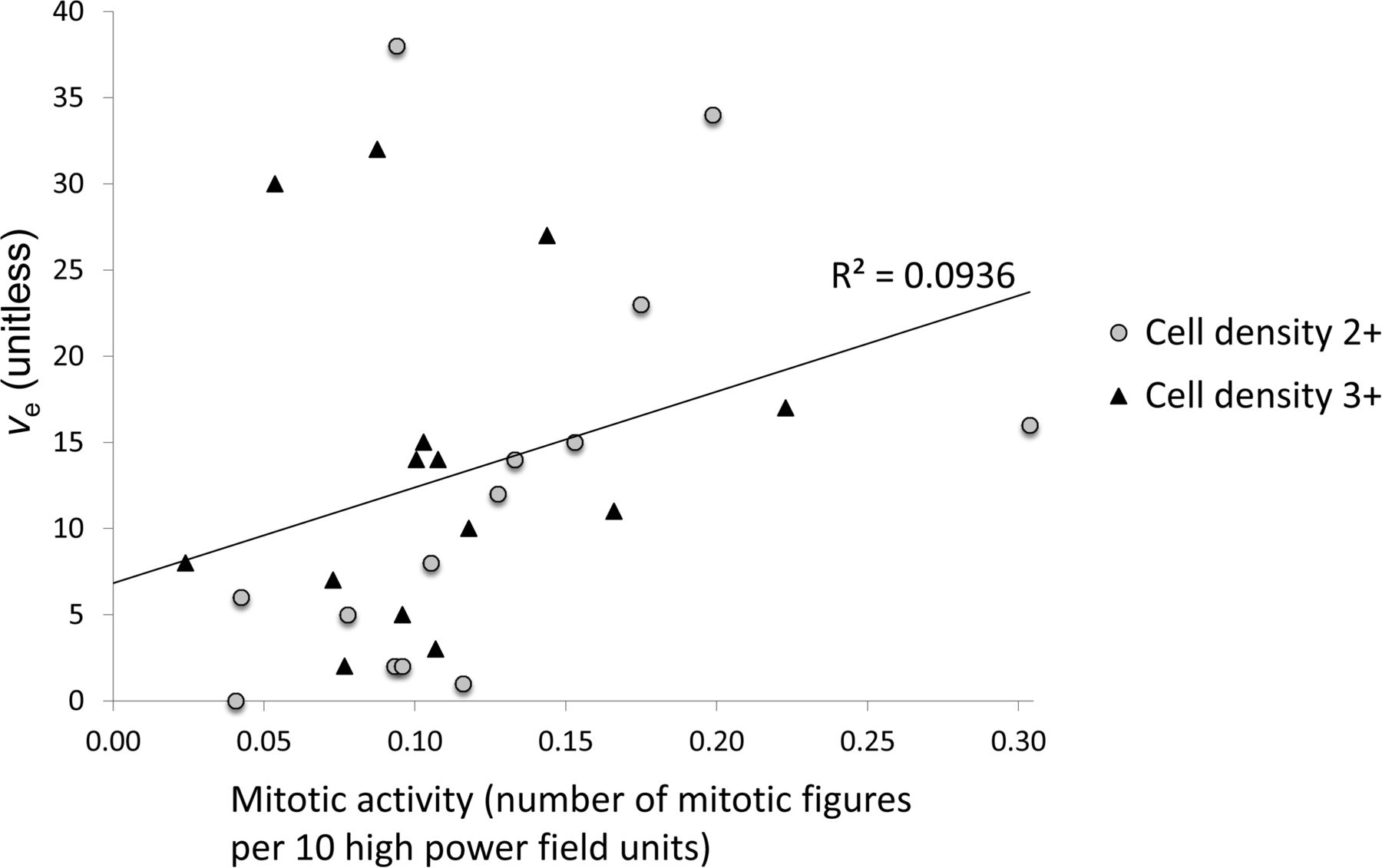
  • Fig 2.
    • Download figure
    • Open in new tab
    • Download powerpoint
    Fig 2.

    Scatterplot of mitotic activity versus ve (P = .012, ρ = 0.470), marker shapes depict separate scores of cell density measures.

Tables

  • Figures
    • View popup
    Table 1:

    Cross-correlations of MRI parametersa

    Untreated GBM (n = 28)
    vevpEnFIAUC60>0EnFIAUC60>2.5
    Ktrans0.016,b ρ = 0.4500.006,c ρ = 0.5070.005,c ρ = 0.513<0.001,c ρ = 0.784
    ve0.105, ρ = 0.3130.888, ρ = 0.0280.059, ρ = 0.361
    vp0.02,b ρ = 0.437<0.001,c ρ = 0.620
    EnFIAUC60>0<0.001,c ρ = 0.825
    • ↵a Significance levels and Spearman ρ are shown.

    • ↵b Significance was P < .05.

    • ↵c Significance was P < .01.

    • View popup
    Table 2:

    Cross-correlations of quantitative histologic measuresa

    Untreated GBM (n = 28)
    ESA RatioVSA RatioVPC/mm2
    Mitotic activity0.098, ρ = 0.3250.066, ρ = 0.3580.737, ρ = 0.068
    ESA ratio<0.001,b ρ = 0.9930.04,c ρ = 0.530
    VSA ratio0.04,c ρ = 0.532
    • Note:—VPC indicates vascular profile count per square millimeter; VSA, vascular surface area; ESA, endothelial surface area.

    • ↵a Significance levels and Spearman ρ are shown.

    • ↵b Significance was P < .01.

    • ↵c Significance was P < .05.

PreviousNext
Back to top

In this issue

American Journal of Neuroradiology: 37 (5)
American Journal of Neuroradiology
Vol. 37, Issue 5
1 May 2016
  • Table of Contents
  • Index by author
  • Complete Issue (PDF)
Advertisement
Print
Download PDF
Email Article

Thank you for your interest in spreading the word on American Journal of Neuroradiology.

NOTE: We only request your email address so that the person you are recommending the page to knows that you wanted them to see it, and that it is not junk mail. We do not capture any email address.

Enter multiple addresses on separate lines or separate them with commas.
Mitotic Activity in Glioblastoma Correlates with Estimated Extravascular Extracellular Space Derived from Dynamic Contrast-Enhanced MR Imaging
(Your Name) has sent you a message from American Journal of Neuroradiology
(Your Name) thought you would like to see the American Journal of Neuroradiology web site.
CAPTCHA
This question is for testing whether or not you are a human visitor and to prevent automated spam submissions.
Cite this article
S.J. Mills, D. du Plessis, P. Pal, G. Thompson, G. Buonacorrsi, C. Soh, G.J.M. Parker, A. Jackson
Mitotic Activity in Glioblastoma Correlates with Estimated Extravascular Extracellular Space Derived from Dynamic Contrast-Enhanced MR Imaging
American Journal of Neuroradiology May 2016, 37 (5) 811-817; DOI: 10.3174/ajnr.A4623

Citation Manager Formats

  • BibTeX
  • Bookends
  • EasyBib
  • EndNote (tagged)
  • EndNote 8 (xml)
  • Medlars
  • Mendeley
  • Papers
  • RefWorks Tagged
  • Ref Manager
  • RIS
  • Zotero
0 Responses
Respond to this article
Share
Bookmark this article
Mitotic Activity in Glioblastoma Correlates with Estimated Extravascular Extracellular Space Derived from Dynamic Contrast-Enhanced MR Imaging
S.J. Mills, D. du Plessis, P. Pal, G. Thompson, G. Buonacorrsi, C. Soh, G.J.M. Parker, A. Jackson
American Journal of Neuroradiology May 2016, 37 (5) 811-817; DOI: 10.3174/ajnr.A4623
del.icio.us logo Twitter logo Facebook logo Mendeley logo
  • Tweet Widget
  • Facebook Like
  • Google Plus One
Purchase

Jump to section

  • Article
    • Abstract
    • ABBREVIATIONS:
    • Materials and Methods
    • Results
    • Discussion
    • Conclusions
    • Footnotes
    • References
  • Figures & Data
  • Supplemental
  • Info & Metrics
  • Responses
  • References
  • PDF

Related Articles

  • PubMed
  • Google Scholar

Cited By...

  • No citing articles found.
  • Crossref (22)
  • Google Scholar

This article has been cited by the following articles in journals that are participating in Crossref Cited-by Linking.

  • Brain Gliomas: Multicenter Standardized Assessment of Dynamic Contrast-enhanced and Dynamic Susceptibility Contrast MR Images
    Nicoletta Anzalone, Antonella Castellano, Marcello Cadioli, Gian Marco Conte, Valeria Cuccarini, Alberto Bizzi, Marco Grimaldi, Antonella Costa, Giovanni Grillea, Paolo Vitali, Domenico Aquino, Maria Rosa Terreni, Valter Torri, Bradley J. Erickson, Massimo Caulo
    Radiology 2018 287 3
  • MRI in Glioma Immunotherapy: Evidence, Pitfalls, and Perspectives
    Domenico Aquino, Andrea Gioppo, Gaetano Finocchiaro, Maria Grazia Bruzzone, Valeria Cuccarini
    Journal of Immunology Research 2017 2017
  • Prognostic value of preoperative dynamic contrast-enhanced MRI perfusion parameters for high-grade glioma patients
    Agne Ulyte, Vasileios K. Katsaros, Evangelia Liouta, Georgios Stranjalis, Christos Boskos, Nickolas Papanikolaou, Jurgita Usinskiene, Sotirios Bisdas
    Neuroradiology 2016 58 12
  • Radiotherapy of glioblastoma 15 years after the landmark Stupp’s trial: more controversies than standards?
    Tomas Kazda, Adam Dziacky, Petr Burkon, Petr Pospisil, Marek Slavik, Zdenek Rehak, Radim Jancalek, Pavel Slampa, Ondrej Slaby, Radek Lakomy
    Radiology and Oncology 2018 52 2
  • Progress in neuro-imaging of brain tumors
    Antonella Castellano, Andrea Falini
    Current Opinion in Oncology 2016 28 6
  • Physiological Imaging Methods for Evaluating Response to Immunotherapies in Glioblastomas
    Sanjeev Chawla, Vanessa Shehu, Pradeep K. Gupta, Kavindra Nath, Harish Poptani
    International Journal of Molecular Sciences 2021 22 8
  • Correlation between amide proton transfer-related signal intensity and diffusion and perfusion magnetic resonance imaging parameters in high-grade glioma
    Masanori Nakajo, Manisha Bohara, Kiyohisa Kamimura, Nayuta Higa, Takashi Yoshiura
    Scientific Reports 2021 11 1
  • Detection of early changes in the post-radiosurgery vestibular schwannoma microenvironment using multinuclear MRI
    Daniel Lewis, Damien J. McHugh, Ka-loh Li, Xiaoping Zhu, Catherine Mcbain, Simon K. Lloyd, Alan Jackson, Omar N. Pathmanaban, Andrew T. King, David J. Coope
    Scientific Reports 2021 11 1
  • Quantitative Assessment of Tumor Cell Proliferation in Brain Gliomas with Dynamic Contrast-Enhanced MRI
    Jia Shen Jiang, Ye Hua, Xue Jun Zhou, Dan Dan Shen, Jin Long Shi, Min Ge, Qi Nan Geng, Zhong Zheng Jia
    Academic Radiology 2019 26 9
  • Application of Distributed Parameter Model to Assessment of Glioma IDH Mutation Status by Dynamic Contrast-Enhanced Magnetic Resonance Imaging
    Zongfang Li, Wei Zhao, Bo He, Tong San Koh, Yanxi Li, Yizhen Zeng, Zhuo Zhang, Jingzhong Zhang, Zujun Hou, Mattia Veronese
    Contrast Media & Molecular Imaging 2020 2020

More in this TOC Section

  • Diagnostic Neuroradiology of Monoclonal Antibodies
  • ML for Glioma Molecular Subtype Prediction
  • NCCT vs. MRI for Brain Atrophy in Acute Stroke
Show more ADULT BRAIN

Similar Articles

Advertisement

Indexed Content

  • Current Issue
  • Accepted Manuscripts
  • Article Preview
  • Past Issues
  • Editorials
  • Editor's Choice
  • Fellows' Journal Club
  • Letters to the Editor
  • Video Articles

Cases

  • Case Collection
  • Archive - Case of the Week
  • Archive - Case of the Month
  • Archive - Classic Case

More from AJNR

  • Trainee Corner
  • Imaging Protocols
  • MRI Safety Corner
  • Book Reviews

Multimedia

  • AJNR Podcasts
  • AJNR Scantastics

Resources

  • Turnaround Time
  • Submit a Manuscript
  • Submit a Video Article
  • Submit an eLetter to the Editor/Response
  • Manuscript Submission Guidelines
  • Statistical Tips
  • Fast Publishing of Accepted Manuscripts
  • Graphical Abstract Preparation
  • Imaging Protocol Submission
  • Evidence-Based Medicine Level Guide
  • Publishing Checklists
  • Author Policies
  • Become a Reviewer/Academy of Reviewers
  • News and Updates

About Us

  • About AJNR
  • Editorial Board
  • Editorial Board Alumni
  • Alerts
  • Permissions
  • Not an AJNR Subscriber? Join Now
  • Advertise with Us
  • Librarian Resources
  • Feedback
  • Terms and Conditions
  • AJNR Editorial Board Alumni

American Society of Neuroradiology

  • Not an ASNR Member? Join Now

© 2025 by the American Society of Neuroradiology All rights, including for text and data mining, AI training, and similar technologies, are reserved.
Print ISSN: 0195-6108 Online ISSN: 1936-959X

Powered by HighWire