Advertisement

Main menu

  • Home
  • Content
    • Current Issue
    • Accepted Manuscripts
    • Article Preview
    • Past Issue Archive
    • Video Articles
    • AJNR Case Collection
    • Case of the Week Archive
    • Case of the Month Archive
    • Classic Case Archive
  • Special Collections
    • AJNR Awards
    • Low-Field MRI
    • Alzheimer Disease
    • ASNR Foundation Special Collection
    • Photon-Counting CT
    • View All
  • Multimedia
    • AJNR Podcasts
    • AJNR SCANtastic
    • Trainee Corner
    • MRI Safety Corner
    • Imaging Protocols
  • For Authors
    • Submit a Manuscript
    • Submit a Video Article
    • Submit an eLetter to the Editor/Response
    • Manuscript Submission Guidelines
    • Statistical Tips
    • Fast Publishing of Accepted Manuscripts
    • Graphical Abstract Preparation
    • Imaging Protocol Submission
    • Author Policies
  • About Us
    • About AJNR
    • Editorial Board
    • Editorial Board Alumni
  • More
    • Become a Reviewer/Academy of Reviewers
    • Subscribers
    • Permissions
    • Alerts
    • Feedback
    • Advertisers
    • ASNR Home

User menu

  • Alerts
  • Log in
  • Log out

Search

  • Advanced search
American Journal of Neuroradiology

American Journal of Neuroradiology

ASHNR American Society of Functional Neuroradiology ASHNR American Society of Pediatric Neuroradiology ASSR
  • Alerts
  • Log in
  • Log out

Advanced Search

  • Home
  • Content
    • Current Issue
    • Accepted Manuscripts
    • Article Preview
    • Past Issue Archive
    • Video Articles
    • AJNR Case Collection
    • Case of the Week Archive
    • Case of the Month Archive
    • Classic Case Archive
  • Special Collections
    • AJNR Awards
    • Low-Field MRI
    • Alzheimer Disease
    • ASNR Foundation Special Collection
    • Photon-Counting CT
    • View All
  • Multimedia
    • AJNR Podcasts
    • AJNR SCANtastic
    • Trainee Corner
    • MRI Safety Corner
    • Imaging Protocols
  • For Authors
    • Submit a Manuscript
    • Submit a Video Article
    • Submit an eLetter to the Editor/Response
    • Manuscript Submission Guidelines
    • Statistical Tips
    • Fast Publishing of Accepted Manuscripts
    • Graphical Abstract Preparation
    • Imaging Protocol Submission
    • Author Policies
  • About Us
    • About AJNR
    • Editorial Board
    • Editorial Board Alumni
  • More
    • Become a Reviewer/Academy of Reviewers
    • Subscribers
    • Permissions
    • Alerts
    • Feedback
    • Advertisers
    • ASNR Home
  • Follow AJNR on Twitter
  • Visit AJNR on Facebook
  • Follow AJNR on Instagram
  • Join AJNR on LinkedIn
  • RSS Feeds

AJNR Awards, New Junior Editors, and more. Read the latest AJNR updates

Research ArticleADULT BRAIN
Open Access

High-Resolution 7T MR Imaging of the Motor Cortex in Amyotrophic Lateral Sclerosis

M. Cosottini, G. Donatelli, M. Costagli, E. Caldarazzo Ienco, D. Frosini, I. Pesaresi, L. Biagi, G. Siciliano and M. Tosetti
American Journal of Neuroradiology March 2016, 37 (3) 455-461; DOI: https://doi.org/10.3174/ajnr.A4562
M. Cosottini
aFrom the Department of Translational Research and New Technologies in Medicine and Surgery (M.Cosottini) and Neurology Unit
  • Find this author on Google Scholar
  • Find this author on PubMed
  • Search for this author on this site
  • ORCID record for M. Cosottini
G. Donatelli
cNeuroradiology Unit (G.D., I.P.), Department of Diagnostic and Interventional Radiology, Azienda Ospedaliero-Universitaria Pisana, Pisa, Italy
  • Find this author on Google Scholar
  • Find this author on PubMed
  • Search for this author on this site
  • ORCID record for G. Donatelli
M. Costagli
dIMAGO7 Foundation (M.Costagli), Pisa, Italy
  • Find this author on Google Scholar
  • Find this author on PubMed
  • Search for this author on this site
  • ORCID record for M. Costagli
E. Caldarazzo Ienco
bDepartment of Clinical and Experimental Medicine (E.C.I., D.F., G.S.), University of Pisa, Pisa, Italy
  • Find this author on Google Scholar
  • Find this author on PubMed
  • Search for this author on this site
  • ORCID record for E. Caldarazzo Ienco
D. Frosini
bDepartment of Clinical and Experimental Medicine (E.C.I., D.F., G.S.), University of Pisa, Pisa, Italy
  • Find this author on Google Scholar
  • Find this author on PubMed
  • Search for this author on this site
  • ORCID record for D. Frosini
I. Pesaresi
cNeuroradiology Unit (G.D., I.P.), Department of Diagnostic and Interventional Radiology, Azienda Ospedaliero-Universitaria Pisana, Pisa, Italy
  • Find this author on Google Scholar
  • Find this author on PubMed
  • Search for this author on this site
  • ORCID record for I. Pesaresi
L. Biagi
eIRCCS Stella Maris (L.B., M.T.), Pisa, Italy.
  • Find this author on Google Scholar
  • Find this author on PubMed
  • Search for this author on this site
  • ORCID record for L. Biagi
G. Siciliano
bDepartment of Clinical and Experimental Medicine (E.C.I., D.F., G.S.), University of Pisa, Pisa, Italy
  • Find this author on Google Scholar
  • Find this author on PubMed
  • Search for this author on this site
  • ORCID record for G. Siciliano
M. Tosetti
eIRCCS Stella Maris (L.B., M.T.), Pisa, Italy.
  • Find this author on Google Scholar
  • Find this author on PubMed
  • Search for this author on this site
  • ORCID record for M. Tosetti
  • Article
  • Figures & Data
  • Supplemental
  • Info & Metrics
  • Responses
  • References
  • PDF
Loading

Abstract

BACKGROUND AND PURPOSE: Amyotrophic lateral sclerosis is a progressive motor neuron disorder that involves degeneration of both upper and lower motor neurons. In patients with amyotrophic lateral sclerosis, pathologic studies and ex vivo high-resolution MR imaging at ultra-high field strength revealed the co-localization of iron and activated microglia distributed in the deep layers of the primary motor cortex. The aims of the study were to measure the cortical thickness and evaluate the distribution of iron-related signal changes in the primary motor cortex of patients with amyotrophic lateral sclerosis as possible in vivo biomarkers of upper motor neuron impairment.

MATERIALS AND METHODS: Twenty-two patients with definite amyotrophic lateral sclerosis and 14 healthy subjects underwent a high-resolution 2D multiecho gradient-recalled sequence targeted on the primary motor cortex by using a 7T scanner. Image analysis consisted of the visual evaluation and quantitative measurement of signal intensity and cortical thickness of the primary motor cortex in patients and controls. Qualitative and quantitative MR imaging parameters were correlated with electrophysiologic and laboratory data and with clinical scores.

RESULTS: Ultra-high field MR imaging revealed atrophy and signal hypointensity in the deep layers of the primary motor cortex of patients with amyotrophic lateral sclerosis with a diagnostic accuracy of 71%. Signal hypointensity of the deep layers of the primary motor cortex correlated with upper motor neuron impairment (r = −0.47; P < .001) and with disease progression rate (r = −0.60; P = .009).

CONCLUSIONS: The combined high spatial resolution and sensitivity to paramagnetic substances of 7T MR imaging demonstrate in vivo signal changes of the cerebral motor cortex that resemble the distribution of activated microglia within the cortex of patients with amyotrophic lateral sclerosis. Cortical thinning and signal hypointensity of the deep layers of the primary motor cortex could constitute a marker of upper motor neuron impairment in patients with amyotrophic lateral sclerosis.

ABBREVIATIONS:

ALS
amyotrophic lateral sclerosis
ALSFRS-R
ALS Functional Rating Scale-Revised
DPR
disease progression rate
HS
healthy subjects
M1
primary motor cortex
UHF
ultra-high field
UMN
upper motor neuron

Amyotrophic lateral sclerosis (ALS) is a progressive motor neuron disorder that entails degeneration of both upper and lower motor neurons,1 producing fasciculation, muscle wasting and weakness, increased spasticity, and hyperreflexia.

Upper motor neuron (UMN) impairment in ALS can be detected from clinical signs such as brisk reflexes and spasticity; however, the masking effect of the lower motor neuron involvement that reduces muscle strength and reflexes may make it hard to reveal clinical evidence of pyramidal involvement. Electrophysiologic investigations, such as prolongation of central motor conduction time in motor-evoked potentials in transcranial magnetic stimulation, may detect the upper motor neuron involvement, but results are conflicting and these measures are not a sensitive tool or a useful indicator of disease severity or prognosis.2⇓–4 Therefore, a robust in vivo biomarker of UMN impairment, though desirable,5 is not available.

The hallmark of UMN pathology in ALS is depopulation of the Betz cells in the motor cortex and axonal loss within the descending motor pathway associated with myelin pallor and gliosis of the corticospinal tracts.6

Conversely, also, characteristic of ALS pathology similar to that in other neurodegenerative disorders is the occurrence of a neuroinflammatory reaction, consisting of activated glial cells, mainly microglia and astrocytes, and T-cells.7 In transgenic mouse models of mutant SOD1-associated familial ALS, reactive microglial cells and astrocytes actively contribute to the death of motor neurons.7

Moreover, a significant association between cortical microgliosis and UMN damage has been described by using PET with a radioligand for microglia.8

Recently, an ex vivo study with Perls' DAB staining detected intracellular and extracellular iron deposits in the motor cortex of patients with ALS. Iron in the form of ferritin is detected in activated microglia (CD68+) in the middle and deep layers of the motor cortex, sparing the superficial layers.9

Gradient recalled-echo T2* sequences, which can provide signal magnitude, phase, and their combination in susceptibility-weighted imaging,10 are considered the most appropriate technique to visualize small amounts of uniformly distributed iron, such as ferritin,11,12 and are the most sensitive sequences for detecting the low signal intensity in the precentral cortex in patients with ALS.13

The introduction of ultra-high field (UHF) MR imaging equipment greatly increases the sensitivity to susceptibility phenomena and allows obtaining in vivo spatial resolution of approximately 200 μm, which enables determination of the cortical layers.14

The aims of our study were to measure the thickness of the primary motor cortex (M1) and to describe the distribution of the iron-related signal changes in the M1 of patients with ALS as a possible in vivo biomarker of UMN impairment by using targeted high-resolution gradient recalled-echo T2* sequences at UHF.

Materials and Methods

Patients and Controls

Twenty-two patients (5 women and 17 men; mean age, 61 ± 10 years) with a diagnosis of definite ALS (limb onset) according to the revised El Escorial diagnostic criteria15 were enrolled (On-line Table 1). Patients with dementia were excluded by administering the Mini-Mental State Examination16 and the Frontal Assessment Battery,17 as short and rapid bedside tests for a screening of cognitive, behavioral, and global executive dysfunction. Clinical history and standard neurologic and psychiatric assessment were used to rule out possible neuropsychiatric comorbidities and other neurodegenerative diseases.

Patients underwent CSF and plasma laboratory analysis to respectively assess concentration of β-amyloid, τ, and p-τ neuronal proteins and some peripheral oxidative stress biomarkers such as advanced oxidation protein products, ferric-reducing ability of plasma, and thiols.18 Neurophysiologic investigation included motor-evoked potentials with calculations of central conduction time, cortical latency, and cortical silent period. Disease severity was evaluated by using the ALS Functional Rating Scale-Revised (ALSFRS-R) (range, 0–48),19 the Medical Research Council scale for segmentary muscle strength (range, 0–130),20 and a composite semiquantitative arbitrary score of UMN burden (UMN score; range, 0–33) (On-line Table 2).8,21,22 Disease duration was expressed in months from symptom appearance to MR imaging acquisition, while disease progression rate (DPR) was calculated according the following formula: (48 − ALSFRS-R)/Disease Duration.23

Fourteen right-handed healthy subjects (HS), age-matched with patients (8 women and 6 men; mean age, 58 ± 12 years), were enrolled to compare the cerebral motor cortex morphology with respect to patients with ALS. HS were volunteers who had no history of psychiatric and neurologic disorders; their neurologic examination findings were normal.

All patients and controls gave their informed consent to the enrollment and diagnostic procedures on the basis of the adherence to an experimental protocol “Clinical Impact of Ultra-High Field MR Imaging in Neurodegenerative Diseases Diagnosis” (RF-2009-1546281) approved and funded by Italian Ministry of Health and cofunded by the Health Service of Tuscany. The study was approved by the local competent ethics committee.

MR Imaging Acquisition

MR imaging experiments were performed on a 7T MR950 scanner (GE Healthcare, Milwaukee, Wisconsin) equipped with a 2CH-TX/32CH-RX head coil (Nova Medical, Wilmington, Massachusetts).

The MR imaging protocol included a high-resolution 2D multiecho gradient-recalled sequence with TE = 10 ms and 20 ms, TR = 500 ms, flip angle = 15°, NEX = 2, section thickness = 2 mm, FOV = 112 mm, and in-plane resolution = 250 μm, targeted to the M1, oriented in the axial plane, and covering the cerebrum from the vertex to the internal capsule. The total acquisition time was 7 minutes 32 seconds.

Image Analysis

Visual Inspection and Subjective Assessment.

Two expert neuroradiologists (M.Cosottini and I.P.) blinded to the clinical status of subjects visually evaluated, independently, images on the basis of morphologic criteria.

The M1 in the precentral gyrus was identified on UHF MR images for the presence of the “hand knob.”24 The anatomy of the cerebral motor cortex at the level of the M1 was evaluated on the basis of accepted cognition sourcing from anatomic atlases25 and MR imaging studies26,27 and atlases28 at UHF. The normal anatomy of the M1 at UHF MR imaging consists of deeper cortical layers identified as a slight hypointense band immediately subjacent to a thinner superficial hyperintense layer. The subcortical hypointense line corresponding to U-fibers appears smooth, and the gray-white junction, poorly demarcated (Fig 1A, -B).

Fig 1.
  • Download figure
  • Open in new tab
  • Download powerpoint
Fig 1.

Normal anatomy. In vivo UHF high-resolution gradient recalled-echo T2* sequence acquisition of the pre- and postcentral gyri (A) and its schematic representation (B) in healthy subjects. The postcentral gyrus resembles the classic ex vivo UHF MR imaging appearance of the unimodal sensory cortices: a low signal intensity tier corresponding to heavily myelinated intracortical layer IV (external band of Baillarger) (arrows) and a subcortical hypointensity (arrowheads) corresponding to arcuate U-fibers demarcating the gray-white matter junction.56 The cortical ribbon of M1 (bracket) as the functional counterpart of Brodmann area 435 is thicker than S157because it is agranular36 and contains the giant Betz pyramidal cells. The superficial hyperintense layer in MR imaging (dots) is due to the lightly myelinated superficial layers relative to the more heavily myelinated deep layers.58 M1 does not have an intermediate sharp hypointense line as in S1 but shows a more widespread slight hypointensity of the deeper cortical layer corresponding to its astriate myeloarchitecture.59,60 The deeper layers of M1 are not easily distinguished from the underlying white matter due to the heavily myelinated M1 cortex.32,58 ALS features. UHF high-resolution gradient recalled-echo T2* sequence acquisition of the pre- and postcentral gyri in a patient with ALS (D) and the corresponding schematic view (E). Beneath a preserved superficial cortical layer (dots), the deep layers in M1 are thinner and more hypointense (bracket) than those in healthy subjects and show an inconstant track appearance. The M1 signal hypointensity in ALS could be due to paramagnetic effects of the iron-containing microglia within the deep motor cortex. The method adopted for quantitative measurement of signal intensity and thickness in a healthy subject (C) and in patients with ALS (F) is reported. Quantitative assessments of the deep layers of M1 are obtained by drawing an oval ROI within them for the signal intensity measurement and a line with an electronic caliper for the thickness quantification.

When the M1 anatomy differed from such configurations, and namely the M1 deeper layers were particularly thin23,29 and/or hypointense9,30 (Fig 1D, -E), images were judged as belonging to patients with ALS.

Sensitivity, specificity, positive predictive value, negative predictive value, and diagnostic accuracy in distinguishing patients and HS were calculated for each reader. Interobserver agreement was calculated with the Cohen κ statistic.

Differences in UMN scores between correctly diagnosed and misdiagnosed patients were evaluated by a Mann-Whitney U test.

Quantitative Assessment.

For each cerebral hemisphere, thickness and signal intensity of the deeper layers of the M1 were measured in 4 anatomic regions corresponding to the Penfield's areas of foot, leg, hand, and face (Fig 1C, -F).31 Signal intensities were measured by using oval ROIs of 4 mm2 and were normalized with respect to those of the superficial cerebral cortex of the left precuneus. Measurement of cortical thickness was obtained by tracing, with an electronic caliper, a line encompassing the deep layer of the M1. The measurements of signal intensity and cortical thickness for each subject were then averaged to obtain mean values from both cerebral hemispheres.

Quantitative data of cortical thickness and the signal intensity of the M1 were compared between patients and HS by using a Mann-Whitney U test. In the group of patients, the relationship between morphometric parameters (mean values of cortical thickness and signal intensity) and clinical scores (ALSFRS-R, Medical Research Council scale, disease duration, and DPR) and the electrophysiologic (central conduction time, cortical latency, cortical silent period) and laboratory (ferric reducing ability of plasma, advanced oxidation protein products, plasma thiols) data was evaluated by the Spearman rank correlation coefficient. Cortical thickness and signal intensity of the M1 for each Penfield's area of hand, leg, and foot were correlated to the correspondent UMN score by using the Spearman rank correlation coefficient.

Using the median of DPR as a cutoff, we divided patients on the basis of the progression rate into 2 groups: “faster” and “slower.” A Mann-Whitney U test was applied to investigate differences in morphometric parameters between the 2 groups.

Results

Four patients and 1 healthy control subject were excluded from the analysis due to the presence of severe motion artifacts.

In the remaining 18 patients, the mean values were 40 ± 5 for ALSFRS-R, 9 ± 10 for the UMN score, 118 ± 13 for the Medical Research Council scale, 16 ± 12 for disease duration, and 0.68 ± 0.47 for DPR.

Visual Inspection and Subjective Assessment

At visual inspection, reader 1 detected hypointensity and thinning of the deeper layers of the M1 in 12 of 18 patients with ALS and in 3 of 13 HS. Sensitivity, specificity, positive predictive value, negative predictive value, and diagnostic accuracy in distinguishing patients and HS on the basis of morphologic criteria were 67%, 77%, 80%, 63%, and 71%, respectively.

Reader 2 detected hypointensity and thinning of the deeper layers of the M1 in 9 of 18 patients with ALS and in zero of 13 HS. Sensitivity, specificity, positive predictive value, negative predictive value, and diagnostic accuracy in distinguishing patients and HS on the basis of morphologic criteria were 50%, 100%, 100%, 59%, and 71%, respectively.

The Interobserver agreement was good (0.61, Cohen κ statistic).

On the basis of both readings, the UMN score was significantly higher (P = .003 and P = .028, respectively) in correctly diagnosed patients (13 ± 10 and 14 ± 10, respectively) than in misdiagnosed ones (1 ± 1 and 4 ± 8, respectively) (On-line Fig 1A, -B and On-line Tables 3–6).

Also, the DPR was higher on the basis of both readings (P = .019 and P = .066, respectively) in correctly diagnosed patients (0.85 ± 0.48 and 0.87 ± 0.49, respectively) than in misdiagnosed ones (0.33 ± 0.20 and 0.48 ± 0.38, respectively).

Quantitative Assessment

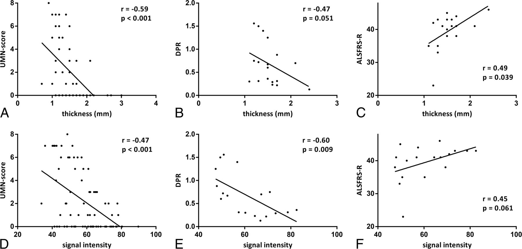
Cortical thickness was significantly (P < .001) lower in patients (mean value = 1.5 mm) than in HS (mean value = 2 mm) (On-line Fig 1C) and showed a moderate correlation with the UMN score (r = −0.59; P < .001) (Fig 2A). Cortical thickness had a weaker correlation with DPR (r = −0.47; P = .051) and ALSFRS-R (r = 0.49; P = .039) (Fig 2B, -C).

Fig 2.
  • Download figure
  • Open in new tab
  • Download powerpoint
Fig 2.

Correlations in patients with ALS between morphometric parameters (thickness and signal intensity of deeper layers of M1) and clinical scores (UMN score, DPR, and ALSFRS-R). The UMN score ranges from zero to 8 because it refers to a single limb and not to the overall evaluation. Correlations between morphometric parameters and UMN score were calculated by taking into account the clinical score of each limb of every patient and signal intensity and thickness of the corresponding areas of M1.

Signal intensity did not differ between patients with ALS and HS (On-line Fig 1D) but correlated with UMN score (r = −0.47; P < .001), DPR (r = −0.60; P = .009), and ALSFRS-R (r = 0.45; P = .061) (Fig 2D–F).

Signal intensity in the subgroup of patients with faster DPR was significantly lower than that in the slower DPR subgroup (P = .024), while no significant difference in cortical thickness was found between subgroups of patients (On-line Fig 2).

No significant correlations were found between morphometric parameters and either motor-evoked potential indices (central motor conduction time and cortical silent period) or the ferric-reducing ability of plasma, advanced oxidation protein products, and thiols.

Discussion

UHF MR Signal in M1 of Controls and Patients with ALS

UHF MR imaging allows an in vivo evaluation of the inner structure of the cerebral motor cortex of patients with ALS and controls. The gradient recalled-echo T2* sequence at 7T provides information on the architectural organization of the human cortex in vivo,32 due to the iron and myelin contribution to T2* contrast.26 Because myelin and iron in the form of ferritin are strongly co-localized in the intracortical myelinated fibers,33 the MR signals across the cortical gray matter reflect myelin content and myeloarchitecture.34 The normal anatomy of the M1 (Brodmann area 4)35 at UHF depends on its peculiar architecture: The large cortical thickness, inconspicuous lamination pattern, low cell density, and the absence of a clear boundary between layer VI and white matter36 correspond to the MR imaging findings of the motor cortex as shown in Fig 1A, -B.

In patients with ALS, in accordance with previous ex vivo UHF images of the affected cerebral cortices,9 we revealed a rim of signal hypointensity with an inconstant track appearance beneath a preserved superficial hyperintense cortical layer (Fig 1D, -E).

The decreased signal intensity of the motor cortex on T2*-weighted images in patients with ALS may represent ferritin because at postmortem examination, the precentral cortex showed intensely stained microglia and macrophages after antiferritin antibody staining.13 Until now, cortical hypointensity in vivo seemed to involve the motor cortex in a full-thickness fashion at both conventional37 and UHF9 MR imaging. Our targeted UHF MR imaging high-resolution study, as well as the ex vivo UHF imaging, demonstrated that in patients with ALS, the signal hypointensity is confined to the deeper layers of the cerebral cortex, the same structures in which several studies have shown an increased number of microglial cells resulting from local proliferation of resident microglia.38

PET with radioligand-binding biomarkers of activated microglia revealed that areas with the highest microglial activation in patients with ALS were the motor cortices,39 thus corresponding to the distribution of signal changes and atrophy that we have detected in our group of patients.

Cortical Thickness in the M1 of Patients with ALS

Cortical atrophy in ALS has been revealed in several voxel-based morphometry works40⇓–42 by manual or automated measurements of the cortical thickness and has been interpreted as a structural surrogate of UMN degeneration.23 In particular, the regional grade of atrophy in different areas of M1 corresponds to a somatotopic functional disability supporting the concepts of cortical focality and motor phenotype heterogeneity.43 In line with previous results, our data confirmed the thinning of the M1 in patients with ALS and the correlation between the cortical atrophy and the UMN burden, but we observed that the alteration is confined to the deeper cortical layers rather than the full-thickness cortex. Moreover, cortical atrophy and signal hypointensity of the M1 in patients with ALS are in line, respectively, with the reduction of N-acetylaspartate and the increase of myo-inositol44 as an expression of the neurodegenerative phenomena and gliosis.

MR Imaging of M1: Biomarker for Diagnosis or Prognosis

Several conventional MR imaging studies subjectively evaluated the cortical morphology and signal changes in patients with ALS.37,45⇓⇓⇓–49 These studies using full-thickness measures of cortical signal changes were limited by low specificity because similar findings are age-related and can also be detected in subjects without ALS.50 Although our approach permitted the evaluation of the inner structure of the cerebral motor cortex, the diagnostic accuracy (71%) and the sensitivity (67% and 50% for reader 1 and reader 2, respectively) of M1 morphologic changes remain unsatisfactory.

Unlike electrophysiologic data, in which a correlation with UMN impairment has never been demonstrated,51 in our study, the quantitative analysis showed that increased image hypointensity and the atrophic deep motor cortex both correspond to greater UMN involvement. The correlation between the signal intensity and the UMN score and the fact that the UMN score was significantly higher in correctly diagnosed patients than in misdiagnosed ones support the hypothesis that hypointensity is a marker of UMN degeneration rather than of ALS.

This relationship influences the diagnostic accuracy of MR imaging, which remains modest because the correct diagnosis of ALS based on cortical signal changes depends on the amount of UMN impairment in sampled patients. The variable impairment of UMNs in ALS explains why decreased signal intensity in ALS has been reported at a variable extent, ranging from 93%37 to 30%.13

The cortical signal intensity correlates with DPR and could constitute a prognostic marker of the disease. Although we interpret signal intensity as an expression of activated microglia, we do not know the pathologic mechanism that regulates the disease progression in our patients.

Translational research in mutant SOD1 transgenic mice shows that microglial cells become activated before motor neurons disappear and well before clinical disease onset.52 Moreover SOD mutant levels in microglia regulate the rate of disease progression.53 The correlation between cortical signal hypointensity as a presumptive marker of activated microglia and DPR could constitute the basis of a longitudinal study of microgliosis in patients with ALS, widening the pathogenic scenario of the causes of this disease and offering novel targets for therapeutic development. To understand whether the neuroinflammatory reaction could be a therapeutic target in ALS, a reliable biomarker able to investigate and monitor the neuroinflammatory response to motor-neuron degeneration with time would be desirable. Besides laboratory inflammatory markers in the CSF54 and PET studies by using microglial ligands, high-resolution UHF MR imaging might provide alternative relevant information on neuroinflammation.

In ALS, biomarkers of disease or prognosis would be paramount for patient management and therapeutic implications.5 Advanced neuroimaging techniques provide an opportunity to assess disease pathophysiology and promise new biomarkers.55 The introduction of the UHF MR imaging of the cerebral cortex in evaluating patients with ALS could be an adjunctive tool in a multimodal approach to the research of biomarkers.5

The main limitation of the present study is the small sample of patients, which affects the statistical power. A further limitation is the impossibility of enrolling patients with advanced disease for logistic reasons related to their disability, which would probably have provided information on the course of the disease.

Conclusions

The combined high spatial resolution and sensitivity to paramagnetic substances of 7T MR imaging demonstrate in vivo signal changes of the cerebral motor cortex that resemble the distribution of activated microglia within the cortex of patients with ALS. Reduced cortical thickness and signal hypointensity of the M1 deep layers could constitute a marker of UMN impairment in patients with ALS. Further studies with a larger sample of patients would be desirable to confirm the UHF MR imaging results in motor neuron disorders.

Footnotes

  • Disclosures: Ilaria Pesaresi—RELATED: Grant: Ministry of Health.* Gabriele Siciliano—UNRELATED: Board Membership: Baxter International, Comments: clinical trial board membership; Expert Testimony: CSL Behring,* Comments: therapeutic trial testimony; Payment for Lectures (including service on Speakers Bureaus): Baxter International, Grunenthal, Grifols; Payment for Manuscript Preparation: Baxter International; Payment for Development of Educational Presentations: Baxter International; Travel/Accommodations/Meeting Expenses Unrelated to Activities Listed: Kedrion, CSL Behring, Comments: meeting expenses. *Money paid to the institution.

  • This work is part of the protocol “Clinical Impact of Ultra-High Field MRI in Neurodegenerative Diseases Diagnosis” (RF RF-2009-1546281) approved and funded by the Italian Ministry of Health and cofunded by the Health Service of Tuscany.

Indicates open access to non-subscribers at www.ajnr.org

References

  1. 1.↵
    1. Strong M,
    2. Rosenfeld J
    . Amyotrophic lateral sclerosis: a review of current concepts. Amyotroph Lateral Scler Other Motor Neuron Disord 2003;4:136–43 doi:10.1080/14660820310011250 pmid:13129799
    CrossRefPubMed
  2. 2.↵
    1. Claus D,
    2. Brunhölzl C,
    3. Kerling FP, et al
    . Transcranial magnetic stimulation as a diagnostic and prognostic test in amyotrophic lateral sclerosis. J Neurol Sci 1995;129(suppl):30–34 doi:10.1016/0022-510X(95)00056-8 pmid:7595614
    CrossRefPubMed
  3. 3.↵
    1. de Carvalho M,
    2. Dengler R,
    3. Eisen A, et al
    . Electrodiagnostic criteria for diagnosis of ALS. Clin Neurophysiol 2008;119:497–503 doi:10.1016/j.clinph.2007.09.143 pmid:18164242
    CrossRefPubMed
  4. 4.↵
    1. Vucic S,
    2. Kiernan MC
    . Utility of transcranial magnetic stimulation in delineating amyotrophic lateral sclerosis pathophysiology. Handb Clin Neurol 2013;116:561–75 doi:10.1016/B978-0-444-53497-2.00045-0 pmid:24112924
    CrossRefPubMed
  5. 5.↵
    1. Chiò A,
    2. Pagani M,
    3. Agosta F, et al
    . Neuroimaging in amyotrophic lateral sclerosis: insights into structural and functional changes. Lancet Neurol 2014;13:1228–40 doi:10.1016/S1474-4422(14)70167-X pmid:25453462
    CrossRefPubMed
  6. 6.↵
    1. Ince PG,
    2. Evans J,
    3. Knopp M, et al
    . Corticospinal tract degeneration in the progressive muscular atrophy variant of ALS. Neurology 2003;60:1252–58 doi:10.1212/01.WNL.0000058901.75728.4E pmid:12707426
    Abstract/FREE Full Text
  7. 7.↵
    1. Philips T,
    2. Robberecht W
    . Neuroinflammation in amyotrophic lateral sclerosis: role of glial activation in motor neuron disease. Lancet Neurol 2011;10:253–63 doi:10.1016/S1474-4422(11)70015-1 pmid:21349440
    CrossRefPubMed
  8. 8.↵
    1. Turner MR,
    2. Cagnin A,
    3. Turkheimer FE, et al
    . Evidence of widespread cerebral microglial activation in amyotrophic lateral sclerosis: an [11C](R)-PK11195 positron emission tomography study. Neurobiol Dis 2004;15:601–09 doi:10.1016/j.nbd.2003.12.012 pmid:15056468
    CrossRefPubMed
  9. 9.↵
    1. Kwan JY,
    2. Jeong SY,
    3. Van Gelderen P, et al
    . Iron accumulation in deep cortical layers accounts for MRI signal abnormalities in ALS: correlating 7 Tesla MRI and pathology. PLoS One 2012;7:e35241 doi:10.1371/journal.pone.0035241 pmid:22529995
    CrossRefPubMed
  10. 10.↵
    1. Haacke EM,
    2. Mittal S,
    3. Wu Z, et al
    . Susceptibility-weighted imaging: technical aspects and clinical applications, part 1. AJNR Am J Neuroradiol 2009;30:19–30 pmid:19039041
    Abstract/FREE Full Text
  11. 11.↵
    1. Haacke EM,
    2. Reichenbach JR
    1. Liu M,
    2. Habib C,
    3. Miao Y, et al
    . Measuring iron content with phase. In: Haacke EM, Reichenbach JR, eds. Susceptibility Weighted Imaging in MRI: Basic Concept and Clinical Applications. 10th ed. Hoboken: John Wiley & Sons; 2011:369–401
  12. 12.↵
    1. Duyn JH
    . High-field MRI of brain iron. Methods Mol Biol 2011;711:239–49 doi:10.1007/978-1-61737-992-5_11 pmid:21279605
    CrossRefPubMed
  13. 13.↵
    1. Adachi Y,
    2. Sato N,
    3. Saito Y, et al
    . Usefulness of SWI for the detection of iron in the motor cortex in amyotrophic lateral sclerosis. J Neuroimaging 2015;25:443–51 doi:10.1111/jon.12127 pmid:24888543
    CrossRefPubMed
  14. 14.↵
    1. Geyer S,
    2. Turner R
    1. Fischl B
    . Estimating the location of Brodmann areas from cortical folding patterns using histology and ex vivo MRI. In: Geyer S, Turner R, eds. Microstructural Parcellation of the Human Cerebral Cortex: From Brodmann's Post-Mortem Map to in Vivo Mapping with High-Field Magnetic Resonance Imaging. Berlin: Springer-Verlag; 2013:129–56
  15. 15.↵
    1. Brooks BR,
    2. Miller RG,
    3. Swash M, et al
    ; World Federation of Neurology Research Group on Motor Neuron Disease. El Escorial revisited: revised criteria for the diagnosis of amyotrophic lateral sclerosis. Amyotroph Lateral Scler Other Motor Neuron Disord 2000;1:293–99 doi:10.1080/146608200300079536 pmid:11464847
    CrossRefPubMed
  16. 16.↵
    1. Folstein MF,
    2. Folstein SE,
    3. McHugh PR
    . “Mini-mental state”: a practical method for grading the cognitive state of patients for the clinician. J Psychiatr Res 1975;12:189–98 doi:10.1016/0022-3956(75)90026-6 pmid:1202204
    CrossRefPubMed
  17. 17.↵
    1. Appollonio I,
    2. Leone M,
    3. Isella V, et al
    . The frontal assessment battery (FAB): normative values in an Italian population sample. Neurol Sci 2005;26:108–16 doi:10.1007/s10072-005-0443-4 pmid:15995827
    CrossRefPubMed
  18. 18.↵
    1. Siciliano G,
    2. Piazza S,
    3. Carlesi C, et al
    . Antioxidant capacity and protein oxidation in cerebrospinal fluid of amyotrophic lateral sclerosis. J Neurol 2007;254:575–80 doi:10.1007/s00415-006-0301-1 pmid:17426914
    CrossRefPubMed
  19. 19.↵
    1. Cedarbaum JM,
    2. Stambler N,
    3. Malta E, et al
    . The ALSFRS-R: a revised ALS functional rating scale that incorporates assessments of respiratory function: BDNF ALS Study Group (Phase III). J Neurol Sci 1999;169:13–21 doi:10.1016/S0022-510X(99)00210-5 pmid:10540002
    CrossRefPubMed
  20. 20.↵
    1. Florence JM,
    2. Pandya S,
    3. King WM, et al
    . Intrarater reliability of manual muscle test (Medical Research Council scale) grades in Duchenne's muscular dystrophy. Phys Ther 1992;72:115–22; discussion 122–26 pmid:1549632
    Abstract/FREE Full Text
  21. 21.↵
    1. Ashworth B
    . Preliminary trial of carisoprodol in multiple sclerosis. Practitioner 1964;192:540–42 pmid:14143329
    PubMed
  22. 22.↵
    1. Woo JH,
    2. Wang S,
    3. Melhem ER, et al
    . Linear associations between clinically assessed upper motor neuron disease and diffusion tensor imaging metrics in amyotrophic lateral sclerosis. PLoS One 2014;9:e105753 doi:10.1371/journal.pone.0105753 pmid:25144708
    CrossRefPubMed
  23. 23.↵
    1. Walhout R,
    2. Westeneng HJ,
    3. Verstraete E, et al
    . Cortical thickness in ALS: towards a marker for upper motor neuron involvement. J Neurol Neurosurg Psychiatry 2015;86:288–94 doi:10.1136/jnnp-2013-306839 pmid:25121571
    Abstract/FREE Full Text
  24. 24.↵
    1. Yousry TA,
    2. Schmid UD,
    3. Alkadhi H, et al
    . Localization of the motor hand area to a knob on the precentral gyrus: a new landmark. Brain 1997;120:141–57 doi:10.1093/brain/120.1.141 pmid:9055804
    Abstract/FREE Full Text
  25. 25.↵
    1. Mai JK,
    2. Paxinos G
    1. Zilles K,
    2. Amunts K
    . Architecture of the cerebral cortex. In: Mai JK, Paxinos G, eds. The Human Nervous System. 3rd ed. London: Elsevier; 2012:836–95
  26. 26.↵
    1. Duyn JH,
    2. van Gelderen P,
    3. Li TQ, et al
    . High-field MRI of brain cortical substructure based on signal phase. Proc Natl Acad Sci U S A 2007;104:11796–801 doi:10.1073/pnas.0610821104 pmid:17586684
    Abstract/FREE Full Text
  27. 27.↵
    1. Geyer S,
    2. Weiss M,
    3. Reimann K, et al
    . Microstructural parcellation of the human cerebral cortex: from Brodmann's post-mortem map to in vivo mapping with high-field magnetic resonance imaging. Front Hum Neurosci 2011;5:19 doi:10.3389/fnhum.2011.00019 pmid:21373360
    CrossRefPubMed
  28. 28.↵
    1. Geyer S,
    2. Turner R
    . Microstructural Parcellation of the Human Cerebral Cortex: From Brodmann's Post-Mortem Map to in Vivo Mapping with High-Field Magnetic Resonance Imaging. Berlin: Springer-Verlag; 2013
  29. 29.↵
    1. Verstraete E,
    2. Veldink JH,
    3. Hendrikse J, et al
    . Structural MRI reveals cortical thinning in amyotrophic lateral sclerosis. J Neurol Neurosurg Psychiatry 2012;83:383–88 doi:10.1136/jnnp-2011-300909 pmid:21965521
    Abstract/FREE Full Text
  30. 30.↵
    1. Ignjatović A,
    2. Stević Z,
    3. Lavrnić S, et al
    . Brain iron MRI: a biomarker for amyotrophic lateral sclerosis. J Magn Reson Imaging 2013;38:1472–79 doi:10.1002/jmri.24121 pmid:23564606
    CrossRefPubMed
  31. 31.↵
    1. Penfield W,
    2. Rasmussen T
    . The Cerebral Cortex of Man: A Clinical Study of Localization of Function. New York: Macmillan; 1950
  32. 32.↵
    1. Cohen-Adad J,
    2. Polimeni JR,
    3. Helmer KG, et al
    . T2* mapping and B0 orientation-dependence at 7 T reveal cyto- and myeloarchitecture organization of the human cortex. Neuroimage 2012;60:1006–14 doi:10.1016/j.neuroimage.2012.01.053 pmid:22270354
    CrossRefPubMed
  33. 33.↵
    1. Fukunaga M,
    2. Li TQ,
    3. van Gelderen P, et al
    . Layer-specific variation of iron content in cerebral cortex as a source of MRI contrast. Proc Natl Acad Sci U S A 2010;107:3834–39 doi:10.1073/pnas.0911177107
    Abstract/FREE Full Text
  34. 34.↵
    1. Geyer S,
    2. Turner R
    1. Turner R
    . MRI methods for in-vivo cortical parcellation. In: Geyer S, Turner R, eds. Microstructural Parcellation of the Human Cerebral Cortex: From Brodmann's Post-Mortem Map to in Vivo Mapping with High-Field Magnetic Resonance Imaging. Berlin: Springer-Verlag; 2013:197–220
  35. 35.↵
    1. Brodmann K
    . Vergleichende Lokalisationslehre der Grosshirnrinde. Leipzig: Barth; 1909
  36. 36.↵
    1. Brodmann K
    . Beiträge zur histologischen Lokalisation der Grosshirnrinde: Erste Mitteilung—Die Regio Rolandica. J Psychol Neurol 1903;2:79–107
  37. 37.↵
    1. Oba H,
    2. Araki T,
    3. Ohtomo K, et al
    . Amyotrophic lateral sclerosis: T2 shortening in motor cortex at MR imaging. Radiology 1993;189:843–46 doi:10.1148/radiology.189.3.8234713 pmid:8234713
    CrossRefPubMed
  38. 38.↵
    1. Gowing G,
    2. Philips T,
    3. Van Wijmeersch B, et al
    . Ablation of proliferating microglia does not affect motor neuron degeneration in amyotrophic lateral sclerosis caused by mutant superoxide dismutase. J Neurosci 2008;28:10234–44 doi:10.1523/JNEUROSCI.3494-08.2008 pmid:18842883
    Abstract/FREE Full Text
  39. 39.↵
    1. Corcia P,
    2. Tauber C,
    3. Vercoullie J, et al
    . Molecular imaging of microglial activation in amyotrophic lateral sclerosis. PLoS One 2012;7:e52941 doi:10.1371/journal.pone.0052941 pmid:23300829
    CrossRefPubMed
  40. 40.↵
    1. Agosta F,
    2. Pagani E,
    3. Rocca MA, et al
    . Voxel-based morphometry study of brain volumetry and diffusivity in amyotrophic lateral sclerosis patients with mild disability. Hum Brain Mapp 2007;28:1430–38 doi:10.1002/hbm.20364 pmid:17370339
    CrossRefPubMed
  41. 41.↵
    1. Mezzapesa DM,
    2. Ceccarelli A,
    3. Dicuonzo F, et al
    . Whole-brain and regional brain atrophy in amyotrophic lateral sclerosis. AJNR Am J Neuroradiol 2007;28:255–59 pmid:17296989
    Abstract/FREE Full Text
  42. 42.↵
    1. Cosottini M,
    2. Pesaresi I,
    3. Piazza S, et al
    . Structural and functional evaluation of cortical motor areas in amyotrophic lateral sclerosis. Exp Neurol 2012;234:169–80 doi:10.1016/j.expneurol.2011.12.024 pmid:22226599
    CrossRefPubMed
  43. 43.↵
    1. Bede P,
    2. Bokde A,
    3. Elamin M, et al
    . Grey matter correlates of clinical variables in amyotrophic lateral sclerosis (ALS): a neuroimaging study of ALS motor phenotype heterogeneity and cortical focality. J Neurol Neurosurg Psychiatry 2013;84:766–73 doi:10.1136/jnnp-2012-302674 pmid:23085933
    Abstract/FREE Full Text
  44. 44.↵
    1. Foerster BR,
    2. Pomper MG,
    3. Callaghan BC, et al
    . An imbalance between excitatory and inhibitory neurotransmitters in amyotrophic lateral sclerosis revealed by use of 3-T proton magnetic resonance spectroscopy. JAMA Neurol 2013;70:1009–16 doi:10.1001/jamaneurol.2013.234 pmid:23797905
    CrossRefPubMed
  45. 45.↵
    1. Goodin DS,
    2. Rowley HA,
    3. Olney RK
    . Magnetic resonance imaging in amyotrophic lateral sclerosis. Ann Neurol 1988;23:418–20 doi:10.1002/ana.410230424 pmid:3382182
    CrossRefPubMed
  46. 46.↵
    1. Ishikawa K,
    2. Nagura H,
    3. Yokota T, et al
    . Signal loss in the motor cortex on magnetic resonance images in amyotrophic lateral sclerosis. Ann Neurol 1993;33:218–22 doi:10.1002/ana.410330214 pmid:8434885
    CrossRefPubMed
  47. 47.↵
    1. Cheung G,
    2. Gawel MJ,
    3. Cooper PW, et al
    . Amyotrophic lateral sclerosis: correlation of clinical and MR imaging findings. Radiology 1995;194:263–70 doi:10.1148/radiology.194.1.7997565 pmid:7997565
    CrossRefPubMed
  48. 48.↵
    1. Thorpe JW,
    2. Moseley IF,
    3. Hawkes CH, et al
    . Brain and spinal cord MRI in motor neuron disease. J Neurol Neurosurg Psychiatry 1996;61:314–17 doi:10.1136/jnnp.61.3.314 pmid:8795607
    Abstract/FREE Full Text
  49. 49.↵
    1. Hecht MJ,
    2. Fellner F,
    3. Fellner C, et al
    . Hyperintense and hypointense MRI signals of the precentral gyrus and corticospinal tract in ALS: a follow-up examination including FLAIR images. J Neurol Sci 2002;199:59–65 doi:10.1016/S0022-510X(02)00104-1 pmid:12084444
    CrossRefPubMed
  50. 50.↵
    1. Cosottini M,
    2. Cecchi P,
    3. Piazza S, et al
    . Mapping cortical degeneration in ALS with magnetization transfer ratio and voxel-based morphometry. PLoS One 2013;8:e68279 doi:10.1371/journal.pone.0068279 pmid:23874570
    CrossRefPubMed
  51. 51.↵
    1. Yokota T,
    2. Yoshino A,
    3. Inaba A, et al
    . Double cortical stimulation in amyotrophic lateral sclerosis. J Neurol Neurosurg Psychiatry 1996;61:596–600 doi:10.1136/jnnp.61.6.596 pmid:8971106
    Abstract/FREE Full Text
  52. 52.↵
    1. Hall ED,
    2. Oostveen JA,
    3. Gurney ME
    . Relationship of microglial and astrocytic activation to disease onset and progression in a transgenic model of familial ALS. Glia 1998;23:249–56 doi:10.1002/(SICI)1098-1136(199807)23:3<249::AID-GLIA7>3.0.CO;2-# pmid:9633809
    CrossRefPubMed
  53. 53.↵
    1. Boillée S,
    2. Yamanaka K,
    3. Lobsiger CS, et al
    . Onset and progression in inherited ALS determined by motor neurons and microglia. Science 2006;312:1389–92 doi:10.1126/science.1123511 pmid:16741123
    Abstract/FREE Full Text
  54. 54.↵
    1. Mitchell RM,
    2. Freeman WM,
    3. Randazzo WT, et al
    . A CSF biomarker panel for identification of patients with amyotrophic lateral sclerosis. Neurology 2009;72:14–19 doi:10.1212/01.wnl.0000333251.36681.a5 pmid:18987350
    Abstract/FREE Full Text
  55. 55.↵
    1. Foerster BR,
    2. Welsh RC,
    3. Feldman EL
    . 25 years of neuroimaging in amyotrophic lateral sclerosis. Nat Rev Neurol 2013;9:513–24 doi:10.1038/nrneurol.2013.153 pmid:23917850
    CrossRefPubMed
  56. 56.↵
    1. Fatterpekar GM,
    2. Naidich TP,
    3. Delman BN, et al
    . Cytoarchitecture of the human cerebral cortex: MR microscopy of excised specimens at 9.4 Tesla. AJNR Am J Neuroradiol 2002;23:1313–21 pmid:12223371
    Abstract/FREE Full Text
  57. 57.↵
    1. Meyer J,
    2. Roychowdhury S,
    3. Russell EJ, et al
    . Location of the central sulcus via cortical thickness of the precentral and postcentral gyri on MR. AJNR Am J Neuroradiol 1996;17:1699–706 pmid:8896626
    Abstract
  58. 58.↵
    1. Glasser MF,
    2. Van Essen DC
    . Mapping human cortical areas in vivo based on myelin content as revealed by T1- and T2-weighted MRI. J Neurosci 2011;31:11597–616 doi:10.1523/JNEUROSCI.2180-11.2011 pmid:21832190
    Abstract/FREE Full Text
  59. 59.↵
    1. Vogt O
    . Die myeloarchitektonische Felderung des Menschlichen Stirnhirns. J Psychol Neurol 1910;15:221–32
  60. 60.↵
    1. Rehwald E
    1. Braitenberg V
    . Die Gliederung der Stirnhirnrinde auf Grund ihres Markfaserbaus (Myeloarchitektonik). In: Rehwald E, ed. Das Hirntrauma. Stuttgart: Thieme; 1956:183–203
  • Received June 26, 2015.
  • Accepted after revision August 12, 2015.
  • © 2016 by American Journal of Neuroradiology
View Abstract
PreviousNext
Back to top

In this issue

American Journal of Neuroradiology: 37 (3)
American Journal of Neuroradiology
Vol. 37, Issue 3
1 Mar 2016
  • Table of Contents
  • Index by author
  • Complete Issue (PDF)
Advertisement
Print
Download PDF
Email Article

Thank you for your interest in spreading the word on American Journal of Neuroradiology.

NOTE: We only request your email address so that the person you are recommending the page to knows that you wanted them to see it, and that it is not junk mail. We do not capture any email address.

Enter multiple addresses on separate lines or separate them with commas.
High-Resolution 7T MR Imaging of the Motor Cortex in Amyotrophic Lateral Sclerosis
(Your Name) has sent you a message from American Journal of Neuroradiology
(Your Name) thought you would like to see the American Journal of Neuroradiology web site.
CAPTCHA
This question is for testing whether or not you are a human visitor and to prevent automated spam submissions.
Cite this article
M. Cosottini, G. Donatelli, M. Costagli, E. Caldarazzo Ienco, D. Frosini, I. Pesaresi, L. Biagi, G. Siciliano, M. Tosetti
High-Resolution 7T MR Imaging of the Motor Cortex in Amyotrophic Lateral Sclerosis
American Journal of Neuroradiology Mar 2016, 37 (3) 455-461; DOI: 10.3174/ajnr.A4562

Citation Manager Formats

  • BibTeX
  • Bookends
  • EasyBib
  • EndNote (tagged)
  • EndNote 8 (xml)
  • Medlars
  • Mendeley
  • Papers
  • RefWorks Tagged
  • Ref Manager
  • RIS
  • Zotero
0 Responses
Respond to this article
Share
Bookmark this article
High-Resolution 7T MR Imaging of the Motor Cortex in Amyotrophic Lateral Sclerosis
M. Cosottini, G. Donatelli, M. Costagli, E. Caldarazzo Ienco, D. Frosini, I. Pesaresi, L. Biagi, G. Siciliano, M. Tosetti
American Journal of Neuroradiology Mar 2016, 37 (3) 455-461; DOI: 10.3174/ajnr.A4562
del.icio.us logo Twitter logo Facebook logo Mendeley logo
  • Tweet Widget
  • Facebook Like
  • Google Plus One
Purchase

Jump to section

  • Article
    • Abstract
    • ABBREVIATIONS:
    • Materials and Methods
    • Results
    • Discussion
    • Conclusions
    • Footnotes
    • References
  • Figures & Data
  • Supplemental
  • Info & Metrics
  • Responses
  • References
  • PDF

Related Articles

  • PubMed
  • Google Scholar

Cited By...

  • Precise MRI-Histology Coregistration of Paraffin-Embedded Tissue with Blockface Imaging
  • Individualized Phenotyping of Functional ALS Pathology in Sensorimotor Cortex
  • Motor band sign: a magnetic resonance imaging clue in motor neurone disease
  • Multimodal layer modelling reveals in-vivo pathology in ALS
  • Black Line Sign in Focal Cortical Dysplasia IIB: A 7T MRI and Electroclinicopathologic Study
  • Ultra-high field (7T) functional magnetic resonance imaging in amyotrophic lateral sclerosis: a pilot study
  • Introduction of Ultra-High-Field MR Imaging in Infants: Preparations and Feasibility
  • Ultra-High-Field Targeted Imaging of Focal Cortical Dysplasia: The Intracortical Black Line Sign in Type IIb
  • Semiautomated Evaluation of the Primary Motor Cortex in Patients with Amyotrophic Lateral Sclerosis at 3T
  • Crossref (42)
  • Google Scholar

This article has been cited by the following articles in journals that are participating in Crossref Cited-by Linking.

  • Genetic Convergence Brings Clarity to the Enigmatic Red Line in ALS
    Casey Cook, Leonard Petrucelli
    Neuron 2019 101 6
  • The role of high‐field magnetic resonance imaging in parkinsonian disorders: Pushing the boundaries forward
    Stéphane Lehericy, David E. Vaillancourt, Klaus Seppi, Oury Monchi, Irena Rektorova, Angelo Antonini, Martin J. McKeown, Mario Masellis, Daniela Berg, James B. Rowe, Simon J. G. Lewis, Caroline H. Williams‐Gray, Alessandro Tessitore, Hartwig R. Siebner
    Movement Disorders 2017 32 4
  • Magnetic susceptibility in the deep layers of the primary motor cortex in Amyotrophic Lateral Sclerosis
    M. Costagli, G. Donatelli, L. Biagi, E. Caldarazzo Ienco, G. Siciliano, M. Tosetti, M. Cosottini
    NeuroImage: Clinical 2016 12
  • Brain signal intensity changes as biomarkers in amyotrophic lateral sclerosis
    Juan F. Vázquez‐Costa, Miguel Mazón, Joan Carreres‐Polo, David Hervás, Jordi Pérez‐Tur, Luis Martí‐Bonmatí, Teresa Sevilla
    Acta Neurologica Scandinavica 2018 137 2
  • Breached Barriers: A Scoping Review of Blood-Central Nervous System Barrier Pathology in Amyotrophic Lateral Sclerosis
    Ario Mirian, Alexander Moszczynski, Serena Soleimani, Isabelle Aubert, Lorne Zinman, Agessandro Abrahao
    Frontiers in Cellular Neuroscience 2022 16
  • Diagnostic and Prognostic Value of Conventional Brain MRI in the Clinical Work-Up of Patients with Amyotrophic Lateral Sclerosis
    Giovanni Rizzo, Anna Marliani, Stella Battaglia, Luca Albini Riccioli, Silvia De Pasqua, Veria Vacchiano, Rossella Infante, Patrizia Avoni, Vincenzo Donadio, Massimiliano Passaretti, Ilaria Bartolomei, Fabrizio Salvi, Rocco Liguori
    Journal of Clinical Medicine 2020 9 8
  • MRI cortical feature of bulbar impairment in patients with amyotrophic lateral sclerosis
    Graziella Donatelli, Elena Caldarazzo Ienco, Mauro Costagli, Gianmichele Migaleddu, Paolo Cecchi, Gabriele Siciliano, Mirco Cosottini
    NeuroImage: Clinical 2019 24
  • Manganese Enhanced MRI for Use in Studying Neurodegenerative Diseases
    Galit Saar, Alan P. Koretsky
    Frontiers in Neural Circuits 2019 12
  • Quantitative susceptibility mapping of the motor cortex: a comparison of susceptibility among patients with amyotrophic lateral sclerosis, cerebrovascular disease, and healthy controls
    Ji Young Lee, Young-Jun Lee, Dong Woo Park, Yoonho Nam, Seung Hyun Kim, Jinseok Park, Young Seo Kim, Hyun Young Kim, Ki-Wook Oh
    Neuroradiology 2017 59 12
  • Toward a marker of upper motor neuron impairment in amyotrophic lateral sclerosis: A fully automatic investigation of the magnetic susceptibility in the precentral cortex
    Valeria Elisa Contarino, Giorgio Conte, Claudia Morelli, Francesca Trogu, Elisa Scola, Sonia Francesca Calloni, Luis Carlos Sanmiguel Serpa, Chunlei Liu, Vincenzo Silani, Fabio Triulzi
    European Journal of Radiology 2020 124

More in this TOC Section

  • Diagnostic Neuroradiology of Monoclonal Antibodies
  • Clinical Outcomes After Chiari I Decompression
  • Segmentation of Brain Metastases with BLAST
Show more ADULT BRAIN

Similar Articles

Advertisement

Indexed Content

  • Current Issue
  • Accepted Manuscripts
  • Article Preview
  • Past Issues
  • Editorials
  • Editor's Choice
  • Fellows' Journal Club
  • Letters to the Editor
  • Video Articles

Cases

  • Case Collection
  • Archive - Case of the Week
  • Archive - Case of the Month
  • Archive - Classic Case

More from AJNR

  • Trainee Corner
  • Imaging Protocols
  • MRI Safety Corner
  • Book Reviews

Multimedia

  • AJNR Podcasts
  • AJNR Scantastics

Resources

  • Turnaround Time
  • Submit a Manuscript
  • Submit a Video Article
  • Submit an eLetter to the Editor/Response
  • Manuscript Submission Guidelines
  • Statistical Tips
  • Fast Publishing of Accepted Manuscripts
  • Graphical Abstract Preparation
  • Imaging Protocol Submission
  • Evidence-Based Medicine Level Guide
  • Publishing Checklists
  • Author Policies
  • Become a Reviewer/Academy of Reviewers
  • News and Updates

About Us

  • About AJNR
  • Editorial Board
  • Editorial Board Alumni
  • Alerts
  • Permissions
  • Not an AJNR Subscriber? Join Now
  • Advertise with Us
  • Librarian Resources
  • Feedback
  • Terms and Conditions
  • AJNR Editorial Board Alumni

American Society of Neuroradiology

  • Not an ASNR Member? Join Now

© 2025 by the American Society of Neuroradiology All rights, including for text and data mining, AI training, and similar technologies, are reserved.
Print ISSN: 0195-6108 Online ISSN: 1936-959X

Powered by HighWire