Advertisement

Main menu

  • Home
  • Content
    • Current Issue
    • Accepted Manuscripts
    • Article Preview
    • Past Issue Archive
    • Video Articles
    • AJNR Case Collection
    • Case of the Week Archive
    • Case of the Month Archive
    • Classic Case Archive
  • Special Collections
    • AJNR Awards
    • Low-Field MRI
    • Alzheimer Disease
    • ASNR Foundation Special Collection
    • Photon-Counting CT
    • View All
  • Multimedia
    • AJNR Podcasts
    • AJNR SCANtastic
    • Trainee Corner
    • MRI Safety Corner
    • Imaging Protocols
  • For Authors
    • Submit a Manuscript
    • Submit a Video Article
    • Submit an eLetter to the Editor/Response
    • Manuscript Submission Guidelines
    • Statistical Tips
    • Fast Publishing of Accepted Manuscripts
    • Graphical Abstract Preparation
    • Imaging Protocol Submission
    • Author Policies
  • About Us
    • About AJNR
    • Editorial Board
    • Editorial Board Alumni
  • More
    • Become a Reviewer/Academy of Reviewers
    • Subscribers
    • Permissions
    • Alerts
    • Feedback
    • Advertisers
    • ASNR Home

User menu

  • Alerts
  • Log in

Search

  • Advanced search
American Journal of Neuroradiology

American Journal of Neuroradiology

ASHNR American Society of Functional Neuroradiology ASHNR American Society of Pediatric Neuroradiology ASSR
  • Alerts
  • Log in

Advanced Search

  • Home
  • Content
    • Current Issue
    • Accepted Manuscripts
    • Article Preview
    • Past Issue Archive
    • Video Articles
    • AJNR Case Collection
    • Case of the Week Archive
    • Case of the Month Archive
    • Classic Case Archive
  • Special Collections
    • AJNR Awards
    • Low-Field MRI
    • Alzheimer Disease
    • ASNR Foundation Special Collection
    • Photon-Counting CT
    • View All
  • Multimedia
    • AJNR Podcasts
    • AJNR SCANtastic
    • Trainee Corner
    • MRI Safety Corner
    • Imaging Protocols
  • For Authors
    • Submit a Manuscript
    • Submit a Video Article
    • Submit an eLetter to the Editor/Response
    • Manuscript Submission Guidelines
    • Statistical Tips
    • Fast Publishing of Accepted Manuscripts
    • Graphical Abstract Preparation
    • Imaging Protocol Submission
    • Author Policies
  • About Us
    • About AJNR
    • Editorial Board
    • Editorial Board Alumni
  • More
    • Become a Reviewer/Academy of Reviewers
    • Subscribers
    • Permissions
    • Alerts
    • Feedback
    • Advertisers
    • ASNR Home
  • Follow AJNR on Twitter
  • Visit AJNR on Facebook
  • Follow AJNR on Instagram
  • Join AJNR on LinkedIn
  • RSS Feeds

AJNR Awards, New Junior Editors, and more. Read the latest AJNR updates

Research ArticleADULT BRAIN
Open Access

Diagnostic Accuracy of Transcranial Doppler for Brain Death Confirmation: Systematic Review and Meta-Analysis

J.J. Chang, G. Tsivgoulis, A.H. Katsanos, M.D. Malkoff and A.V. Alexandrov
American Journal of Neuroradiology March 2016, 37 (3) 408-414; DOI: https://doi.org/10.3174/ajnr.A4548
J.J. Chang
aFrom the Department of Neurology (J.J.C., G.T., M.D.M., A.V.A.), University of Tennessee Health Science Center, Memphis, Tennessee
  • Find this author on Google Scholar
  • Find this author on PubMed
  • Search for this author on this site
  • ORCID record for J.J. Chang
G. Tsivgoulis
aFrom the Department of Neurology (J.J.C., G.T., M.D.M., A.V.A.), University of Tennessee Health Science Center, Memphis, Tennessee
bSecond Department of Neurology (G.T., A.H.K.), Attikon University Hospital, School of Medicine, University of Athens, Athens, Greece
cInternational Clinical Research Center (G.T.), St. Anne's University Hospital in Brno, Czech Republic
  • Find this author on Google Scholar
  • Find this author on PubMed
  • Search for this author on this site
  • ORCID record for G. Tsivgoulis
A.H. Katsanos
bSecond Department of Neurology (G.T., A.H.K.), Attikon University Hospital, School of Medicine, University of Athens, Athens, Greece
dDepartment of Neurology (A.H.K.), University of Ioannina, School of Medicine, Ioannina, Epirus, Greece.
  • Find this author on Google Scholar
  • Find this author on PubMed
  • Search for this author on this site
  • ORCID record for A.H. Katsanos
M.D. Malkoff
aFrom the Department of Neurology (J.J.C., G.T., M.D.M., A.V.A.), University of Tennessee Health Science Center, Memphis, Tennessee
  • Find this author on Google Scholar
  • Find this author on PubMed
  • Search for this author on this site
  • ORCID record for M.D. Malkoff
A.V. Alexandrov
aFrom the Department of Neurology (J.J.C., G.T., M.D.M., A.V.A.), University of Tennessee Health Science Center, Memphis, Tennessee
  • Find this author on Google Scholar
  • Find this author on PubMed
  • Search for this author on this site
  • ORCID record for A.V. Alexandrov
  • Article
  • Figures & Data
  • Supplemental
  • Info & Metrics
  • Responses
  • References
  • PDF
Loading

Abstract

BACKGROUND AND PURPOSE: Transcranial Doppler is a useful ancillary test for brain death confirmation because it is safe, noninvasive, and done at the bedside. Transcranial Doppler confirms brain death by evaluating cerebral circulatory arrest. Case series studies have generally reported good correlations between transcranial Doppler confirmation of cerebral circulatory arrest and clinical confirmation of brain death. The purpose of this study is to evaluate the utility of transcranial Doppler as an ancillary test in brain death confirmation.

MATERIALS AND METHODS: We conducted a systematic review of the literature and a diagnostic test accuracy meta-analysis to compare the sensitivity and specificity of transcranial Doppler confirmation of cerebral circulatory arrest, by using clinical confirmation of brain death as the criterion standard.

RESULTS: We identified 22 eligible studies (1671 patients total), dating from 1987 to 2014. Pooled sensitivity and specificity estimates from 12 study protocols that reported data for the calculation of both values were 0.90 (95% CI, 0.87–0.92) and 0.98 (95% CI, 0.96–0.99), respectively. Between-study differences in the diagnostic performance of transcranial Doppler were found for both sensitivity (I2 = 76%; P < .001) and specificity (I2 = 74.3%; P < .001). The threshold effect was not significant (Spearman r = −0.173; P = .612). The area under the curve with the corresponding standard error (SE) was 0.964 ± 0.018, while index Q test ± SE was estimated at 0.910 ± 0.028.

CONCLUSIONS: The results of this meta-analysis suggest that transcranial Doppler is a highly accurate ancillary test for brain death confirmation. However, transcranial Doppler evaluates cerebral circulatory arrest rather than brain stem function, and this limitation needs to be taken into account when interpreting the results of this meta-analysis.

ABBREVIATIONS:

CCA
cerebral circulatory arrest
Q*
index Q test
QUADAS
Quality Assessment of Diagnostic Accuracy Studies
sROC
summary receiver operating curve
TCD
transcranial Doppler
SE
standard error

The concept and irreversibility of brain death have evolved in the past 50 years, further differentiating it from “irreversible coma” as initially described in 1968.1 Today, the clinical examination, apnea test, etiology, and ascertainment of irreversibility; radiologic confirmation of a structural lesion; and elimination of confounding laboratory tests remain the criterion standard for brain death diagnosis.2

Ancillary testing for brain death confirmation remains controversial. The latest guidelines from the American Academy of Neurology and American Academy of Pediatrics report insufficient evidence for determining brain death with ancillary tests.3,4 However, ancillary tests remain essential in brain death confirmation when clinical instability prevents safe use of an apnea test5,6 or barbiturate therapy or hypothermia precludes proper brain death confirmation.7 In such circumstances, ancillary testing may complement criterion standard testing and confirm brain death. Transcranial Doppler (TCD) is useful for ancillary brain death confirmation because it is safe and noninvasive. TCD confirms brain death by evaluating cerebral circulatory arrest (CCA), which has distinctive flow patterns: oscillatory flow representing reversal of diastolic flow and systolic spikes representing lack of net forward flow.

Case reports and case series have generally reported good correlations between TCD confirmation of CCA and clinical confirmation of brain death, with sensitivities ranging from 70.5% to 100%. Most recently, a meta-analysis of 10 studies demonstrated TCD having a sensitivity of 89% and specificity of 99% compared with the criterion standard clinical confirmation of brain death.8 However, this review omitted key methodologies, including estimation of publication bias and the relative strength of each study, and did not adopt Quality Assessment of Diagnostic Accuracy Studies (QUADAS) methodology that standardizes quality assessment and diagnostic accuracy of individual studies in systematic reviews.9

In view of these former considerations, we conducted a systematic review of the literature and a meta-analysis adopting the Preferred Reporting Items for Systematic Reviews and Meta-Analyses guidelines10 and by using QUADAS methodology9 for quality assessment of included studies. The aim of this meta-analysis was to compare the sensitivity and specificity of TCD confirmation of CCA with clinical confirmation of brain death.

Materials and Methods

Data Sources and Study Selection

We searched PubMed, Scopus, and the Cochrane Central data bases for all published studies by using the terms “transcranial Doppler,” “cerebral circulatory arrest,” and “brain death.” Three reviewers (J.J.C., G.T., and A.H.K.) examined the references independently to exclude duplicates. The last literature search was conducted on October 16, 2014. Relevant review articles were examined to identify those that might have been missed in the data base search. We imposed no language limitations on the literature search. Case reports, studies not offering a comparison criterion standard clinical examination, and studies consisting of an exclusively pediatric population or primarily focusing on TCD analysis in populations with structural defects of the cranium were excluded.

Study Eligibility

All retrieved studies were independently examined by 3 reviewers (J.J.C., G.T., and A.H.K.) to determine overall eligibility. Prospectively collected, retrospective, and case series were included. Inclusion criteria for studies were as follows: 1) clinical confirmation of brain death serving as a criterion standard; 2) insonation window that included a transtemporal, suboccipital, or transorbital approach; 3) studies in which quantitative numbers could be extrapolated to calculate sensitivities and specificities; and 4) ages ranging from neonate to 100 years of age while excluding studies focused solely on pediatric populations.

Data Extraction

The following information from each study was extracted by the 3 investigators independently: true-positives, false-negatives, true-negatives, false-positives, etiology of neurologic injury, and window used for insonation. When identifying the accuracy of TCD waveforms for sensitivity and specificity analysis, we used the following rules: First, waveforms that could not be obtained through the necessary bone windows on TCD were marked as a false-negative result, ultimately lowering the sensitivity for the study and, in some cases, resulting in a lower sensitivity than that reported by the article. Second, for serial TCD examinations, sensitivity was calculated by using the last TCD examination completed because this would have been closest to CCA and would yield the highest sensitivity.

As recommended by the Cochrane Collaboration (http://www.cochrane.org/), the QUADAS-2 tool (http://www.bris.ac.uk/quadas/quadas-2/) was used to assess the risk of bias of each primary study that reported both sensitivity and specificity measures.9,11 The 3 reviewers independently evaluated QUADAS-2 items, and conflicts were resolved by consensus.

Data Synthesis and Statistical Analysis

Details regarding data synthesis and statistical analyses are provided in the On-line Appendix. In brief, sensitivity and specificity were calculated for individual studies, and a random-effects analysis model (DerSimonian and Laird) was used for the estimation of both pooled sensitivity and specificity.

Heterogeneity between studies was assessed by the Cochran Q test and I2 statistic. Summary receiver operating curves were also constructed by using the random-effects model. The area under the curve and point of the curve where sensitivity equals specificity index Q test were used to assess and summarize the discriminating ability of the summary receiver operating curve (sROC).

Results

Study Selection and Study Characteristics

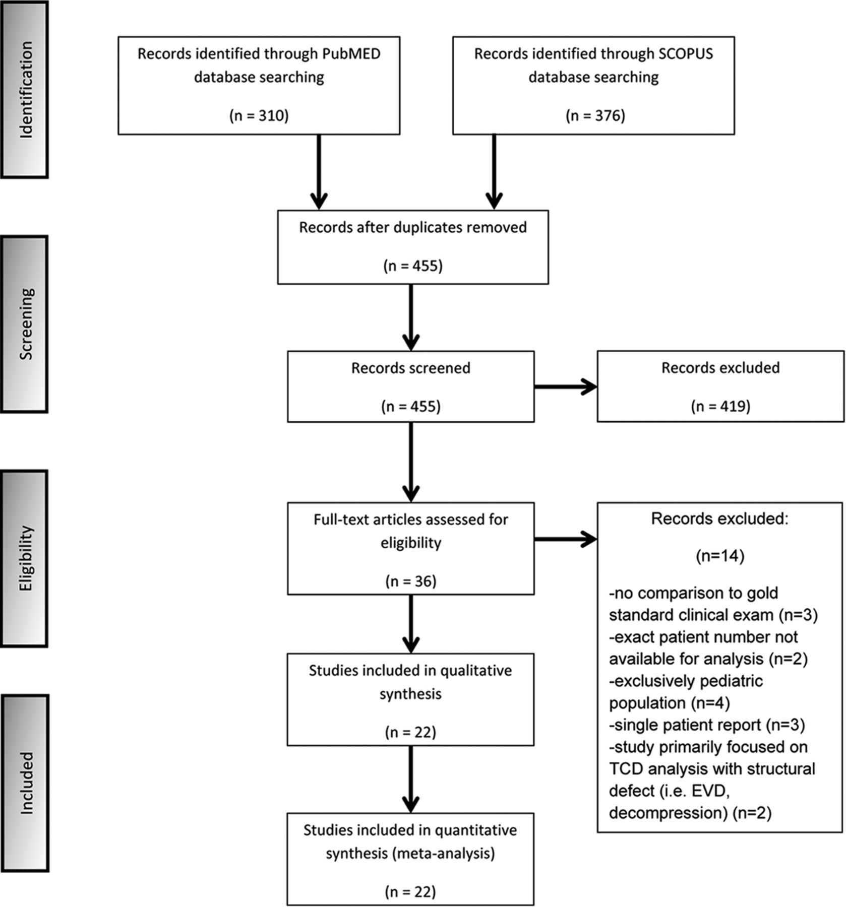
We searched PubMed and Scopus data bases, initially identifying 310 and 376 studies, respectively, that dated from 1987 to 2014. After removing duplicates, we screened the titles and abstracts from the remaining 455 studies, yielding 36 potentially eligible studies for the meta-analysis. The Cochrane Central data base search retrieved no additional studies. Full-text versions of these 36 studies were obtained, and 14 studies were excluded for the following reasons: 1) They did not offer a comparison criterion standard clinical examination,12⇓–14 2) the exact number of patients could not be extrapolated from the methodology or results for analysis,15,16 3) the study consisted of an exclusively pediatric population,17⇓⇓–20 4) the article was a single-patient case report,21⇓–23 or 5) the study primarily focused on TCD analysis in populations with structural defects of the cranium (ie, patients with external ventricular devices or postdecompressive hemicraniectomy).24,25

In the final presentation of the literature search results, there was no conflict or disagreement between 2 reviewers (J.J.C. and G.T.), and the 22 studies that met the protocol inclusion criteria of the study were included in the present systematic review (Fig 1). The characteristics of the included studies,26⇓⇓⇓⇓⇓⇓⇓⇓⇓⇓⇓⇓⇓⇓⇓⇓⇓⇓⇓⇓–47 comprising 1596 total patients, are shown in the On-line Table.

Fig 1.
  • Download figure
  • Open in new tab
  • Download powerpoint
Fig 1.

Flow chart diagram presenting the selection of eligible studies. EVD indicates external ventricular drain.

Quality Assessment

The quality assessment of included studies by using QUADAS-2 is presented in the Table. High bias was introduced in all 22 studies when using the risk of bias/index test asking the question “could the conduct or interpretation of the index test have introduced bias?” because TCDs were always performed after confirmation of clinical brain death. Risk of bias/patient selection was introduced in 4 studies because patients were excluded due to either TCD windows being unobtainable27 or, by study design only, a portion of the patients who were clinically brain dead underwent TCDs.34,38,43 High bias was introduced in risk of bias/flow and timing in 3 studies because patients were excluded from analysis due to TCD waveforms being unobtainable despite vasopressor use,28 bone windows or clinical brain death not being determined,30 and clinical brain death and even ancillary tests not being performed because of severe clinical instability.41 Unclear bias was introduced in applicability concerns/patient selection in 6 studies because pediatric patients were included in the analysis.33,34,37,42,45,47 Finally, high bias was introduced in 1 study in applicability concerns/index test because in addition to the standard TCD transtemporal window, contrast-induced TCD was used.26 No study had >2 high risks of bias noted.

View this table:
  • View inline
  • View popup

Quality assessment of eligible studies

Data Synthesis and Statistical Analysis

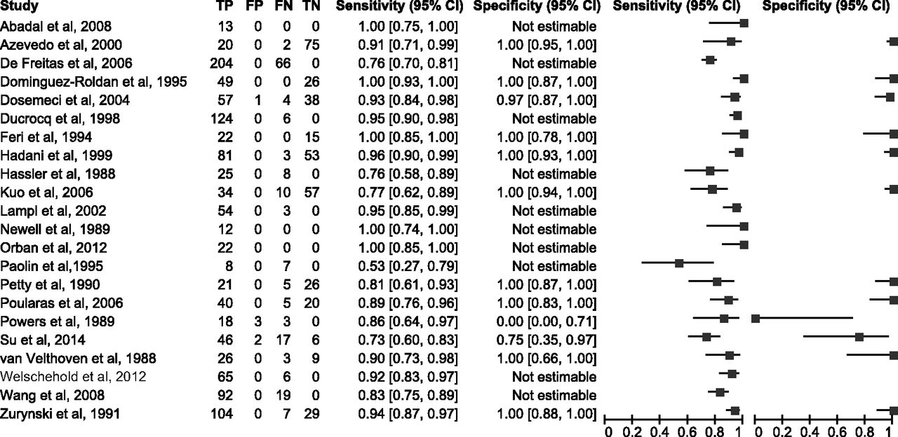
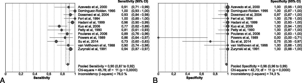
The sensitivity and specificity values, with the corresponding 95% CIs, were calculated for all included studies. However, overall sensitivity and specificity estimates were pooled from 12 of the 22 study protocols because only these studies reported data for the calculation of both sensitivity and specificity values (Fig 2).

Fig 2.
  • Download figure
  • Open in new tab
  • Download powerpoint
Fig 2.

Sensitivity and specificity ratios of individual studies, with their corresponding 95% confidence intervals. TP indicates true-positive; FP, positive; FN, false-negative; TN, true-negative.

In the pooled analysis of 859 patients (56.1% with clinically confirmed brain death), overall sensitivity was 0.90 (95% CI, 0.87–0.92; Fig 3A) and overall specificity was 0.98 (95% CI, 0.96–0.99; Fig 3B). Between-study differences in the diagnostic performance of TCDs were found for both sensitivity (I2 = 76%, P < .001, Fig 3A) and specificity (I2 = 74.3%, P < .001, Fig 3B). The threshold effect was not significant (Spearman r = −0.173, P = .612). The sROC analysis, displaying the individual studies that reported data for the calculation of both sensitivity and specificity and the sROC curve (with its corresponding 95% CI), is presented in Fig 4. The area under the curve of sROC with the corresponding standard error (SE) was 0.964 ± 0.018, while the Q* ± SE was estimated at 0.910 ± 0.028.

Fig 3.
  • Download figure
  • Open in new tab
  • Download powerpoint
Fig 3.

Pooled sensitivity (A) and specificity (B) of eligible studies reporting both diagnostic accuracy testing parameters.

Fig 4.
  • Download figure
  • Open in new tab
  • Download powerpoint
Fig 4.

The summary receiver operating characteristic curve with its corresponding 95% confidence interval of the diagnostic threshold of transcranial Doppler in the diagnosis of brain death.

Discussion

This study provides the most comprehensive review evaluating the sensitivity and specificity of TCD in confirming brain death. The results of this meta-analysis suggest that TCD is a highly accurate ancillary test for brain death confirmation with a sensitivity and specificity of 90% and 98%, respectively. Moreover, the area under the curve of sROC was estimated at 0.964, underscoring the very satisfactory diagnostic yield of TCD for diagnosing CCA.

The findings of this meta-analysis are roughly in line with the prior meta-analysis of Monteiro et al,8 with a sensitivity of 89% and specificity of 99% when using TCD for brain death confirmation. In our meta-analysis, we included all 12 studies used in that article and 10 additional studies. We also improved on the study design by systematically evaluating potential sources of bias and the relative strengths of each study. Moreover, sROC analyses were not performed in the study of Monteiro et al, while Q* was also not estimated. Finally, the risk of bias was not systematically addressed by QUADAS methodology. The findings of the present meta-analysis when combined with the study of Monteiro et al argue in favor of using TCD as a standard ancillary test for brain death diagnosis, because TCD remains an inexpensive, easily repeatable, and noninvasive examination.

Quality of Study

Evaluation of QUADAS elicited several concerns about the quality of the studies that are largely unavoidable given the nonrandomized, observational nature of these individual studies. First, high bias was introduced in all studies regarding the risk of bias/index test. This was inevitable because TCD and any ancillary test will always be measured against and completed after clinical brain death confirmation. Regarding the risk of bias/patient selection, high bias was introduced in 4 studies and unclear bias was introduced in 4 studies. Azevedo et al27 introduced bias because they did not enroll patients in whom TCDs could not be obtained, while they failed to describe the number of excluded patients. The other 3 articles introduced high bias in this category because they were primarily designed to use and compare various ancillary tests in brain death confirmation (TCD being used in addition to angiography, electroencephalography, intracranial pressure monitoring, and somatosensory-evoked potentials).34,38,43 Although high bias was introduced in 3 studies regarding risk of bias/flow and timing, the exclusion of these patients was unavoidable due to clinical instability. In all 3 studies, these patients were likely excluded because they had reached an advanced state of brain herniation that left them in multiorgan dysfunction with extreme hemodynamic instability.28,30,41 Unclear bias was introduced in 5 studies regarding applicability concerns/patient selection because pediatric populations were incorporated and analyzed. Enough details were not provided to individually remove the pediatric patients. Although unfortunate, as evidenced by mean ages, these populations were primarily adult.33,34,37,42,45,47 Finally, 1 study introduced high bias in applicability concerns/index test because while using tests that incorporated a standard TCD window (transtemporal), this study also introduced contrast-induced TCD to increase sensitivity.26

Limitations

Several limitations must be addressed when using TCDs for confirmation of brain death: 1) worse sensitivity of TCD compared with the clinical examination, 2) association between the mechanism of neurologic injury and the sensitivity of TCD, 3) the impossibility of having perfect sensitivity with TCDs, and 4) the impossibility of preventing false-positive results. First, TCD will ultimately have lower sensitivity than clinical confirmation of brain death at earlier time points because blood vessel velocities and patterns demonstrated on TCDs for brain death confirmation depend on CCA. Although isolated case reports exist demonstrating clinical confirmation of brain death, followed by CCA, and then a rebound improvement in the clinical examination (usually via resumption of respiratory drive),21 CCA represents a more severe pathologic state than brain death because it is characterized by refractory high intracranial pressures preventing cerebral perfusion. Therefore, CCA should usually temporally occur after brain stem dysfunction. This was shown because serial TCDs conducted on patients who were clinically brain dead always resulted in the best sensitivities occurring at later time points.30,33,38,41 In addition, the accuracy of TCD evaluation for brain death may also be subject to brain death criteria used in different countries, the largest difference being inclusion of apnea testing which was present in 59% of countries surveyed48 and would likely result in lower TCD sensitivities.

Second, confirmation of brain death via TCD is influenced by the mechanism of neurologic injury. The classic neurologic injury leading to brain death is a supratentorial mass lesion with downward herniation into the brain stem.49 Because herniation continues due to increased intracranial pressure, cerebral perfusion will wane and finally be absent, resulting in CCA and corresponding TCD waveforms. Other etiologies that result in global cerebral edema (diffuse anoxic brain injury, leukoencephalopathy from toxin ingestion, poor-grade subarachnoid hemorrhage, and diffuse traumatic brain injury) will also lead to refractory intracranial pressures and CCA whose waveform patterns can be detected by TCD.

Third, TCDs will never offer perfect sensitivities because structural abnormalities will inevitably arise, primarily with obtaining adequate bone windows. Sixty-four patients were reported to have unobtainable bone windows during TCD examination in 8 studies of this meta-analysis (On-line Table), giving a corresponding insufficient insonation rate of 8.4% (range, 2.8%–16%). These patients were evaluated as false-negatives, and this decreased the sensitivity measure. In addition, due to unilateral supratentorial lesions, cases will occur in which 1 large vessel will demonstrate a CCA pattern on TCD without this occurring in the contralateral vessel.39 In addition, abnormalities of the cranium, primarily manifested by postsurgical procedures such as decompressive hemicraniectomy24,25 or external ventricular device placement,24 or the more malleable cranium of infants and young children,23 will lead to decreased sensitivities. The most likely mechanism for this was increased pulsatile arterial blood flow introduced by relief of intracranial pressure.

Fourth, although having an extremely high specificity, false-positive results with TCDs are still possible and occurred in 3 studies. In Powers et al,42 2 of these false-positive cases occurred because though TCD confirmation did precede clinical brain death, unanticipated cardiopulmonary arrest prevented the inevitable confirmation of clinical brain death. The last false-positive result occurred in a 31-year-old woman with a gunshot wound to the head who had retrograde diastolic flow velocities noted on TCD but who also had extremely high net flow velocities. Her neurologic function improved, and she was ultimately discharged from the hospital.42 In Su et al, 43 2 false-positives occurred; however, details behind these results were not evaluated in the article. Finally, Dosemeci et al30 had 1 false-positive brain death result with TCD, which was associated with a patient with TCD waveforms suggestive of CCA but who still had weak respiratory movements elicited by an apnea test.

Finally, no adjustment was reported in the individual study protocols, thus providing only the unadjusted sensitivity/specificity measures in the published reports of the included studies. Therefore pooling of unadjusted sensitivity/specificity measures can neither eliminate the risk of potential confounder existence in the included study protocols nor exclude their accession to the pooled estimates.

Topics of Future Study

Several topics may help clinicians better understand the limitations associated with TCD use in brain death confirmation. One such topic may be trying to better understand the mechanism and pathophysiology associated with CCA. This would include gaining a better understanding of the time lag that occurs from brain death to CCA. Such knowledge may assist in determining the earliest time to use TCD as ancillary confirmation of brain death. Other future studies would evaluate how TCD velocity changes relative to skull defects or abnormalities. Future studies would likely need to segregate infants and young children from adults because of their malleable skulls.

Conclusions

In our meta-analysis, TCD as ancillary testing for brain death was found to be highly sensitive and specific with rates of 89% and 98%, respectively. However, the main limitation of TCD is that it detects CCA rather than brain stem dysfunction, which will inevitably result in less sensitivity than the clinical examination. As a result, TCD is still best suited for an ancillary test to be performed when clinical conditions or medications make the clinical examination unsuitable.

Footnotes

  • Disclosures: Georgios Tsivgoulis—UNRELATED: Grants/Grants Pending: I was supported by the European Regional Development Fund–Project St. Anne's University Hospital, Brno International Clinical Research Center (FNUSA-ICRC) (No. CZ.1.05/1.1.00/02.0123).

Indicates open access to non-subscribers at www.ajnr.org

References

  1. 1.↵
    A definition of irreversible coma: Report of the Ad Hoc Committee of the Harvard Medical School to Examine the Definition of Brain Death. JAMA 1968;205:337–40 doi:10.1001/jama.205.6.337 pmid:5694976
    CrossRefPubMed
  2. 2.↵
    1. Wijdicks EF
    . The diagnosis of brain death. N Engl J Med 2001;344:1215–21 doi:10.1056/NEJM200104193441606 pmid:11309637
    CrossRefPubMed
  3. 3.↵
    1. Wijdicks EF,
    2. Varelas PN,
    3. Gronseth GS, et al
    ; American Academy of Neurology. Evidence-based guideline update: determining brain death in adults: report of the Quality Standards Subcommittee of the American Academy of Neurology. Neurology 2010;74:1911–18 doi:10.1212/WNL.0b013e3181e242a8 pmid:20530327
    Abstract/FREE Full Text
  4. 4.↵
    1. Nakagawa TA,
    2. Ashwal S,
    3. Mathur M, et al
    ; Society of Critical Care Medicine, Section on Critical Care and Section on Neurology of the American Academy of Pediatrics, Child Neurology Society. Guidelines for the determination of brain death in infants and children: an update of the 1987 Task Force recommendations. Crit Care Med 2011;39:2139–55 doi:10.1097/CCM.0b013e31821f0d4f pmid:21849823
    CrossRefPubMed
  5. 5.↵
    1. Datar S,
    2. Fugate J,
    3. Rabinstein A, et al
    . Completing the apnea test: decline in complications. Neurocrit Care 2014;21:392–96 doi:10.1007/s12028-014-9958-y pmid:24522760
    CrossRefPubMed
  6. 6.↵
    1. Goudreau JL,
    2. Wijdicks EF,
    3. Emery SF
    . Complications during apnea testing in the determination of brain death: predisposing factors. Neurology 2000;55:1045–48 pmid:11061269
    Abstract/FREE Full Text
  7. 7.↵
    1. Bayliff CD,
    2. Schwartz ML,
    3. Hardy BG
    . Pharmacokinetics of high-dose pentobarbital in severe head trauma. Clin Pharmacol Ther 1985;38:457–61 doi:10.1038/clpt.1985.204 pmid:4042529
    CrossRefPubMed
  8. 8.↵
    1. Monteiro LM,
    2. Bollen CW,
    3. van Huffelen AC, et al
    . Transcranial Doppler ultrasonography to confirm brain death: a meta-analysis. Intensive Care Med 2006;32:1937–44 doi:10.1007/s00134-006-0353-9 pmid:17019556
    CrossRefPubMed
  9. 9.↵
    1. Whiting PF,
    2. Rutjes AW,
    3. Westwood ME, et al
    ; QUADAS-2 Group. QUADAS-2: a revised tool for the quality assessment of diagnostic accuracy studies. Ann Intern Med 2011;155:529–36 doi:10.7326/0003-4819-155-8-201110180-00009 pmid:22007046
    CrossRefPubMed
  10. 10.↵
    1. Liberati A,
    2. Altman DG,
    3. Tetzlaff J, et al
    . The PRISMA statement for reporting systematic reviews and meta-analyses of studies that evaluate health care interventions: explanation and elaboration. J Clin Epidemiol 2009;62:e1–34 doi:10.1016/j.jclinepi.2009.06.006 pmid:19631507
    CrossRefPubMed
  11. 11.↵
    1. Deeks JJ,
    2. Bossuyt PM,
    3. Gatsonis C
    1. Reitsma JB,
    2. Rutjes AW,
    3. Whiting P, et al
    . Chapter 9: assessing methodological quality. In: Deeks JJ, Bossuyt PM, Gatsonis C, eds. Cochrane Handbook for Systematic Reviews of Diagnostic Test Accuracy. The Cochrane Collaboration, 2009. http://srdta.cochrane.org/. Accessed September 30, 2014.
  12. 12.↵
    1. Dominguez-Roldan JM,
    2. Garcia-Alfaro C,
    3. Jimenez-Gonzalez PI, et al
    . Brain death due to supratentorial masses: diagnosis using transcranial Doppler sonography. Transplant Proc 2004;36:2898–900 doi:10.1016/j.transproceed.2004.11.017 pmid:15686656
    CrossRefPubMed
  13. 13.↵
    1. Sharma D,
    2. Souter MJ,
    3. Moore AE, et al
    . Clinical experience with transcranial Doppler ultrasonography as a confirmatory test for brain death: a retrospective analysis. Neurocrit Care 2011;14:370–76 doi:10.1007/s12028-010-9415-5 pmid:20694525
    CrossRefPubMed
  14. 14.↵
    1. Shiogai T,
    2. Sato E,
    3. Tokitsu M, et al
    . Transcranial Doppler monitoring in severe brain damage: relationships between intracranial haemodynamics, brain dysfunction and outcome. Neurol Res 1990;12:205–13 pmid:1982161
    PubMed
  15. 15.↵
    1. Vakilian A,
    2. Iranmanesh F
    . Comparison of cerebral blood flow pattern by transcranial Doppler in patients with diffuse and focal causes of brain death. J Res Med Sci 2012;17:1156–60 pmid:23853634
    PubMed
  16. 16.↵
    1. Llompart-Pou JA,
    2. Abadal JM,
    3. Velasco J, et al
    . Contrast-enhanced transcranial color sonography in the diagnosis of cerebral circulatory arrest. Transplant Proc 2009;41:1466–68 doi:10.1016/j.transproceed.2008.12.036 pmid:19545657
    CrossRefPubMed
  17. 17.↵
    1. Kirkham FJ,
    2. Levin SD,
    3. Padayachee TS, et al
    . Transcranial pulsed Doppler ultrasound findings in brain stem death. J Neurol Neurosurg Psychiatry 1987;50:1504–13 doi:10.1136/jnnp.50.11.1504 pmid:3320276
    Abstract/FREE Full Text
  18. 18.↵
    1. Qian SY,
    2. Fan XM,
    3. Yin HH
    . Transcranial Doppler assessment of brain death in children. Singapore Med J 1998;39:247–50 pmid:9803811
    PubMed
  19. 19.↵
    1. Vicenzini E,
    2. Pulitano P,
    3. Cicchetti R, et al
    . Transcranial Doppler for brain death in infants: the role of the fontanelles. Eur Neurol 2010;63:164–69 doi:10.1159/000286232 pmid:20173326
    CrossRefPubMed
  20. 20.↵
    1. Rodriguez RA,
    2. Cornel G,
    3. Alghofaili F, et al
    . Transcranial Doppler during suspected brain death in children: potential limitation in patients with cardiac “shunt.” Pediatr Crit Care Med 2002;3:153–57 doi:10.1097/00130478-200204000-00012 pmid:12780986
    CrossRefPubMed
  21. 21.↵
    1. Budohoski KP,
    2. Aries MJ,
    3. Kirkpatrick PJ, et al
    . Protracted cerebral circulatory arrest and cortical electrical silence coexisting with preserved respiratory drive and flexor motor response. Br J Anaest 2012;109:293–94 doi:10.1093/bja/aes243 pmid:22782987
    FREE Full Text
  22. 22.↵
    1. Eder KE,
    2. Haussen DC,
    3. Searls DE, et al
    . Reverberating TCD flow pattern in brain death. Neurology 2012;79:e79 doi:10.1212/WNL.0b013e318266fc89 pmid:22927685
    FREE Full Text
  23. 23.↵
    1. Mata-Zubillaga D,
    2. Oulego-Erroz I
    . Persistent cerebral blood flow by transcranial Doppler ultrasonography in an asphyxiated newborn meeting brain death diagnosis: case report and review of the literature. J Perinatal 2012;32:473–75 doi:10.1038/jp.2011.147 pmid:22643293
    CrossRefPubMed
  24. 24.↵
    1. Thompson BB,
    2. Wendell LC,
    3. Potter NS, et al
    . The use of transcranial Doppler ultrasound in confirming brain death in the setting of skull defects and extraventricular drains. Neurocrit Care 2014;21:534–38 doi:10.1007/s12028-014-9979-6 pmid:24718963
    CrossRefPubMed
  25. 25.↵
    1. Cabrer C,
    2. Dominguez-Roldan JM,
    3. Manyalich M, et al
    . Persistence of intracranial diastolic flow in transcranial Doppler sonography exploration of patients in brain death. Transplant Proc 2003;35:1642–43 doi:10.1016/S0041-1345(03)00692-4 pmid:12962741
    CrossRefPubMed
  26. 26.↵
    1. Abadal JM,
    2. Llompart-Pou JA,
    3. Homar J, et al
    . Ultrasonographic cerebral perfusion in assessment of brain death: a preliminary study. J Ultrasound Med 2008;27:791–94 pmid:18424656
    Abstract/FREE Full Text
  27. 27.↵
    1. Azevedo E,
    2. Teixeira J,
    3. Neves JC, et al
    . Transcranial Doppler and brain death. Transplant Proc 2000;32:2579–81 doi:10.1016/S0041-1345(00)01794-2 pmid:11134714
    CrossRefPubMed
  28. 28.↵
    1. de Freitas GR,
    2. André C
    . Sensitivity of transcranial Doppler for confirming brain death: a prospective study of 270 cases. Acta Neurol Scand 2006;113:426–32 doi:10.1111/j.1600-0404.2006.00645.x pmid:16674610
    CrossRefPubMed
  29. 29.↵
    1. Dominguez-Roldan JM,
    2. Murillo-Cabezas F,
    3. Muñoz-Sanchez A, et al
    . Changes in the Doppler waveform of intracranial arteries in patients with brain-death status. Transplant Proc 1995;27:2391–92 pmid:7652849
    PubMed
  30. 30.↵
    1. Dosemeci L,
    2. Dora B,
    3. Yilmaz M, et al
    . Utility of transcranial Doppler ultrasonography for confirmatory diagnosis of brain death: two sides of the coin. Transplantation 2004;77:71–75 doi:10.1097/01.TP.0000092305.00155.72 pmid:14724438
    CrossRefPubMed
  31. 31.↵
    1. Ducrocq X,
    2. Braun M,
    3. Debouverie M, et al
    . Brain death and transcranial Doppler: experience in 130 cases of brain dead patients. J Neurol Sci 1998;160:41–46 doi:10.1016/S0022-510X(98)00188-9 pmid:9804115
    CrossRefPubMed
  32. 32.↵
    1. Feri M,
    2. Ralli L,
    3. Felici M, et al
    . Transcranial Doppler and brain death diagnosis. Crit Care Med 1994;22:1120–26 doi:10.1097/00003246-199407000-00012 pmid:7913008
    CrossRefPubMed
  33. 33.↵
    1. Hadani M,
    2. Bruk B,
    3. Ram Z, et al
    . Application of transcranial Doppler ultrasonography for the diagnosis of brain death. Intensive Care Med 1999;25:822–28 doi:10.1007/s001340050958 pmid:10447539
    CrossRefPubMed
  34. 34.↵
    1. Hassler W,
    2. Steinmetz H,
    3. Gawlowski J
    . Transcranial Doppler ultrasonography in raised intracranial pressure and in intracranial circulatory arrest. J Neurosurg 1988;68:745–51 pmid:3282040
    PubMed
  35. 35.↵
    1. Kuo JR,
    2. Chen CF,
    3. Chio CC, et al
    . Time dependent validity in the diagnosis of brain death using transcranial Doppler sonography. J Neurol Neurosurg Psychiatry 2006;77:646–49 doi:10.1136/jnnp.2005.076406 pmid:16614026
    Abstract/FREE Full Text
  36. 36.↵
    1. Lampl Y,
    2. Gilad R,
    3. Eschel Y, et al
    . Diagnosing brain death using the transcranial Doppler with a transorbital approach. Arch Neurol 2002;59:58–60 doi:10.1001/archneur.59.1.58 pmid:11790231
    CrossRefPubMed
  37. 37.↵
    1. Newell DW,
    2. Grady MS,
    3. Sirotta P, et al
    . Evaluation of brain death using transcranial Doppler. Neurosurgery 1989;24:509–13 doi:10.1227/00006123-198904000-00004 pmid:2651959
    CrossRefPubMed
  38. 38.↵
    1. Orban JC,
    2. El-Mahjoub A,
    3. Rami L, et al
    . Transcranial Doppler shortens the time between clinical brain death and angiographic confirmation: a randomized trial. Transplantation 2012;94:585–88 doi:10.1097/TP.0b013e3182612947 pmid:22918218
    CrossRefPubMed
  39. 39.↵
    1. Paolin A,
    2. Manuali A,
    3. Di Paola F, et al
    . Reliability in diagnosis of brain death. Intensive Care Med 1995;21:657–62 doi:10.1007/BF01711544 pmid:8522670
    CrossRefPubMed
  40. 40.↵
    1. Petty GW,
    2. Mohr JP,
    3. Pedley TA, et al
    . The role of transcranial Doppler in confirming brain death: sensitivity, specificity, and suggestions for performance and interpretation. Neurology 1990;40:300–03 doi:10.1212/WNL.40.2.300 pmid:2405294
    Abstract/FREE Full Text
  41. 41.↵
    1. Poularas J,
    2. Karakitsos D,
    3. Kouraklis G, et al
    . Comparison between transcranial color Doppler ultrasonography and angiography in the confirmation of brain death. Transplant Proc 2006;38:1213–17 doi:10.1016/j.transproceed.2006.02.127 pmid:16797266
    CrossRefPubMed
  42. 42.↵
    1. Powers AD,
    2. Graeber MC,
    3. Smith RR
    . Transcranial Doppler ultrasonography in the determination of brain death. Neurosurgery 1989;24:884–89 doi:10.1227/00006123-198906000-00015 pmid:2747862
    CrossRefPubMed
  43. 43.↵
    1. Su Y,
    2. Yang Q,
    3. Liu G, et al
    . Diagnosis of brain death: confirmatory tests after clinical test. Chin Med J (Engl) 2014;127:1272–77 pmid:24709179
    PubMed
  44. 44.↵
    1. Van Velthoven V,
    2. Calliauw L
    . Diagnosis of brain death: transcranial Doppler sonography as an additional method. Acta Neurochir (Wien) 1988;95:57–60 doi:10.1007/BF01793084 pmid:3064557
    CrossRefPubMed
  45. 45.↵
    1. Wang K,
    2. Yuan Y,
    3. Xu ZQ, et al
    . Benefits of combination of electroencephalography, short latency somatosensory evoked potentials, and transcranial Doppler techniques for confirming brain death. J Zhejiang Univ Sci B 2008;9:916–20 doi:10.1631/jzus.B0820123 pmid:18988311
    CrossRefPubMed
  46. 46.↵
    1. Welschehold S,
    2. Boor S,
    3. Reuland K, et al
    . Technical aids in the diagnosis of brain death: a comparison of SEP, AEP, EEG, TCD and CT angiography. Dtsch Arztebl Int 2012;109:624–30 doi:10.3238/arztebl.2012.0624 pmid:23093994
    CrossRefPubMed
  47. 47.↵
    1. Zurynski Y,
    2. Dorsch N,
    3. Pearson I, et al
    . Transcranial Doppler ultrasound in brain death: experience in 140 patients. Neurol Res 1991;13:248–52 pmid:1687335
    PubMed
  48. 48.↵
    1. Wijdicks EF
    . Brain death worldwide: accepted fact but no global consensus in diagnostic criteria. Neurology 2002;58:20–25 doi:10.1212/WNL.58.1.20 pmid:11781400
    Abstract/FREE Full Text
  49. 49.↵
    1. Ropper AH
    . A preliminary MRI study of the geometry of brain displacement and level of consciousness with acute intracranial masses. Neurology 1989;39:622–27 doi:10.1212/WNL.39.5.622 pmid:2710350
    Abstract/FREE Full Text
  • Received May 4, 2015.
  • Accepted after revision July 11, 2015.
  • © 2016 by American Journal of Neuroradiology
View Abstract
PreviousNext
Back to top

In this issue

American Journal of Neuroradiology: 37 (3)
American Journal of Neuroradiology
Vol. 37, Issue 3
1 Mar 2016
  • Table of Contents
  • Index by author
  • Complete Issue (PDF)
Advertisement
Print
Download PDF
Email Article

Thank you for your interest in spreading the word on American Journal of Neuroradiology.

NOTE: We only request your email address so that the person you are recommending the page to knows that you wanted them to see it, and that it is not junk mail. We do not capture any email address.

Enter multiple addresses on separate lines or separate them with commas.
Diagnostic Accuracy of Transcranial Doppler for Brain Death Confirmation: Systematic Review and Meta-Analysis
(Your Name) has sent you a message from American Journal of Neuroradiology
(Your Name) thought you would like to see the American Journal of Neuroradiology web site.
CAPTCHA
This question is for testing whether or not you are a human visitor and to prevent automated spam submissions.
Cite this article
J.J. Chang, G. Tsivgoulis, A.H. Katsanos, M.D. Malkoff, A.V. Alexandrov
Diagnostic Accuracy of Transcranial Doppler for Brain Death Confirmation: Systematic Review and Meta-Analysis
American Journal of Neuroradiology Mar 2016, 37 (3) 408-414; DOI: 10.3174/ajnr.A4548

Citation Manager Formats

  • BibTeX
  • Bookends
  • EasyBib
  • EndNote (tagged)
  • EndNote 8 (xml)
  • Medlars
  • Mendeley
  • Papers
  • RefWorks Tagged
  • Ref Manager
  • RIS
  • Zotero
0 Responses
Respond to this article
Share
Bookmark this article
Diagnostic Accuracy of Transcranial Doppler for Brain Death Confirmation: Systematic Review and Meta-Analysis
J.J. Chang, G. Tsivgoulis, A.H. Katsanos, M.D. Malkoff, A.V. Alexandrov
American Journal of Neuroradiology Mar 2016, 37 (3) 408-414; DOI: 10.3174/ajnr.A4548
del.icio.us logo Twitter logo Facebook logo Mendeley logo
  • Tweet Widget
  • Facebook Like
  • Google Plus One
Purchase

Jump to section

  • Article
    • Abstract
    • ABBREVIATIONS:
    • Materials and Methods
    • Results
    • Discussion
    • Conclusions
    • Footnotes
    • References
  • Figures & Data
  • Supplemental
  • Info & Metrics
  • Responses
  • References
  • PDF

Related Articles

  • PubMed
  • Google Scholar

Cited By...

  • Pediatric and Adult Brain Death/Death by Neurologic Criteria Consensus Guideline: Report of the AAN Guidelines Subcommittee, AAP, CNS, and SCCM
  • Crossref (78)
  • Google Scholar

This article has been cited by the following articles in journals that are participating in Crossref Cited-by Linking.

  • Determination of Brain Death/Death by Neurologic Criteria
    David M. Greer, Sam D. Shemie, Ariane Lewis, Sylvia Torrance, Panayiotis Varelas, Fernando D. Goldenberg, James L. Bernat, Michael Souter, Mehmet Akif Topcuoglu, Anne W. Alexandrov, Marie Baldisseri, Thomas Bleck, Giuseppe Citerio, Rosanne Dawson, Arnold Hoppe, Stephen Jacobe, Alex Manara, Thomas A. Nakagawa, Thaddeus Mason Pope, William Silvester, David Thomson, Hussain Al Rahma, Rafael Badenes, Andrew J. Baker, Vladimir Cerny, Cherylee Chang, Tiffany R. Chang, Elena Gnedovskaya, Moon-Ku Han, Stephen Honeybul, Edgar Jimenez, Yasuhiro Kuroda, Gang Liu, Uzzwal Kumar Mallick, Victoria Marquevich, Jorge Mejia-Mantilla, Michael Piradov, Sarah Quayyum, Gentle Sunder Shrestha, Ying-ying Su, Shelly D. Timmons, Jeanne Teitelbaum, Walter Videtta, Kapil Zirpe, Gene Sung
    JAMA 2020 324 11
  • Brain ultrasonography: methodology, basic and advanced principles and clinical applications. A narrative review
    Chiara Robba, Alberto Goffi, Thomas Geeraerts, Danilo Cardim, Gabriele Via, Marek Czosnyka, Soojin Park, Aarti Sarwal, Llewellyn Padayachy, Frank Rasulo, Giuseppe Citerio
    Intensive Care Medicine 2019 45 7
  • Basic ultrasound head-to-toe skills for intensivists in the general and neuro intensive care unit population: consensus and expert recommendations of the European Society of Intensive Care Medicine 
    Chiara Robba, Adrian Wong, Daniele Poole, Ashraf Al Tayar, Robert T. Arntfield, Michelle S. Chew, Francesco Corradi, Ghislaine Douflé, Alberto Goffi, Massimo Lamperti, Paul Mayo, Antonio Messina, Silvia Mongodi, Mangala Narasimhan, Corina Puppo, Aarti Sarwal, Michel Slama, Fabio S. Taccone, Philippe Vignon, Antoine Vieillard-Baron, Chiara Robba, Adrian Wong, Daniele Poole, Ashraf Al Tayar, Robert T Arntfield, Michelle S Chew, Francesco Corradi, Ghislaine Douflé, Alberto Goffi, Massimo Lamperti, Paul Mayo, Antonio Messina, Silvia Mongodi, Mangala Narasimhan, Corina Puppo, Aarti Sarwal, Michel Slama, Fabio S Taccone, Philippe Vignon, Antoine Vieillard-Baron
    Intensive Care Medicine 2021 47 12
  • Pediatric and Adult Brain Death/Death by Neurologic Criteria Consensus Guideline
    David M. Greer, Matthew P. Kirschen, Ariane Lewis, Gary S. Gronseth, Alexander Rae-Grant, Stephen Ashwal, Maya A. Babu, David F. Bauer, Lori Billinghurst, Amanda Corey, Sonia Partap, Michael A. Rubin, Lori Shutter, Courtney Takahashi, Robert C. Tasker, Panayiotis Nicolaou Varelas, Eelco Wijdicks, Amy Bennett, Scott R. Wessels, John J. Halperin
    Neurology 2023 101 24
  • Quantitative pupillometry and transcranial Doppler measurements in patients treated with hypothermia after cardiac arrest
    Delphine Heimburger, Michel Durand, Lucie Gaide-Chevronnay, Geraldine Dessertaine, Pierre-Henri Moury, Pierre Bouzat, Pierre Albaladejo, Jean-Francois Payen
    Resuscitation 2016 103
  • Extracardiac Signs of Fluid Overload in the Critically Ill Cardiac Patient: A Focused Evaluation Using Bedside Ultrasound
    William Beaubien-Souligny, Josée Bouchard, Georges Desjardins, Yoan Lamarche, Mark Liszkowski, Pierre Robillard, André Denault
    Canadian Journal of Cardiology 2017 33 1
  • When is “brainstem death” brain death? The case for ancillary testing in primary infratentorial brain lesion
    Uwe Walter, José Luis Fernández-Torre, Timo Kirschstein, Steven Laureys
    Clinical Neurophysiology 2018 129 11
  • Ultrasound Guidelines: Emergency, Point-of-Care, and Clinical Ultrasound Guidelines in Medicine
    Annals of Emergency Medicine 2023 82 3
  • Neuromonitoring in Critically Ill Patients
    Swarna Rajagopalan, Aarti Sarwal
    Critical Care Medicine 2023 51 4
  • How to use cerebral ultrasound in the ICU
    Rita Bertuetti, Paolo Gritti, Paolo Pelosi, Chiara Robba
    Minerva Anestesiologica 2020 86 3

More in this TOC Section

  • Diagnostic Neuroradiology of Monoclonal Antibodies
  • Segmentation of Brain Metastases with BLAST
  • Cerebral ADC Changes in Fabry Disease
Show more ADULT BRAIN

Similar Articles

Advertisement

Indexed Content

  • Current Issue
  • Accepted Manuscripts
  • Article Preview
  • Past Issues
  • Editorials
  • Editor's Choice
  • Fellows' Journal Club
  • Letters to the Editor
  • Video Articles

Cases

  • Case Collection
  • Archive - Case of the Week
  • Archive - Case of the Month
  • Archive - Classic Case

More from AJNR

  • Trainee Corner
  • Imaging Protocols
  • MRI Safety Corner
  • Book Reviews

Multimedia

  • AJNR Podcasts
  • AJNR Scantastics

Resources

  • Turnaround Time
  • Submit a Manuscript
  • Submit a Video Article
  • Submit an eLetter to the Editor/Response
  • Manuscript Submission Guidelines
  • Statistical Tips
  • Fast Publishing of Accepted Manuscripts
  • Graphical Abstract Preparation
  • Imaging Protocol Submission
  • Evidence-Based Medicine Level Guide
  • Publishing Checklists
  • Author Policies
  • Become a Reviewer/Academy of Reviewers
  • News and Updates

About Us

  • About AJNR
  • Editorial Board
  • Editorial Board Alumni
  • Alerts
  • Permissions
  • Not an AJNR Subscriber? Join Now
  • Advertise with Us
  • Librarian Resources
  • Feedback
  • Terms and Conditions
  • AJNR Editorial Board Alumni

American Society of Neuroradiology

  • Not an ASNR Member? Join Now

© 2025 by the American Society of Neuroradiology All rights, including for text and data mining, AI training, and similar technologies, are reserved.
Print ISSN: 0195-6108 Online ISSN: 1936-959X

Powered by HighWire