Advertisement

Main menu

  • Home
  • Content
    • Current Issue
    • Accepted Manuscripts
    • Article Preview
    • Past Issue Archive
    • Video Articles
    • AJNR Case Collection
    • Case of the Week Archive
    • Case of the Month Archive
    • Classic Case Archive
  • Special Collections
    • AJNR Awards
    • Low-Field MRI
    • Alzheimer Disease
    • ASNR Foundation Special Collection
    • Photon-Counting CT
    • View All
  • Multimedia
    • AJNR Podcasts
    • AJNR SCANtastic
    • Trainee Corner
    • MRI Safety Corner
    • Imaging Protocols
  • For Authors
    • Submit a Manuscript
    • Submit a Video Article
    • Submit an eLetter to the Editor/Response
    • Manuscript Submission Guidelines
    • Statistical Tips
    • Fast Publishing of Accepted Manuscripts
    • Graphical Abstract Preparation
    • Imaging Protocol Submission
    • Author Policies
  • About Us
    • About AJNR
    • Editorial Board
    • Editorial Board Alumni
  • More
    • Become a Reviewer/Academy of Reviewers
    • Subscribers
    • Permissions
    • Alerts
    • Feedback
    • Advertisers
    • ASNR Home

User menu

  • Alerts
  • Log in

Search

  • Advanced search
American Journal of Neuroradiology

American Journal of Neuroradiology

ASHNR American Society of Functional Neuroradiology ASHNR American Society of Pediatric Neuroradiology ASSR
  • Alerts
  • Log in

Advanced Search

  • Home
  • Content
    • Current Issue
    • Accepted Manuscripts
    • Article Preview
    • Past Issue Archive
    • Video Articles
    • AJNR Case Collection
    • Case of the Week Archive
    • Case of the Month Archive
    • Classic Case Archive
  • Special Collections
    • AJNR Awards
    • Low-Field MRI
    • Alzheimer Disease
    • ASNR Foundation Special Collection
    • Photon-Counting CT
    • View All
  • Multimedia
    • AJNR Podcasts
    • AJNR SCANtastic
    • Trainee Corner
    • MRI Safety Corner
    • Imaging Protocols
  • For Authors
    • Submit a Manuscript
    • Submit a Video Article
    • Submit an eLetter to the Editor/Response
    • Manuscript Submission Guidelines
    • Statistical Tips
    • Fast Publishing of Accepted Manuscripts
    • Graphical Abstract Preparation
    • Imaging Protocol Submission
    • Author Policies
  • About Us
    • About AJNR
    • Editorial Board
    • Editorial Board Alumni
  • More
    • Become a Reviewer/Academy of Reviewers
    • Subscribers
    • Permissions
    • Alerts
    • Feedback
    • Advertisers
    • ASNR Home
  • Follow AJNR on Twitter
  • Visit AJNR on Facebook
  • Follow AJNR on Instagram
  • Join AJNR on LinkedIn
  • RSS Feeds

AJNR Awards, New Junior Editors, and more. Read the latest AJNR updates

Research ArticleADULT BRAIN

Ultra-High-Field MRI Visualization of Cortical Multiple Sclerosis Lesions with T2 and T2*: A Postmortem MRI and Histopathology Study

L.E. Jonkman, R. Klaver, L. Fleysher, M. Inglese and J.J.G. Geurts
American Journal of Neuroradiology November 2015, 36 (11) 2062-2067; DOI: https://doi.org/10.3174/ajnr.A4418
L.E. Jonkman
aFrom the Department of Anatomy and Neurosciences (L.E.J., R.K., J.J.G.G.), VU University Medical Center, Amsterdam, the Netherlands
  • Find this author on Google Scholar
  • Find this author on PubMed
  • Search for this author on this site
R. Klaver
aFrom the Department of Anatomy and Neurosciences (L.E.J., R.K., J.J.G.G.), VU University Medical Center, Amsterdam, the Netherlands
  • Find this author on Google Scholar
  • Find this author on PubMed
  • Search for this author on this site
L. Fleysher
bDepartments of Radiology (L.F., M.I.)
  • Find this author on Google Scholar
  • Find this author on PubMed
  • Search for this author on this site
M. Inglese
bDepartments of Radiology (L.F., M.I.)
cNeurology (M.I.)
dNeurosciences (M.I.), Mount Sinai School of Medicine, New York, New York
eDepartments of Neuroscience, Rehabilitation, Ophthalmology, Genetics, Maternal and Child Health (M.I.), University of Genoa, Genoa, Italy.
  • Find this author on Google Scholar
  • Find this author on PubMed
  • Search for this author on this site
J.J.G. Geurts
aFrom the Department of Anatomy and Neurosciences (L.E.J., R.K., J.J.G.G.), VU University Medical Center, Amsterdam, the Netherlands
  • Find this author on Google Scholar
  • Find this author on PubMed
  • Search for this author on this site
  • Article
  • Figures & Data
  • Info & Metrics
  • Responses
  • References
  • PDF
Loading

Article Figures & Data

Figures

  • Tables
  • Fig 1.
    • Download figure
    • Open in new tab
    • Download powerpoint
    Fig 1.

    Section stained with anti-proteolipid protein antibodies (A), matched with T2*WI (B) and T2WI (C and D). Note that the histologic section corresponds with multiple slices of the MR image; the top part of image B and D corresponds to the top part of image A, and the bottom part of image C corresponds to the bottom part of image A. The border between successive MR imaging slices is depicted by the blue dotted line. Lesions are indicated with arrows (WML is blue; GML is red). The type of GM lesion is indicated by I–IV. Also indicated is whether histologic lesions were retrospectively seen on MR imaging (asterisk) or missed on MR imaging (number sign). All other histologic lesions were prospectively detected. Degree of magnification: 50×.
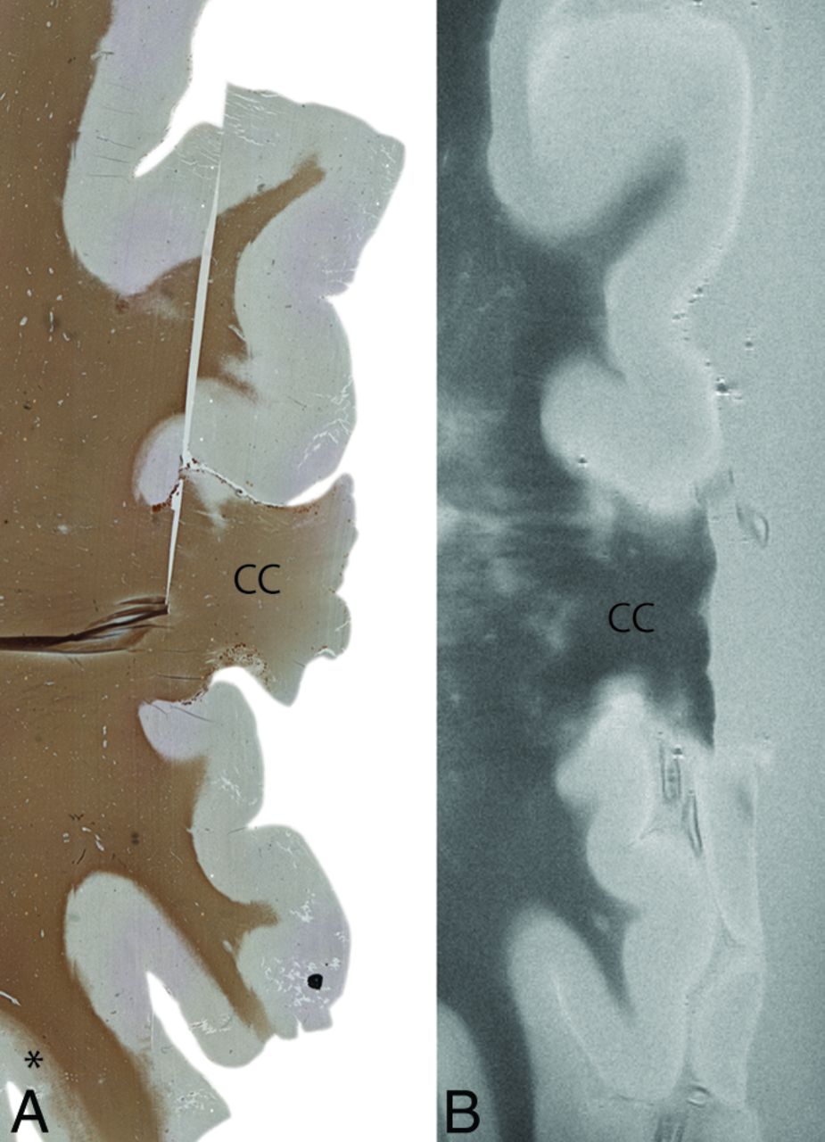
  • Fig 2.
    • Download figure
    • Open in new tab
    • Download powerpoint
    Fig 2.

    An example of extensive cortical demyelination in an MS case. Histologic section with anti-proteolipid protein antibody (left) and a matched T2WI (right). The histologic section shows extensive cortical demyelination (lack of proteolipid protein) in the cortex, except for a small section at the left bottom (asterisk). This extensive demyelination makes it difficult to differentiate lesions and normal-appearing gray matter on MR imaging (right). In this particular case, as a result, prospective MR imaging scoring was negative. CC indicates corpus callosum. Degree of magnification: 50×.

Tables

  • Figures
    • View popup
    Table 1:

    Demographic and neuropathologic data of subjects

    Case No.NoaSexAge (yr)PMD (h:min)bDD (yr)MS TypeCOD
    MS
        1M806:0545SPMSPneumonia
        2F813:3027PPMSPneumonia
        3M7510:1050NAPneumonia
        4F667:3017NAPulmonary hypertension
        5M714:0015SPMSPulmonary carcinoma
        6F546:0016SPMSLiver cancer
        7M634:3025SPMSPneumonia
        8M783:0033SPMSEuthanasia
        9M595:0021SPMSEuthanasia
        10M5610:1013NASuicide
        11F568:2532SPMSPneumonia
        12F543:3031SPMSHeart failure
        13M584:0027SPMSPneumonia
        14F956:3055SPMSUnknown
        15F816:3021SPMSHeart failure
        Mean68.5 ± 12.75:56 ± 2:2728.5 ± 12.9
    Control
        204F72>24:00––Myocardial infarct
        213F58<24:00––Breast cancer
        223F76<24:00––Pneumonia
        232F76<8:00––Pneumonia
        Mean70.5 ± 8.5
    • Note:—PMD indicates postmortem delay; DD, disease duration since diagnosis; SPMS, secondary-progressive MS; PPMS, primary-progressive MS; COD, cause of death; NA, unavailable/unknown; –, not applicable.

    • ↵a The numbers indicate number of hemispheric sections included per case.

    • ↵b Control cases are not part of the rapid postmortem examination program and therefore have a longer PMD.

    • View popup
    Table 2:

    Lesion count and sensitivity of prospective and retrospective MRI scoringa

    Lesion TypeHistologyProspective MRIRetrospective MRI
    CountT2WIT2*WIP ValueT2WIT2*WIP Value
    I148 (57)2 (14)–14 (100)13 (93)–
    II163 (19)5 (31)–13 (81)10 (63)–
    III435 (12)5 (12)–32 (74)36 (84)–
    IV2511 (44)4 (16)–22 (88)23 (92)–
    GML (I–IV)9827 (28)16 (16).05481 (83)82 (84).803
    GML (II–IV)8419 (23)14 (17).38067 (80)69 (82)0.608
    WML76 (86)3 (43)–7 (100)7 (100)–
    Total10533 (31)19 (18).018b88 (84)89 (85).803
    • Note:— – indicates not statistically assessed.

    • ↵a Sensitivity (in percentages between parentheses) was calculated by dividing the number of lesions scored in the prospective or retrospective ratings by the number of lesions assessed on histopathology, times 100%.

    • ↵b Significant.

    • View popup
    Table 3:

    Contrast-to-noise ratio (±SD)

    CNRT2WIT2*WI
    WM-WML12.07 (0.90)5.03 (1.68)
    GM-GML2.01 (0.74)1.7 (1.37)
    GM-WM7.5 (1.58)4.96 (3.79)
    • Note:—WM-WML indicates white matter-to-white matter lesion CNR; GM-GML, gray matter-to-gray matter lesion CNR; GM-WM, gray matter-to-white matter CNR.

PreviousNext
Back to top

In this issue

American Journal of Neuroradiology: 36 (11)
American Journal of Neuroradiology
Vol. 36, Issue 11
1 Nov 2015
  • Table of Contents
  • Index by author
  • Complete Issue (PDF)
Advertisement
Print
Download PDF
Email Article

Thank you for your interest in spreading the word on American Journal of Neuroradiology.

NOTE: We only request your email address so that the person you are recommending the page to knows that you wanted them to see it, and that it is not junk mail. We do not capture any email address.

Enter multiple addresses on separate lines or separate them with commas.
Ultra-High-Field MRI Visualization of Cortical Multiple Sclerosis Lesions with T2 and T2*: A Postmortem MRI and Histopathology Study
(Your Name) has sent you a message from American Journal of Neuroradiology
(Your Name) thought you would like to see the American Journal of Neuroradiology web site.
CAPTCHA
This question is for testing whether or not you are a human visitor and to prevent automated spam submissions.
Cite this article
L.E. Jonkman, R. Klaver, L. Fleysher, M. Inglese, J.J.G. Geurts
Ultra-High-Field MRI Visualization of Cortical Multiple Sclerosis Lesions with T2 and T2*: A Postmortem MRI and Histopathology Study
American Journal of Neuroradiology Nov 2015, 36 (11) 2062-2067; DOI: 10.3174/ajnr.A4418

Citation Manager Formats

  • BibTeX
  • Bookends
  • EasyBib
  • EndNote (tagged)
  • EndNote 8 (xml)
  • Medlars
  • Mendeley
  • Papers
  • RefWorks Tagged
  • Ref Manager
  • RIS
  • Zotero
0 Responses
Respond to this article
Share
Bookmark this article
Ultra-High-Field MRI Visualization of Cortical Multiple Sclerosis Lesions with T2 and T2*: A Postmortem MRI and Histopathology Study
L.E. Jonkman, R. Klaver, L. Fleysher, M. Inglese, J.J.G. Geurts
American Journal of Neuroradiology Nov 2015, 36 (11) 2062-2067; DOI: 10.3174/ajnr.A4418
del.icio.us logo Twitter logo Facebook logo Mendeley logo
  • Tweet Widget
  • Facebook Like
  • Google Plus One
Purchase

Jump to section

  • Article
    • Abstract
    • ABBREVIATIONS:
    • Materials and Methods
    • Results
    • Discussion
    • Conclusions
    • Footnotes
    • REFERENCES
  • Figures & Data
  • Info & Metrics
  • Responses
  • References
  • PDF

Related Articles

  • No related articles found.
  • PubMed
  • Google Scholar

Cited By...

  • Imaging cortical multiple sclerosis lesions with ultra-high field MRI
  • Comparison of Multiple Sclerosis Cortical Lesion Types Detected by Multicontrast 3T and 7T MRI
  • Improved Visualization of Cortical Lesions in Multiple Sclerosis Using 7T MP2RAGE
  • Crossref (25)
  • Google Scholar

This article has been cited by the following articles in journals that are participating in Crossref Cited-by Linking.

  • Deep Learning Based Noise Reduction for Brain MR Imaging: Tests on Phantoms and Healthy Volunteers
    Masafumi Kidoh, Kensuke Shinoda, Mika Kitajima, Kenzo Isogawa, Masahito Nambu, Hiroyuki Uetani, Kosuke Morita, Takeshi Nakaura, Machiko Tateishi, Yuichi Yamashita, Yasuyuki Yamashita
    Magnetic Resonance in Medical Sciences 2020 19 3
  • Iron in Multiple Sclerosis and Its Noninvasive Imaging with Quantitative Susceptibility Mapping
    Carsten Stüber, David Pitt, Yi Wang
    International Journal of Molecular Sciences 2016 17 1
  • Improved Visualization of Cortical Lesions in Multiple Sclerosis Using 7T MP2RAGE
    E.S. Beck, P. Sati, V. Sethi, T. Kober, B. Dewey, P. Bhargava, G. Nair, I.C. Cortese, D.S. Reich
    American Journal of Neuroradiology 2018 39 3
  • Comparison of Multiple Sclerosis Cortical Lesion Types Detected by Multicontrast 3T and 7T MRI
    J. Maranzano, M. Dadar, D.A. Rudko, D. De Nigris, C. Elliott, J.S. Gati, S.A. Morrow, R.S. Menon, D.L. Collins, D.L. Arnold, S. Narayanan
    American Journal of Neuroradiology 2019 40 7
  • Iron related changes in MS lesions and their validity to characterize MS lesion types and dynamics with Ultra‐high field magnetic resonance imaging
    Simon Hametner, Assunta Dal Bianco, Siegfried Trattnig, Hans Lassmann
    Brain Pathology 2018 28 5
  • Present and future of the diagnostic work-up of multiple sclerosis: the imaging perspective
    Massimo Filippi, Paolo Preziosa, Douglas L. Arnold, Frederik Barkhof, Daniel M. Harrison, Pietro Maggi, Caterina Mainero, Xavier Montalban, Elia Sechi, Brian G. Weinshenker, Maria A. Rocca
    Journal of Neurology 2023 270 3
  • The substrate of increased cortical FA in MS: A 7T post-mortem MRI and histopathology study
    Laura E Jonkman, Roel Klaver, Lazar Fleysher, Matilde Inglese, Jeroen JG Geurts
    Multiple Sclerosis Journal 2016 22 14
  • Clinical Neuroimaging Using 7 T MRI: Challenges and Prospects
    Maria Isabel Vargas, Pascal Martelli, Lijing Xin, Ozlem Ipek, Frederic Grouiller, Francesca Pittau, Robert Trampel, Rolf Gruetter, Serge Vulliemoz, Francois Lazeyras
    Journal of Neuroimaging 2018 28 1
  • Imaging cortical multiple sclerosis lesions with ultra-high field MRI
    Mads A.J. Madsen, Vanessa Wiggermann, Stephan Bramow, Jeppe Romme Christensen, Finn Sellebjerg, Hartwig R. Siebner
    NeuroImage: Clinical 2021 32
  • Ultra-high field MTR and qR2* differentiates subpial cortical lesions from normal-appearing gray matter in multiple sclerosis
    Laura E Jonkman, Lazar Fleysher, Martijn D Steenwijk, Jan A Koeleman, Teun-Pieter de Snoo, Frederik Barkhof, Matilde Inglese, Jeroen JG Geurts
    Multiple Sclerosis Journal 2016 22 10

More in this TOC Section

  • Diagnostic Neuroradiology of Monoclonal Antibodies
  • Clinical Outcomes After Chiari I Decompression
  • Segmentation of Brain Metastases with BLAST
Show more ADULT BRAIN

Similar Articles

Advertisement

Indexed Content

  • Current Issue
  • Accepted Manuscripts
  • Article Preview
  • Past Issues
  • Editorials
  • Editor's Choice
  • Fellows' Journal Club
  • Letters to the Editor
  • Video Articles

Cases

  • Case Collection
  • Archive - Case of the Week
  • Archive - Case of the Month
  • Archive - Classic Case

More from AJNR

  • Trainee Corner
  • Imaging Protocols
  • MRI Safety Corner
  • Book Reviews

Multimedia

  • AJNR Podcasts
  • AJNR Scantastics

Resources

  • Turnaround Time
  • Submit a Manuscript
  • Submit a Video Article
  • Submit an eLetter to the Editor/Response
  • Manuscript Submission Guidelines
  • Statistical Tips
  • Fast Publishing of Accepted Manuscripts
  • Graphical Abstract Preparation
  • Imaging Protocol Submission
  • Evidence-Based Medicine Level Guide
  • Publishing Checklists
  • Author Policies
  • Become a Reviewer/Academy of Reviewers
  • News and Updates

About Us

  • About AJNR
  • Editorial Board
  • Editorial Board Alumni
  • Alerts
  • Permissions
  • Not an AJNR Subscriber? Join Now
  • Advertise with Us
  • Librarian Resources
  • Feedback
  • Terms and Conditions
  • AJNR Editorial Board Alumni

American Society of Neuroradiology

  • Not an ASNR Member? Join Now

© 2025 by the American Society of Neuroradiology All rights, including for text and data mining, AI training, and similar technologies, are reserved.
Print ISSN: 0195-6108 Online ISSN: 1936-959X

Powered by HighWire