
Article Figures & Data
Figures
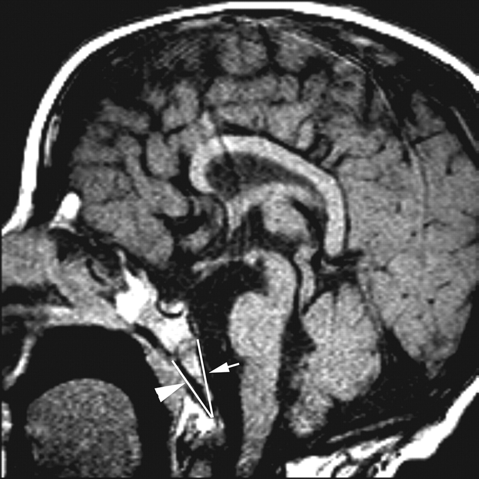
- Fig 1.
Measurement of the lengths of Ba-Es (arrow) and Ba-Xs (arrowhead) on a T1-weighted sagittal image (patient 3).
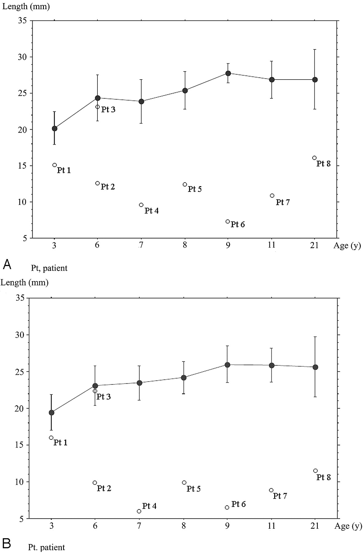
- Fig 2.
Line graph and bars show means and SDs of the length of Ba-Es (A) or Ba-Xs (B) for each age group of healthy controls. White dots show the length of Ba-Es or Ba-Xs of patients with CHARGE syndrome. Pt indicates patient.
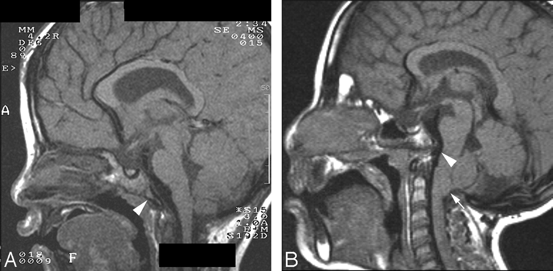
- Fig 3.
T1-weighted sagittal images of patients 1 (A) and 4 (B). A, Mild hypoplastic basiocciput (arrowhead). B, Severe hypoplastic basiocciput (arrowhead) with basilar invagination and Chiari type I malformation (arrow).
- Fig 4.
Contribution of the basiocciput, formed by 4 occipital sclerotomes (I-IV), to the lower portion of clivus. The upper potion is formed by the basisphenoid. The spheno-occipital synchondrosis (open arrow) lies in between.


Tables
Criteria Major Coloboma Coloboma of iris, retina, choroid, disk; microphthalmia Choanal atresia Unilateral/bilateral, membranous/bony, stenosis/atresia Characteristic ear abnormalities External ear (loop or cup-shaped), middle ear (ossicular malformations, chronic serous otitis), mixed deafness; cochlear defects Cranial nerve dysfunction I, Anosmia; VII, facial palsy (unilateral or bilateral); VIII, sensorineural deafness and vestibular problems; IX and/or X, swallowing problems Minor Genital hypoplasia Males: micropenis, cryptorchidism; Females: hypoplastic labia; Both: delayed incomplete pubertal development Developmental delay Delayed motor milestones, hypotonia, mental retardation Cardiovascular malformations All types: conotruncal defects (eg, tetralogy of Fallot), arteriovenous canal defects, and aortic arch anomalies Growth deficiency Short stature Orofacial cleft Cleft lip and/or palate Tracheoesophageal fistula Tracheoesophageal defects of all types Distinctive face Characteristic facial features Reprinted with permission from Clinical Pediatrics (1998;37:159–73). Copyright 1998, Sage Publications.
Patient No. 1 2 3 4 5 6 7 8 Sex Male Male Male Female Female Female Male Female CHARGE features C + + – + – + + – H + + – – + + – + A – – – – – – – – R + + + + + + + + G + + + – – – + + E + + + + + + + + Others, CL – – + + + – + – Genetics Mutation 7367C→G 550C→T 4171delC 4480C→T 5050G→A 4036C→T 5355G→A NE Amino acid S2456X Q184X 1391 fs X1403 R1494X G1684S Q1346X W1785X Age at occipital evaluation (yr) 3.7 6.7 6.9 7.6 8.5 9.6 11.0 21.0 Basiocciput* Mild hypoplasia Severe hypoplasia Normal Severe hypoplasia Severe hypoplasia Severe hypoplasia Severe hypoplasia Severe hypoplasia Basilar invagination – – – + + + + + Chiari I malformation – – – + – – – – Syringomyelia – – – + – – – – Neurologic sequelae – – – + – – – – Length (mm) Ba-Es (SD) 15.0 (−2.2) 12.5 (−3.7) 23.1 (−0.4) 9.5 (−5.0) 12.1 (−5.1) 7.3 (−15.8) 10.9 (−6.1) 16.0 (−2.7) Ba-Xs (SD) 16.0 (−1.4) 9.9 (−4.8) 22.5 (−0.2) 6.0 (−6.8) 9.8 (−6.5) 6.4 (−7.8) 8.8 (−7.4) 11.3 (−3.5) Note:—C indicates coloboma; H, congenital heart defect; A, atresia or stenosis of choanae; R, retarded growth or development and/or central nervous system anomalies; G, genital hypoplasia; E, ear anomalies and/or deafness; CL, cleft lip and/or palate; +, present; –, absent; NE, not evaluated; Ba, basion; Es, endo-sphenobasion; Xs, exo-sphenobasion.
* Mild hypoplasia, length of either Ba-Es or Ba-Xs <2 SDs with a maintained triangular shape; moderate hypoplasia, length of either Ba-Es or Ba-Xs <2 SDs without a maintained triangular shape; severe hypoplasia, length of either Ba-Es and Ba-Xs <2 SDs compared with age-matched control values without a maintained triangular shape.
Age Group (yr) 3 6 7 8 9 11 21 Mean (yr) 3.5 6.6 7.3 8.4 9.6 11.5 21.4 Range (yr) 3.2–3.9 6.2–6.9 7.0–7.9 8.0–8.9 9.3–9.9 11.1–11.8 21.1–21.8 Number 10 10 10 10 10 10 10 Length (mean ± SD) Ba-Es (mm) 20.2 ± 2.3 24.4 ± 3.2 24.0 ± 2.9 25.4 ± 2.6 27.8 ± 1.3 26.9 ± 2.6 26.9 ± 4.1 Ba-Xs (mm) 19.4 ± 2.4 23.1 ± 2.7 23.1 ± 2.5 24.2 ± 2.2 26.0 ± 2.5 25.9 ± 2.3 25.7 ± 4.1
In this issue


Jump to section
Related Articles
Cited By...
- Coronal Clival Cleft in CHARGE Syndrome: Fetal MRI Series
- Cerebellar Heterotopias: Expanding the Phenotype of Cerebellar Dysgenesis in CHARGE Syndrome
- Imaging of Clival Hypoplasia in CHARGE Syndrome and Hypothesis for Development: A Case-Control Study
- Clival Malformations in CHARGE Syndrome
- Head and Neck MRI Findings in CHARGE Syndrome
- Spectrum of Clinical and Associated MR Imaging Findings in Children with Olfactory Anomalies