
Article Figures & Data
Figures
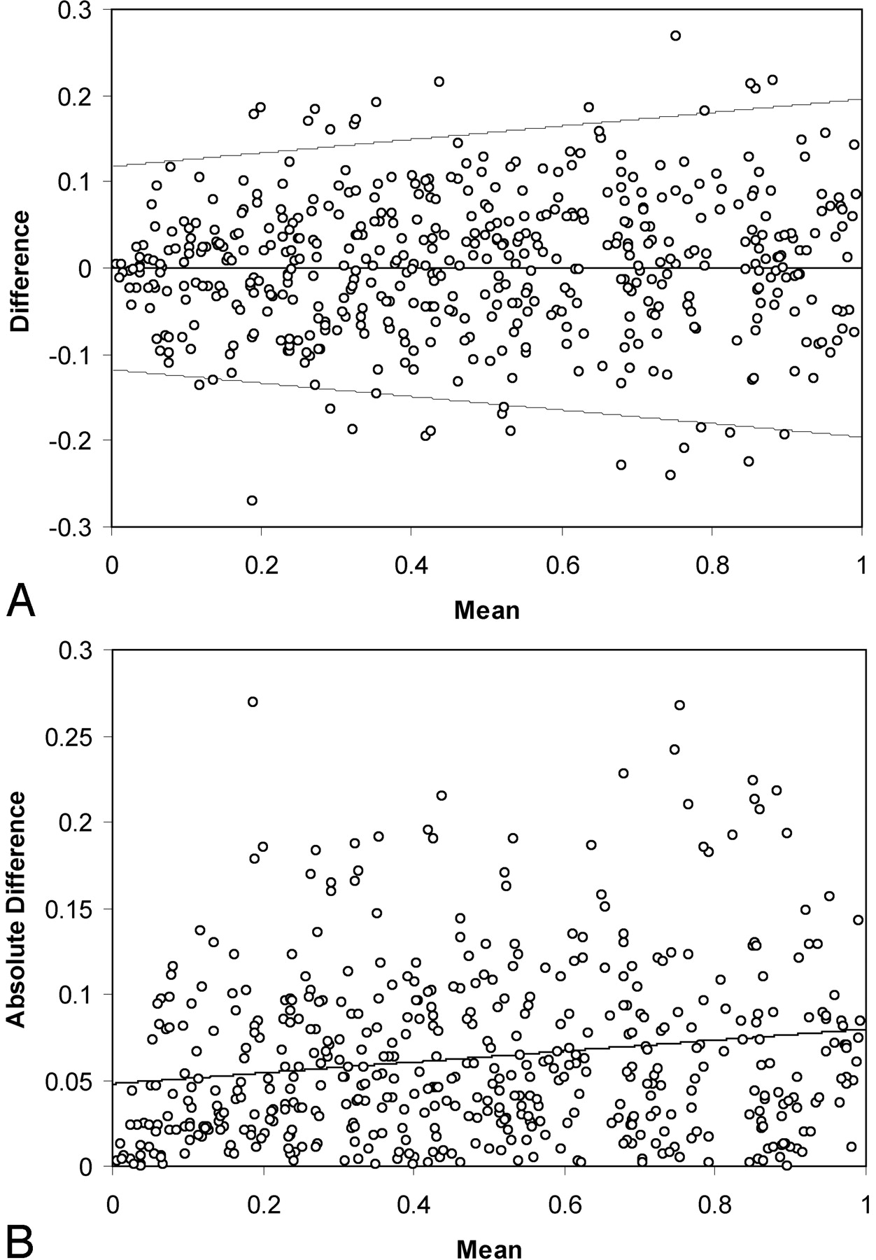
- Fig 1.
Graphs show summary of 500 randomly selected measurements from the simulation run. In A, differences between 2 simulated measurements are plotted versus mean value of s, the fractional residual lumen. B displays absolute difference versus mean for the same data. The line in B represents the linear regression fit. Although not strictly valid because the standard deviation is derived from a half-normal distribution, 95% limits of agreement (21) are indicated in A for reference. These plots illustrate the range of error as well as the mild dependence of standard deviation on stenosis grade in the simulation. The standard deviation tends to decrease for higher percentage diameter stenosis.
- Fig 2.
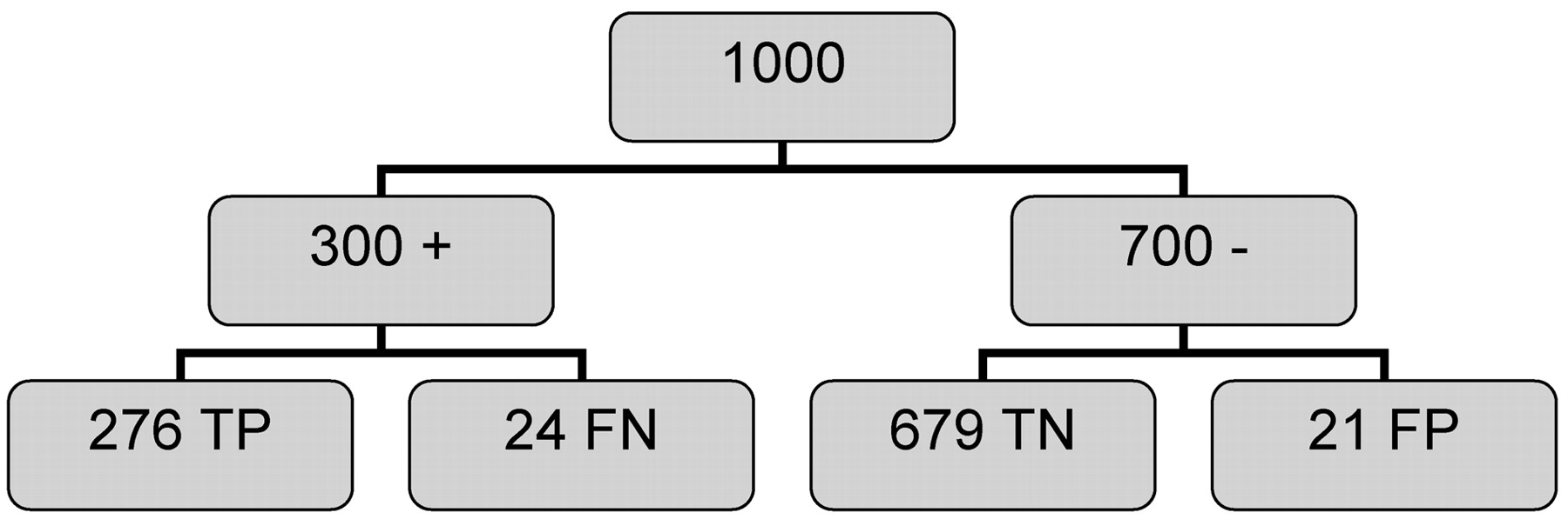
Analysis of 1000 individuals evaluated for 60% diameter carotid stenosis using carotid angiography, is from data generated using the simulation described in the text. Prevalence is taken to be 30%, and the data from Table 3 are used to estimate outcomes, assuming these data are independent of prevalence. Of the 300 individuals with stenosis, 24 (8%) were false-negative (FN), and of the 700 individuals without stenosis, 21 (3%) were false-positive (FP). The overall misclassification rate is approximately 4%. TP indicates, true-positive; TN, indicates true-negative.


Tables
Study Source Method No. of Arteries No. of Readers Weighting* Eliasziw et al, 1994 (6) Fig 5 (upper) NASCET 105 1 Uniform Fig 5 (lower) NASCET 105 2 Uniform Young et al, 1996 (12) Fig 3 CC 101 1 Uniform Fig 4 CC 101 1 Uniform Fig 5 CC 101 2 Uniform Stapf et al, 2000 (15) Table 1 ECST 45 3 Mod-sev Table 1 NASCET 45 3 Mod-sev Rothwell et al, 1994 (8) —† ECST 1001 2 Uniform NASCET indicates the North American Symptomatic Carotid Endarterectomy Trial; CC, common carotid; ECST, European Carotid Surgery Trial.
* Refers to the distribution of carotid stenosis values in the dataset.
† From summary SD plot in Rothwell (14).
- TABLE 2:
Summary of slope and intercept parameters derived from linear regression analysis of absolute differences of s, the fractional residual lumen, versus mean from published data
Study Slope Intercept SD Eliasziw et al, 1994: Fig 5 (upper) (6) 0.016 0.11 0.12 Eliasziw et al, 1994: Fig 5 (lower) (6) 0.077 0.047 0.08 Stapf et al, 2000: Table 1 (ECST) (15) 0.000 0.044 0.04 Stapf et al, 2000: Table 1 (NASCET) (15) −0.003 0.047 0.05 Young et al, 1996: Fig 3 (12) 0.034 0.093 0.11 Young et al, 1996: Fig 4 (12) 0.024 0.062 0.07 Young et al, 1996: Fig 5 (12) 0.017 0.079 0.08 Average 0.023 0.072 0.08 Rothwell et al, 1994 (8) 0.16 0.022 0.08 Simulation 0.036 0.062 0.08 The SD is the value predicted by the regression analysis at s = 0.4 (60% diameter stenosis). For Rothwell et al (8), this represents linear regression of SD data pooled by decile obtained from Figure 6 of Rothwell et al (14). The average SD (0.08) was obtained from the unweighted average of the variance for each of the studies above this line. The 95% CI for the average is 0.05–0.11. For the simulation, the values in the table reflect the fit after iteration to match the variance of the simulation to the mean variance for the published data.
- TABLE 3:
False-positive and false-negative classification of carotid stenosis derived from the simulation
% Stenosis True + Fraction True − Fraction 50 0.96 0.96 60 0.92 0.97 70 0.92 0.98 For all entries, 95% CI is approximately ±0.01.
Simulation used SD of 0.08 for selected cutpoints of clinical interest expressed as true-positive and true-negative fractions.
In this issue


Jump to section
Related Articles
Cited By...
- No citing articles found.