
Article Figures & Data
Figures
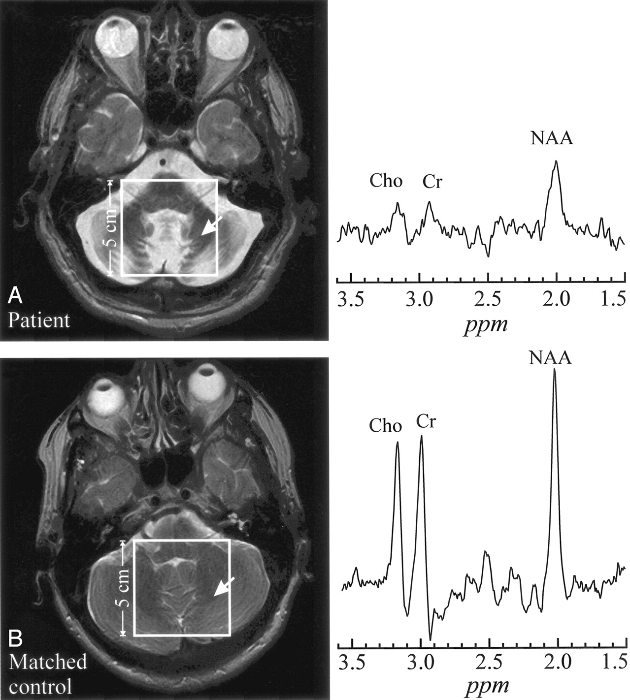
- Fig 1.
A, top left, Axial T2-weighted image from a 54-year-old late-onset GM2G patient superimposed with the 5 × 5 cm2 VOI. Right, 1H spectrum from the cerebellum (arrow).
B, bottom left, Corresponding section from a 52-year-old healthy control superimposed with the 5 × 5 cm2 VOI. Right, Arrow indicates analogous region for metabolite comparison. Both spectra are on the same scale. Note the dramatic atrophy of the cerebellar folia and vermis in the patient and consequent lower metabolites levels reflecting larger partial CSF volume contamination.
- Fig 2.
A, top left, T2-weighted image of a 54-year-old patient with late-onset GM2G, superimposed with the 8 × 8 cm2 1H-MR spectroscopy VOI. Right, Two spectra from thalamus and occipital white matter (arrows 1 and 2).
B, bottom left, Corresponding section from a matched control. The arrows indicate analogous regions to A for metabolite spectral comparisons. All spectra are on common intensity and chemical shift (parts per million) scales.
Note the characteristic lower NAA signal intensity in thalamus and NAWM in the patient (A) compared with the control (B), as well as decreased Cr and Cho.


Tables
- TABLE 1:
Demographics, clinical and genetic information for the patients with late-onset GM2G
Patient No./Age (y)/Sex Disease Onset (Y) Ambulation Index Molecular Defect* 1/20/M 12 0 Exon 7, exon 11 2/32/M 15 2 Exon 7, R504C 3/28/M 13 3 Exon 7, exon 13 4/52/M 12 9 Exon 7, intron 12 5/29/F 17 4 Exon 7, exon 11 6/54/M 36 4 Exon 7, exon 11 7/31/F 12 2 Exon 7, exon 11 8/50/F 12 9 Exon 7, exon 11 9/58/M 17 7 Exon 7, exon 11 * Mutations: The exon 7 mutation involves a G to A transition at position 269 (G269S), which represents the most common disease allele associated with late-onset GM2G. The exon 11 mutation (TATC1278) represents a frameshift mutation resulting from a 4-base insertion; this allele is the most frequent gene defect found either in homozygosity or heterozygosity among patients with the classic infantile form of Tay-Sachs disease.
Patient Thalamus White Matter Cerebellum NAA Cr Cho NAA Cr Cho NAA Cr Cho 1 6.16 3.35 1.06 4.47 2.49 0.75 4.25 3.65 1.30 2 6.35 3.72 1.26 5.38 3.69 1.14 3.77 1.54 0.66 3 6.20 3.09 0.95 4.75 2.49 0.81 5.54 2.14 0.78 4 7.73 4.20 1.16 7.76 4.81 1.01 N/A N/A N/A 5 10.13 5.36 1.45 8.67 6.17 1.93 6.35 4.96 1.93 6 8.10 4.37 1.43 8.76 5.31 1.89 9.38 7.06 2.45 7 4.58 2.69 0.71 3.43 2.14 0.57 2.57 1.30 0.43 8 5.40 3.93 0.74 5.25 3.58 0.47 1.33 1.27 0.52 9 3.77 2.56 0.71 3.29 2.65 0.69 1.37 0.92 0.32 Patients 1–9 6.50 ± 1.9 3.69 ± 0.8 1.03 ± 0.3 5.75 ± 2.1 3.70 ± 1.4 1.05 ± 0.5 4.3 ± 2.7 2.9 ± 2.2 1.05 ± 0.8 Controls (n = 8) 11.0 ± 2.7 5.43 ± 1.2 1.43 ± 0.4 9.92 ± 2.7 5.20 ± 1.6 1.40 ± 0.4 11.1 ± 1.1 8.7 ± 1.3 2.4 ± 0.3 Concentrations of the 3 metabolites are in millimolars. Bold entries (for patients) are significantly different from the controls (P = .005).
In this issue


Jump to section
Related Articles
Cited By...
- Deep Learning Cerebellar Magnetic Resonance Imaging Segmentation in Late-Onset GM2 Gangliosidosis: Implications for Phenotype
- Tay-Sachs and Sandhoff Diseases: Diffusion tensor imaging and correlational fiber tractography findings differentiate late-onset GM2 Gangliosidosis
- Relationship between neurochemical concentrations and neurofunctional measures in late-onset GM2 gangliosidosis
- The landscape of functional brain network impairments in late-onset GM2 gangliosidosis