
Article Figures & Data
Figures
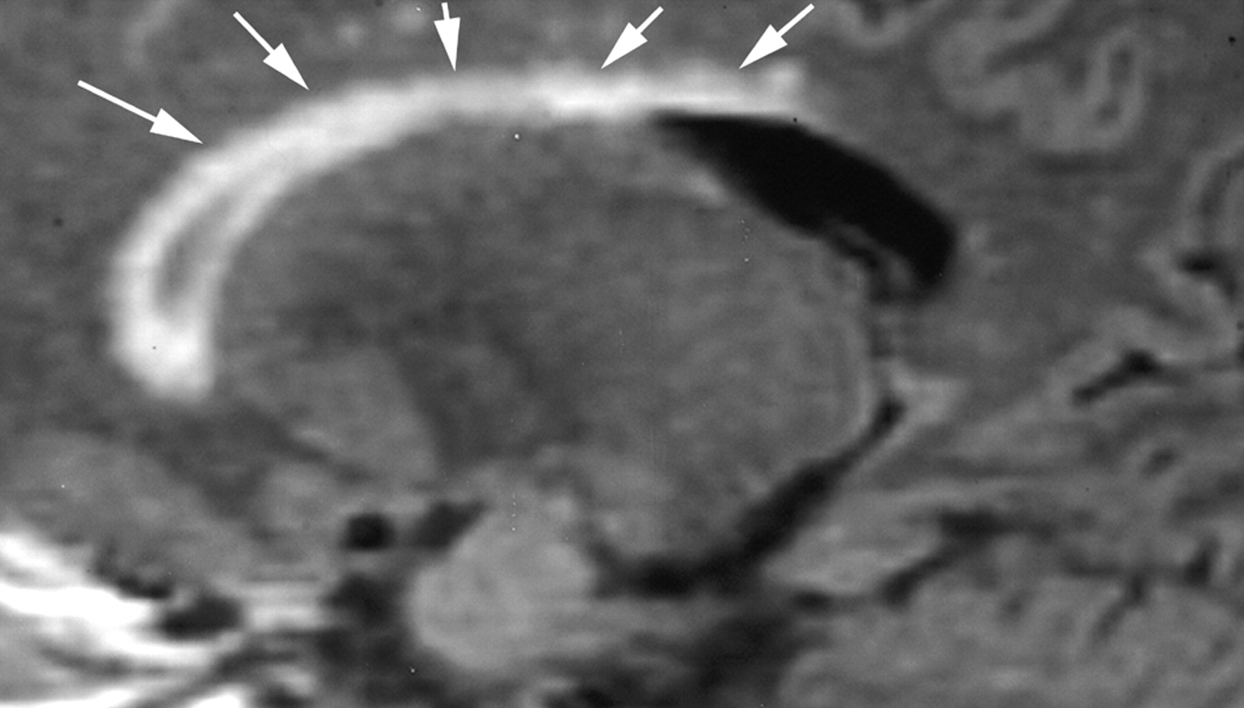
- Fig 1.
Sagittal fluid-attenuated inversion recovery image (TR/TE/TI, 8800/130/2200) shows a normal smooth ependymal stripe (arrows) without irregularity, subcallosal striations, or Dot-Dash sign.
- Fig 2.
Sagittal fluid-attenuated inversion recovery images (TR/TE/TI, 8800/130/2200) from 2 different patients with multiple sclerosis (A, B) show the typical Dot-Dash sign. The arrows indicate the dots, whereas the dashes are the hypointense areas between the dots.
- Fig 3.
Sagittal fluid-attenuated inversion recovery images (TR/TE/TI, 8800/133/2200) are from a patient with recurrent relapsing multiple sclerosis. A shows the typical Dot-Dash sign (arrows). The patient developed worsening neurologic symptoms, and a follow-up image obtained 7 weeks later (B) showed interval development of confluent callosal and white matter lesions (arrowheads) and an additional Dot-Dash sign in the posterior ependyma.
- Fig 4.
Sagittal fluid-attenuated inversion recovery image (TR/TE/TI, 8800/130/2200) shows confluent subependymal and callosal white matter hyperintensity (arrows), which is typical of chronic white matter ischemic changes. There is no evidence of either the Dot-Dash sign or subcallosal striations.


Tables
Comparison of controls and patients (divided by age)
Overall (n = 70) Group I (n = 55) Group II (n = 15) % Sensitivity 91.4 95.7 83.3 Specificity 65.7 71.9 33.3 Positive predictive value 72.7 71.0 83.3 Negative predictive value 88.5 95.8 33.3
In this issue


Jump to section
Related Articles
Cited By...
- An MRI-informed histo-molecular analysis implicates ependymal cells in the pathogenesis of periventricular pathology in multiple sclerosis
- Dynamic 11C-PiB PET Shows Cerebrospinal Fluid Flow Alterations in Alzheimer Disease and Multiple Sclerosis
- Dynamic 11C-PiB PET shows cerebrospinal fluid flow alterations in Alzheimers disease and multiple sclerosis
- Imaging evaluation of demyelinating processes of the central nervous system