
Article Figures & Data
Figures
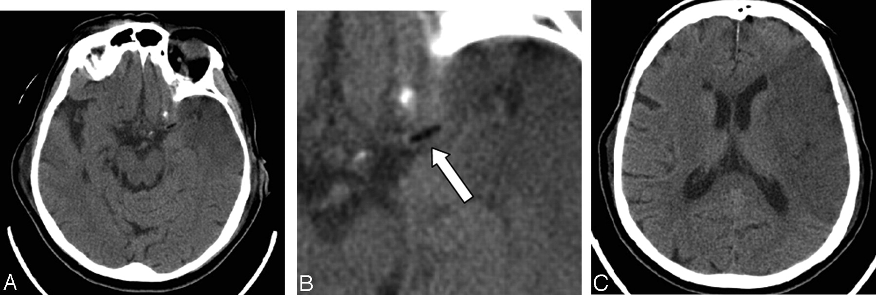
- Fig 1.
A 78-year-old woman who had undergone a mitral valve replacement.
A–C, Axial images from the initial CT head scan obtained on the first postoperative day demonstrate an early left middle cerebral artery territory infarct. There is a fat attenuation filling defect (arrow, B) in the region of the proximal left middle cerebral artery trunk.
- Fig 2.
CT angiography axial image obtained 13 days postoperatively confirms that the fat attenuation lesion (arrow) is within the proximal left middle cerebral artery trunk.
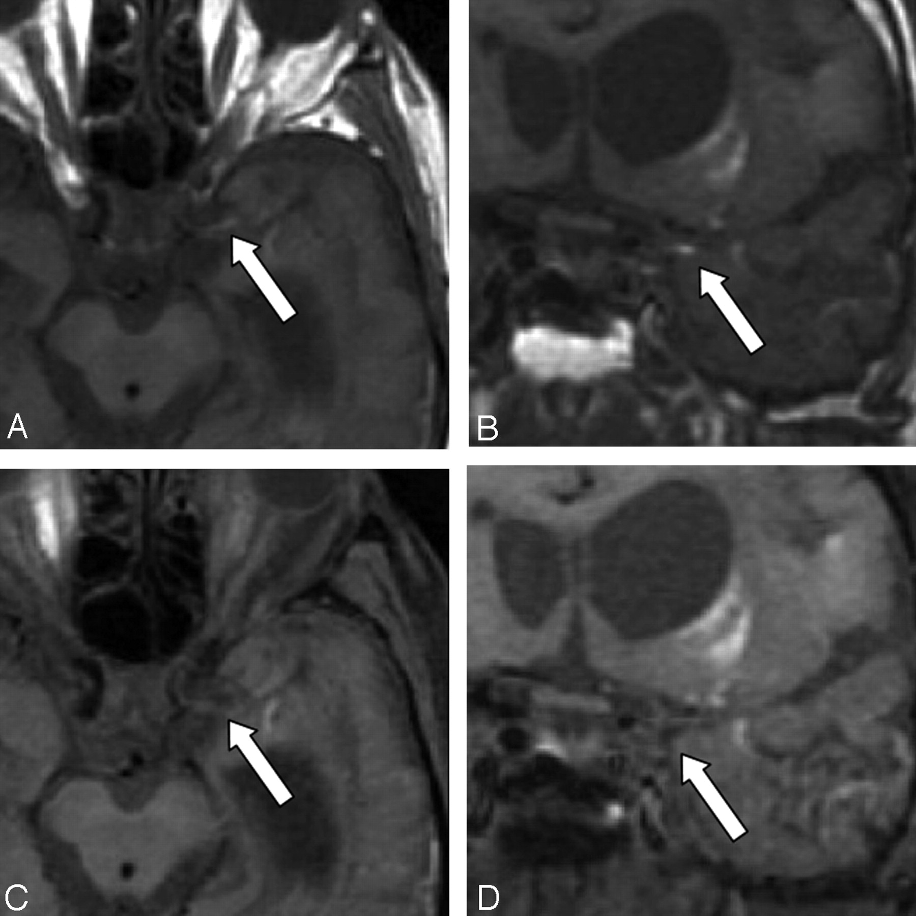
- Fig 3.
Axial and coronal T1-weighted MR images obtained 49 days postoperatively without fat saturation (A,B) and with fat saturation (C,D). The images without fat saturation demonstrate the T1-hyperintense embolus (arrow) in the proximal left M1 trunk. After fat saturation, the T1 hyperintensity of the embolus is saturated (arrow), confirming its fatty composition. High-signal-intensity methemoglobin in the adjacent infarct is unchanged before and after fat saturation.


In this issue


Jump to section
Related Articles
Cited By...
- Teaching NeuroImages: Cerebral liposuction
- Hypodense artery sign in cerebral fat embolism
- Intracranial transthecal subarachnoid fat emboli and subarachnoid haemorrhage arising from a sacral fracture and dural tear
- Teaching NeuroImages: Hypodense artery sign in acute cerebral infarction by contrast-enhanced CT