Advertisement

Main menu

  • Home
  • Content
    • Current Issue
    • Accepted Manuscripts
    • Article Preview
    • Past Issue Archive
    • Video Articles
    • AJNR Case Collection
    • Case of the Week Archive
    • Case of the Month Archive
    • Classic Case Archive
  • Special Collections
    • AJNR Awards
    • Low-Field MRI
    • Alzheimer Disease
    • ASNR Foundation Special Collection
    • Photon-Counting CT
    • View All
  • Multimedia
    • AJNR Podcasts
    • AJNR SCANtastic
    • Trainee Corner
    • MRI Safety Corner
    • Imaging Protocols
  • For Authors
    • Submit a Manuscript
    • Submit a Video Article
    • Submit an eLetter to the Editor/Response
    • Manuscript Submission Guidelines
    • Statistical Tips
    • Fast Publishing of Accepted Manuscripts
    • Graphical Abstract Preparation
    • Imaging Protocol Submission
    • Author Policies
  • About Us
    • About AJNR
    • Editorial Board
    • Editorial Board Alumni
  • More
    • Become a Reviewer/Academy of Reviewers
    • Subscribers
    • Permissions
    • Alerts
    • Feedback
    • Advertisers
    • ASNR Home

User menu

  • Alerts
  • Log in

Search

  • Advanced search
American Journal of Neuroradiology

American Journal of Neuroradiology

ASHNR American Society of Functional Neuroradiology ASHNR American Society of Pediatric Neuroradiology ASSR
  • Alerts
  • Log in

Advanced Search

  • Home
  • Content
    • Current Issue
    • Accepted Manuscripts
    • Article Preview
    • Past Issue Archive
    • Video Articles
    • AJNR Case Collection
    • Case of the Week Archive
    • Case of the Month Archive
    • Classic Case Archive
  • Special Collections
    • AJNR Awards
    • Low-Field MRI
    • Alzheimer Disease
    • ASNR Foundation Special Collection
    • Photon-Counting CT
    • View All
  • Multimedia
    • AJNR Podcasts
    • AJNR SCANtastic
    • Trainee Corner
    • MRI Safety Corner
    • Imaging Protocols
  • For Authors
    • Submit a Manuscript
    • Submit a Video Article
    • Submit an eLetter to the Editor/Response
    • Manuscript Submission Guidelines
    • Statistical Tips
    • Fast Publishing of Accepted Manuscripts
    • Graphical Abstract Preparation
    • Imaging Protocol Submission
    • Author Policies
  • About Us
    • About AJNR
    • Editorial Board
    • Editorial Board Alumni
  • More
    • Become a Reviewer/Academy of Reviewers
    • Subscribers
    • Permissions
    • Alerts
    • Feedback
    • Advertisers
    • ASNR Home
  • Follow AJNR on Twitter
  • Visit AJNR on Facebook
  • Follow AJNR on Instagram
  • Join AJNR on LinkedIn
  • RSS Feeds

AJNR Awards, New Junior Editors, and more. Read the latest AJNR updates

OtherBrain

The Hypodense Artery Sign

Thomas C. Lee, Eric S. Bartlett, Allan J. Fox and Sean P. Symons
American Journal of Neuroradiology September 2005, 26 (8) 2027-2029;
Thomas C. Lee
  • Find this author on Google Scholar
  • Find this author on PubMed
  • Search for this author on this site
Eric S. Bartlett
  • Find this author on Google Scholar
  • Find this author on PubMed
  • Search for this author on this site
Allan J. Fox
  • Find this author on Google Scholar
  • Find this author on PubMed
  • Search for this author on this site
Sean P. Symons
  • Find this author on Google Scholar
  • Find this author on PubMed
  • Search for this author on this site
  • Article
  • Figures & Data
  • Info & Metrics
  • Responses
  • References
  • PDF
Loading

Abstract

Summary: An acute intracranial embolus may be associated with a hyperdense artery sign on CT, related to acute thrombus within the affected artery, a phenomenon that is well known and has been extensively documented. We present an unusual case of a middle cerebral artery territory acute infarct with a hypodense artery on CT. The Hounsfield unit attenuation of the embolic lesion was fat density. CT angiography and MR imaging confirmed the fatty lesion to be within the middle cerebral artery.

CT findings in acute stroke may be subtle or even undetectable on initial examinations, even when a large territory is involved (1). Various signs have been described as markers of early acute stroke on CT. Increased attenuation of the middle cerebral artery due to clot was first described in the early 1980s (2). Since that time, the “hyperdense middle cerebral artery” sign has been described as a marker of thrombus in the middle cerebral artery (3, 4), and several studies have demonstrated that the hyperdense middle cerebral artery sign is a common finding when CT is performed within a few hours from the onset of symptoms (5–8). False-positive hyperdense middle cerebral artery signs have been noted in patients with a high hematocrit level or calcification in the middle cerebral artery, but in these cases, the abnormal attenuation is usually bilateral (5, 8–10). In our patient, a fat embolus caused a hypodense appearance of the middle cerebral artery with an associated acute middle cerebral artery territory infarct.

Case Report

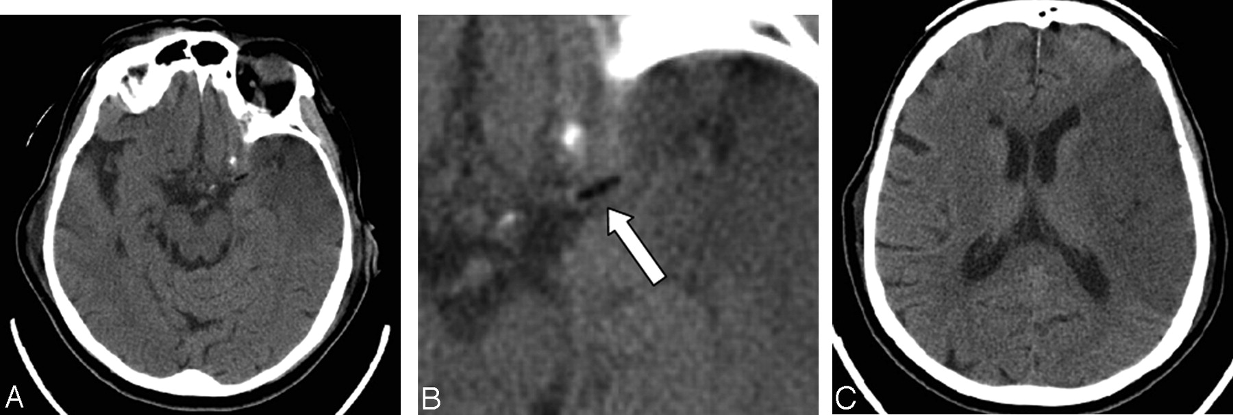
A 78-year-old woman with no significant medical history underwent an abdominoperineal resection for rectal adenocarcinoma in February 2002. At that time, mitral regurgitation was discovered, which progressed clinically to the point where she required surgical treatment. On January 28, 2004, she underwent mitral valve replacement. The surgical anatomy was complicated, but there were no intraoperative complications. In the immediate postoperative period, she was noted to have a decreased level of consciousness, a dense right hemiplegia, and aphasia. CT of the head performed on the first postoperative day demonstrated an early acute large left middle cerebral artery territory infarct. There was a hypodense filling defect in the left M1 proximal trunk, and the Hounsfield unit measurement of it indicated fat attenuation, with the lowest Hounsfield unit measurement of the intraluminal embolus being −25 H (Fig 1). Two subsequent CT scans, obtained during the next few days, demonstrated similar findings. CT angiography performed on the 13th postoperative day confirmed that the fatty lesion was intraluminal (Fig 2). CT angiography also demonstrated patent left common and internal carotid arteries without significant stenosis or atherosclerotic plaque. The fat embolus likely came from the surrounding tissues during the cardiac surgery.

Fig 1.
  • Download figure
  • Open in new tab
  • Download powerpoint
Fig 1.

A 78-year-old woman who had undergone a mitral valve replacement.

A–C, Axial images from the initial CT head scan obtained on the first postoperative day demonstrate an early left middle cerebral artery territory infarct. There is a fat attenuation filling defect (arrow, B) in the region of the proximal left middle cerebral artery trunk.

Fig 2.
  • Download figure
  • Open in new tab
  • Download powerpoint
Fig 2.

CT angiography axial image obtained 13 days postoperatively confirms that the fat attenuation lesion (arrow) is within the proximal left middle cerebral artery trunk.

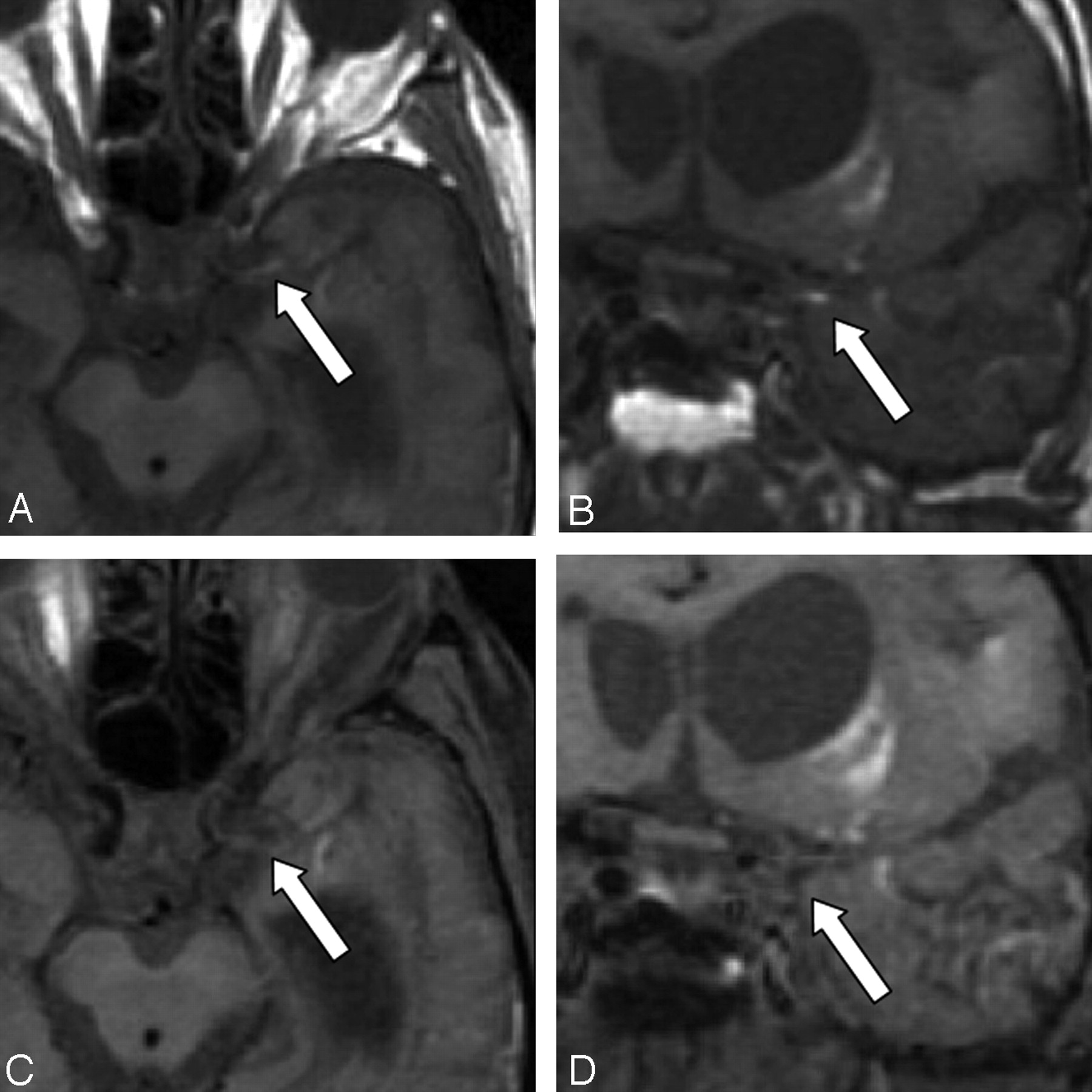
She remained in the cardiovascular intensive care unit with ventricular pacing for nearly 2 months following the operation. MR imaging was performed 49 days after her stroke, once the pacemaker was removed. MR imaging showed interval evolution of the large left middle cerebral artery infarct. The previously seen infarct edema had resolved. There was hemorrhage within the infarcted territory and in the ventricles, which had occurred in the interval since the prior CT scans but was not clinically symptomatic. Imaging through the left proximal middle cerebral artery, without and with fat saturation, showed remnants of the fat embolus within the M1 segment (Fig 3).

Fig 3.
  • Download figure
  • Open in new tab
  • Download powerpoint
Fig 3.

Axial and coronal T1-weighted MR images obtained 49 days postoperatively without fat saturation (A,B) and with fat saturation (C,D). The images without fat saturation demonstrate the T1-hyperintense embolus (arrow) in the proximal left M1 trunk. After fat saturation, the T1 hyperintensity of the embolus is saturated (arrow), confirming its fatty composition. High-signal-intensity methemoglobin in the adjacent infarct is unchanged before and after fat saturation.

Discussion

Emboli to the intracranial circulation can originate from numerous sources. Emboli can originate in the heart, particularly in the setting of atrial fibrillation. Emboli commonly propagate from the carotid arteries as a result of stenosis or complicated atherosclerotic plaque. Thrombus can form at the site of a vessel dissection with subsequent embolization to the intracranial circulation. In these scenarios, the emboli are thrombotic in composition. Fat emboli to the intracranial circulation are uncommon.

Aside from mortality due to other causes, stroke is the most feared complication of cardiac surgery. The incidence of stroke after coronary artery bypass is 1.4%, whereas the risk of perioperative stroke for valve surgery is higher, quoted at 2.5% (11). Persistent atrial fibrillation is the most significant risk factor for stroke after mechanical mitral valve replacement (12).

Fat emboli that usually originate from long bone fractures are more commonly described in the trauma and orthopedic literature. The major clinical criteria for diagnosing fat embolism include respiratory distress and petechiae. Cerebral fat embolism has been described in patients with a venous-to-arterial shunt (13).

The typical fat emboli syndrome is generally thought to be a result of microglobules. The high-resolution chest CT findings of mild fat embolism are nonspecific, consisting of bilateral ground-glass opacities and thickening of the interlobular septa. Centrilobular nodular opacities are present in some patients (13). Therefore, the clinical situation is the most important determinant in diagnosing fat embolus (14, 15).

In our patient, the diagnosis of a fat embolus was suspected on the basis of the negative Hounsfield unit attenuation in the middle cerebral artery. Presumably the mitral valve replacement procedure resulted in dislodgement of a macroscopic portion of fat from the cardiac or surrounding tissue, which subsequently embolized to the left middle cerebral artery. Alternately, fat may have been dislodged during the sternotomy and paradoxically embolized to the brain through an intraoperative atrial defect. Fat containing emboli could also originate from fatty atherosclerotic plaque within the carotid artery or aortic arch. However, there was only minor atherosclerotic plaque in the proximal left internal carotid artery of this patient on CT angiography; therefore, a carotid artery source was excluded. Although the hyperdense middle cerebral artery sign has been widely described as a sign of thromboembolism, a hypodense middle cerebral artery sign associated with acute stroke from a fat embolus is not commonly known.

References

  1. ↵
    von Kummer R, Nolte PN, Schnittger H, Thron A, Ringelstein EB. Detectability of cerebral hemisphere ischaemic infarction by CT within 6 hours of stroke. Neuroradiology 1996;38:31–33
    CrossRefPubMed
  2. ↵
    Gacs G, Fox AJ, Barnett HJM, Vinuela F. CT visualization of intracranial arterial thromboembolism. Stroke 1983;14:756–762
    Abstract/FREE Full Text
  3. ↵
    Pressman BD, Tourje EJ, Thompson JR. An early sign of ischemic infarction: increased density in a cerebral artery. AJNR Am J Neuroradiol 1987;8:645–648
    Abstract/FREE Full Text
  4. ↵
    Schuierer G, Huk W. The unilateral hyperdense middle cerebral artery: an early CT sign of embolism or thrombosis. Neuroradiology 1988;30:120–122
    CrossRefPubMed
  5. ↵
    Bastianello S, Pierallini A, Colonnese C, et al. Hyperdense middle cerebral artery CT sign: comparison with angiography in the acute phase of ischemic supratentorial infarction. Neuroradiology 1991;33:207–211
    CrossRefPubMed
  6. Leys D, Pruvo JP, Godefroy O, Rondepierre PH, Leclerc X. Prevalence and significance of hyperdense middle cerebral artery in acute stroke. Stroke 1992;23:317–324
    Abstract/FREE Full Text
  7. Tomsick T, Brott T, Barsan W, et al. Prognostic value of the hyperdense middle cerebral artery sign and stroke scale score before ultraearly thrombolytic therapy. AJNR Am J Neuroradiol 1996;17:79–85
    Abstract
  8. ↵
    von Kummer R, Meyding-Lamade U, Forsting M, et al. Sensitivity and prognostic value of early CT in occlusion of the middle cerebral artery trunk. AJNR Am J Neuroradiol 1994;15:9–15
    Abstract/FREE Full Text
  9. Tomsick TA, Brott TG, Olinger CP, et al. Hyperdense middle cerebral artery: incidence and quantitative significance. Neuroradiology 1989;31:312–315
    CrossRefPubMed
  10. ↵
    von Kummer R, Holle R, Gizyska U, et al. Interobserver agreement in assessing early CT signs of middle cerebral artery infarction. AJNR Am J Neuroradiol 1996;17:1743–1748
    Abstract
  11. David TE. The Toronto Hospital Cardiac Surgical Program. In: Cheng DCH, David TE, eds. Perioperative care in cardiac anesthesia and surgery. Georgetown, TX: Landes Bioscience;1999;2
  12. ↵
    Bando K, Kobayashi J, Hirata M, et al. Early and late stroke after mitral valve replacement with a mechanical prosthesis: risk factor analysis of a 24-year experience. J Thorac Cardiovasc Surg 2003;126:358–64
    CrossRefPubMed
  13. ↵
    Malagari K, Economopoulos N, Stoupis C, et al. High-resolution CT findings in mild pulmonary fat embolism. Chest 2003;123:1196–1201
    CrossRefPubMed
  14. ↵
    Georgopoulos D, Bouros D. Fat embolism syndrome: clinical examination is still the preferable diagnostic method. Chest 2003;123:982–983
    CrossRefPubMed
  15. ↵
    Riding G, Daly K, Hutchinson S, Rao S, Lovell M, McCollum C. Paradoxical cerebral embolization: an explanation for fat embolism syndrome. J Bone Joint Surg Br 2004;86:95–98
  • Received September 22, 2004.
  • Accepted after revision October 19, 2004.
  • Copyright © American Society of Neuroradiology
View Abstract
PreviousNext
Back to top

In this issue

American Journal of Neuroradiology: 26 (8)
American Journal of Neuroradiology
Vol. 26, Issue 8
1 Sep 2005
  • Table of Contents
  • Index by author
Advertisement
Print
Download PDF
Email Article

Thank you for your interest in spreading the word on American Journal of Neuroradiology.

NOTE: We only request your email address so that the person you are recommending the page to knows that you wanted them to see it, and that it is not junk mail. We do not capture any email address.

Enter multiple addresses on separate lines or separate them with commas.
The Hypodense Artery Sign
(Your Name) has sent you a message from American Journal of Neuroradiology
(Your Name) thought you would like to see the American Journal of Neuroradiology web site.
CAPTCHA
This question is for testing whether or not you are a human visitor and to prevent automated spam submissions.
Cite this article
Thomas C. Lee, Eric S. Bartlett, Allan J. Fox, Sean P. Symons
The Hypodense Artery Sign
American Journal of Neuroradiology Sep 2005, 26 (8) 2027-2029;

Citation Manager Formats

  • BibTeX
  • Bookends
  • EasyBib
  • EndNote (tagged)
  • EndNote 8 (xml)
  • Medlars
  • Mendeley
  • Papers
  • RefWorks Tagged
  • Ref Manager
  • RIS
  • Zotero
0 Responses
Respond to this article
Share
Bookmark this article
The Hypodense Artery Sign
Thomas C. Lee, Eric S. Bartlett, Allan J. Fox, Sean P. Symons
American Journal of Neuroradiology Sep 2005, 26 (8) 2027-2029;
del.icio.us logo Twitter logo Facebook logo Mendeley logo
  • Tweet Widget
  • Facebook Like
  • Google Plus One

Jump to section

  • Article
    • Abstract
    • Case Report
    • Discussion
    • References
  • Figures & Data
  • Info & Metrics
  • Responses
  • References
  • PDF

Related Articles

  • No related articles found.
  • PubMed
  • Google Scholar

Cited By...

  • Teaching NeuroImages: Cerebral liposuction
  • Hypodense artery sign in cerebral fat embolism
  • Intracranial transthecal subarachnoid fat emboli and subarachnoid haemorrhage arising from a sacral fracture and dural tear
  • Teaching NeuroImages: Hypodense artery sign in acute cerebral infarction by contrast-enhanced CT
  • Crossref
  • Google Scholar

This article has not yet been cited by articles in journals that are participating in Crossref Cited-by Linking.

More in this TOC Section

  • Evaluating the Effects of White Matter Multiple Sclerosis Lesions on the Volume Estimation of 6 Brain Tissue Segmentation Methods
  • Quiet PROPELLER MRI Techniques Match the Quality of Conventional PROPELLER Brain Imaging Techniques
  • Predictors of Reperfusion in Patients with Acute Ischemic Stroke
Show more Brain

Similar Articles

Advertisement

Indexed Content

  • Current Issue
  • Accepted Manuscripts
  • Article Preview
  • Past Issues
  • Editorials
  • Editor's Choice
  • Fellows' Journal Club
  • Letters to the Editor
  • Video Articles

Cases

  • Case Collection
  • Archive - Case of the Week
  • Archive - Case of the Month
  • Archive - Classic Case

More from AJNR

  • Trainee Corner
  • Imaging Protocols
  • MRI Safety Corner
  • Book Reviews

Multimedia

  • AJNR Podcasts
  • AJNR Scantastics

Resources

  • Turnaround Time
  • Submit a Manuscript
  • Submit a Video Article
  • Submit an eLetter to the Editor/Response
  • Manuscript Submission Guidelines
  • Statistical Tips
  • Fast Publishing of Accepted Manuscripts
  • Graphical Abstract Preparation
  • Imaging Protocol Submission
  • Evidence-Based Medicine Level Guide
  • Publishing Checklists
  • Author Policies
  • Become a Reviewer/Academy of Reviewers
  • News and Updates

About Us

  • About AJNR
  • Editorial Board
  • Editorial Board Alumni
  • Alerts
  • Permissions
  • Not an AJNR Subscriber? Join Now
  • Advertise with Us
  • Librarian Resources
  • Feedback
  • Terms and Conditions
  • AJNR Editorial Board Alumni

American Society of Neuroradiology

  • Not an ASNR Member? Join Now

© 2025 by the American Society of Neuroradiology All rights, including for text and data mining, AI training, and similar technologies, are reserved.
Print ISSN: 0195-6108 Online ISSN: 1936-959X

Powered by HighWire