
Article Figures & Data
Figures
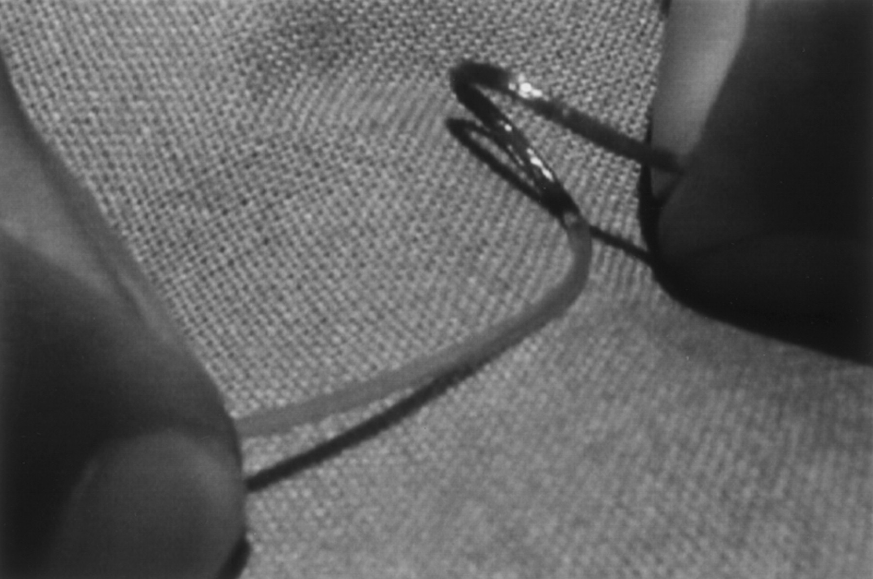
- Fig 1.
Photograph of the self-expanding stent demonstrates the extreme flexibility of the stent system.
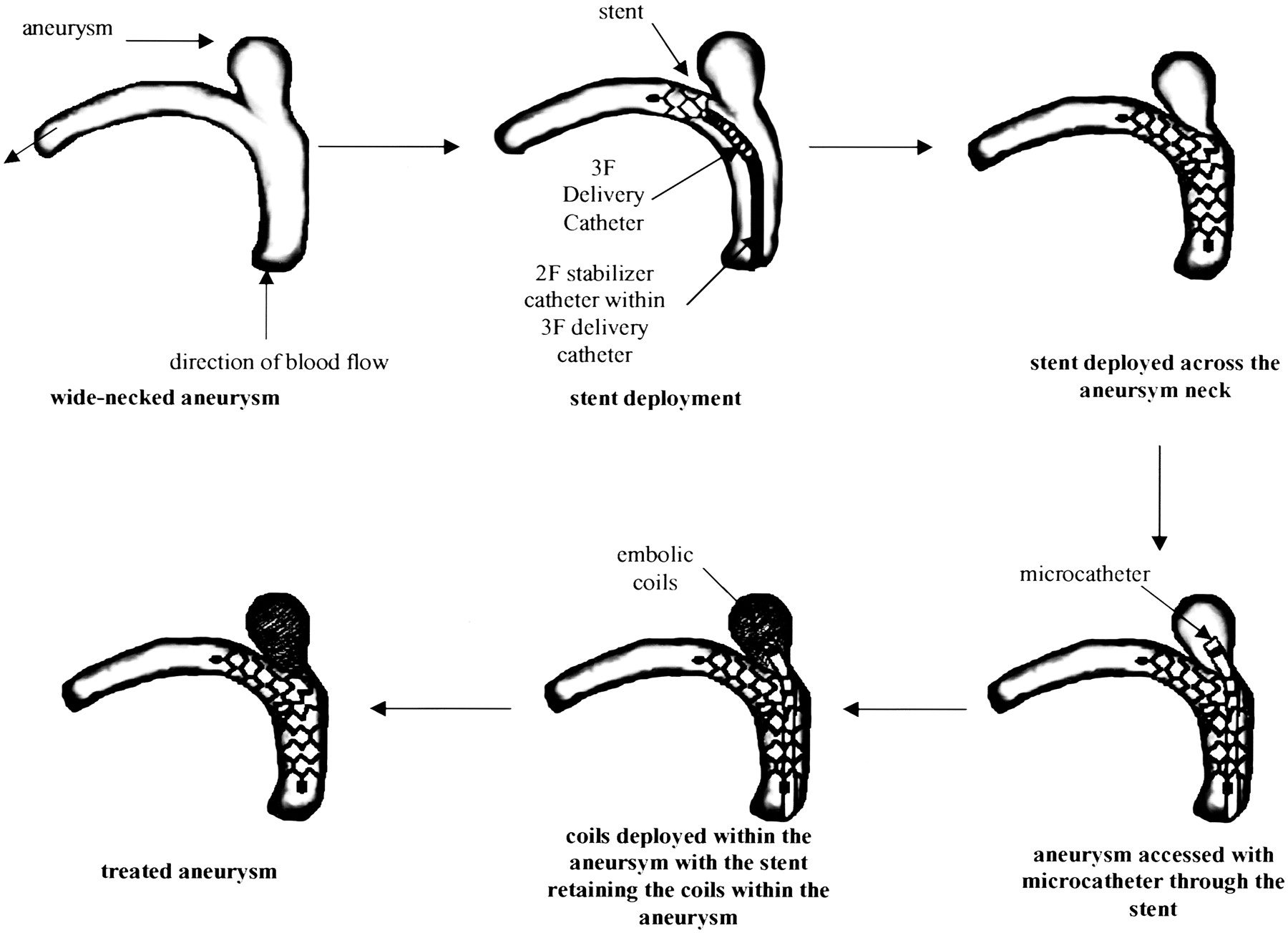
- Fig 2.
Schematic illustrates the combined treatment of primary stent placement and subsequent coil embolization for wide-necked aneurysms.
- Fig 3.
Case 1. Wide-necked paraophthalmic aneurysm (dome, 5.5 × 8 mm; neck, 5 mm).
A and B, DSA images obtained before (A) and after (B) combined therapy with the self-expanding stent and GDCs demonstrate complete obliteration. The patient had two additional aneurysms (at the AcomA and basilar artery) previously treated with GDCs alone.
C and D, Source images of time-of-flight MR angiography reveal no stent-related artifacts and normal flow-void signal intensity of the internal carotid artery. Arrows in D indicate coil mass.
E, Follow-up angiogram after 6 months reveals complete aneurysm occlusion.
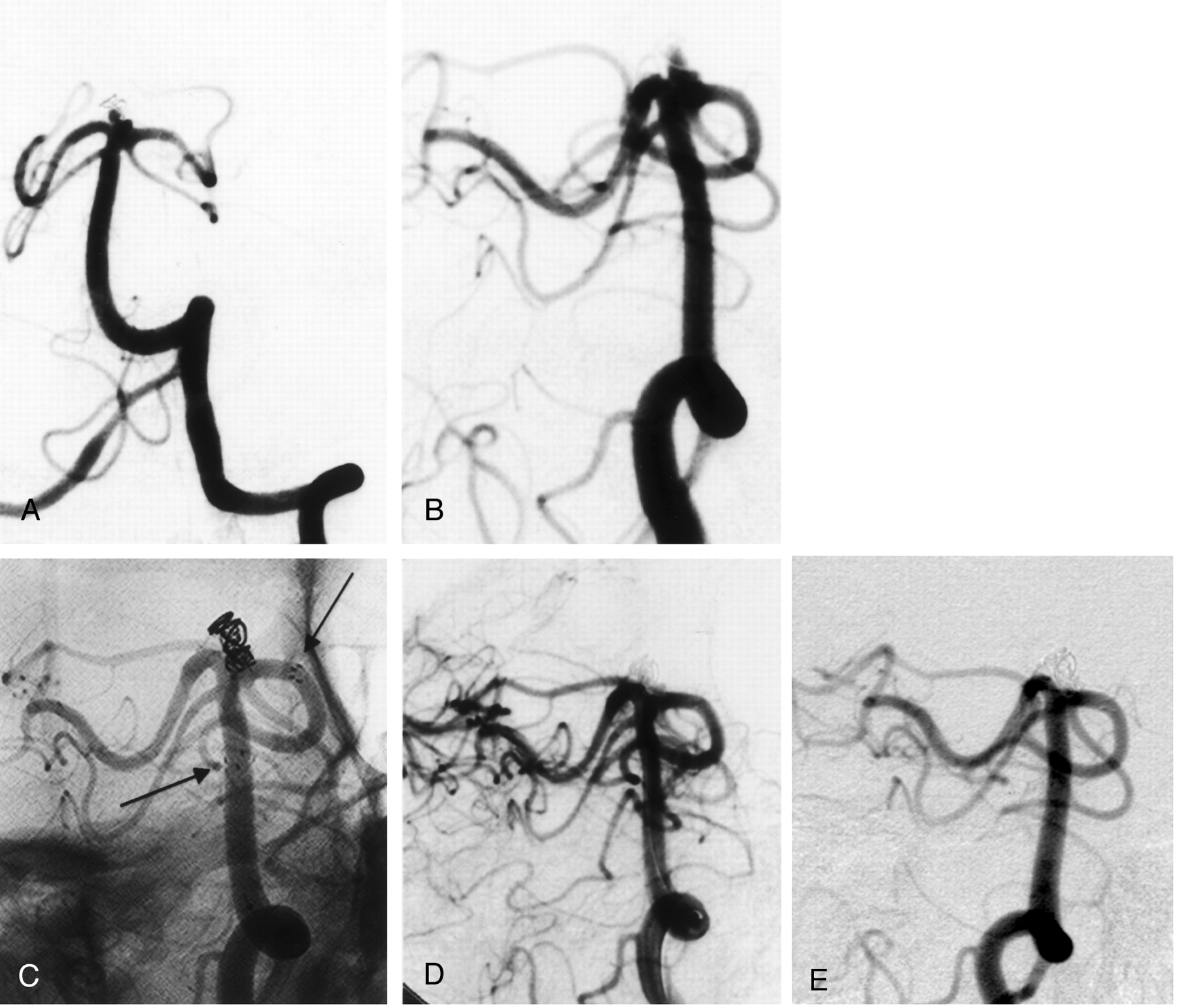
- Fig 4.
Case 2.
A, Angiogram reveals previously coiled recurrent aneurysm (arrows) of the cavernous internal carotid artery.
B, Angiogram obtained after stent deployment shows immediate stasis of the contrast material.
C, Angiogram obtained after coiling through the stent interstices shows that the aneurysm is subtotally occluded.
D, Further thrombosis is noted after 6 months.
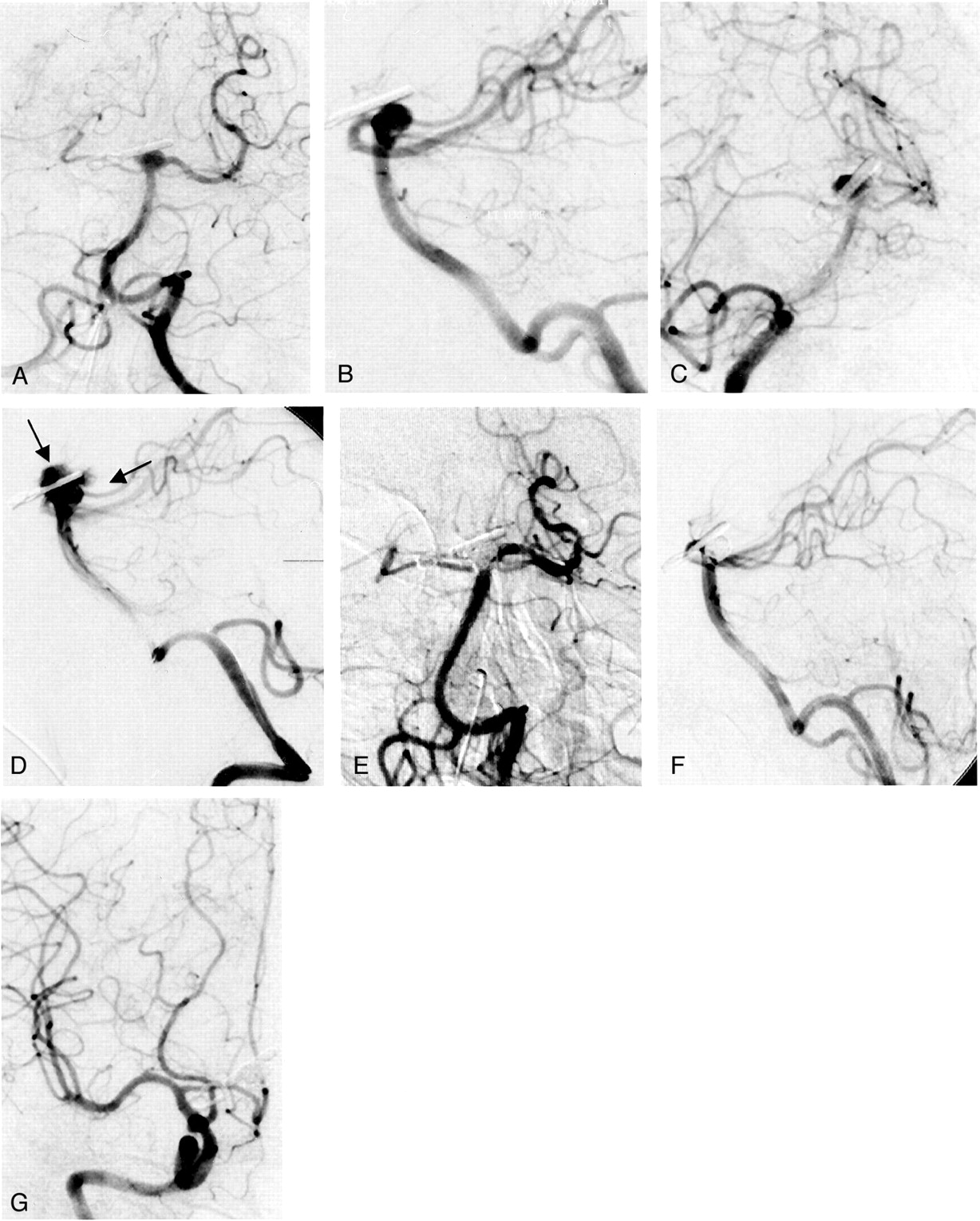
- Fig 5.
Case 3.
A, Anteroposterior and B, lateral DSA views demonstrate a broad-based, previously clipped, recurrent basilar tip aneurysm encroaching on the left P1 segment.
C, Angiogram reveals immediate stasis of contrast material in the aneurysm after stent deployment.
D, Angiogram reveals extravasation of contrast material after aneurysm perforation with the microcatheter (arrows).
E, Posteroanterior and F, lateral final angiograms after treatment show complete obliteration.
G, Angiogram shows that the right posterior cerebral artery is opacified via the internal carotid artery.
- Fig 6.
Case 4.
A, Posteroanterior and B, oblique DSA views show a previously coiled, multilobulated, recurrent basilar tip aneurysm encroaching the left P1 segment.
C, Unsubtracted image demonstrates the proximal and distal markers (arrows) of the self-expanding neurovascular stent positioned with the distal end in the left P1 segment and with the proximal end in the midbasilar artery.
D, Final oblique angiographic view after treatment demonstrates complete obliteration of the aneurysm.
E, Follow-up angiogram reveals that the aneurysm remained occluded 6 months later


In this issue


Jump to section
Related Articles
Cited By...
- Wide-neck bifurcation aneurysms of the middle cerebral artery and basilar apex treated by endovascular techniques: a multicentre, core lab adjudicated study evaluating safety and durability of occlusion (BRANCH)
- Endovascular treatment of complex intracranial aneurysms using Acandis Acclino stents
- Stent-Assisted Coiling of Wide-Neck Intracranial Aneurysms Using Low-Profile LEO Baby Stents: Initial and Midterm Results
- Dual Stenting Using Low-Profile LEO Baby Stents for the Endovascular Management of Challenging Intracranial Aneurysms
- Safety and Efficacy of Neuroform for Treatment of Intracranial Aneurysms: A Prospective, Consecutive, French Multicentric Study
- Treatment of basilar tip aneurysms with horizontal PCA to PCA stent-assisted coiling: case series
- Stent assisted coiling of the ruptured wide necked intracranial aneurysm
- Stent-Assisted Coiling of Complex Middle Cerebral Artery Aneurysms: Initial and Midterm Results
- Bailout Stent Deployment during Coil Embolization of Intracranial Aneurysms
- Stent Conformity in Curved Vascular Models with Simulated Aneurysm Necks Using Flat-Panel CT: An In Vitro Study
- Interventional Neuroradiology