
Article Figures & Data
Figures
- FIG 1.
Flow chart of study participants. Only diagnoses made after MR imaging were considered for this study. PD indicates Parkinson disease; FTD, frontotemporal dementia.
- FIG 2.
Coronal T1 noncontrast MR images with automated segmentation overlay obtained from NeuroQuant showing hippocampal volumes (yellow arrow) for patients with AD (A), mild cognitive impairment (B), and SCD (C). Note the marked hippocampal atrophy for AD compared with MCI and SCD.
- FIG 3.
Boxplot graphs of the variables age at diagnosis (A), NTHV (B), maximum MTA (C), and maximum ERiCA (D) scores against the outcome variables AD, MCI, and SCD. The maximum and minimum values are represented at either end of the whiskers. The box represents the interquartile range (25th percentile to the 75th percentile), with the median represented by the line within the box, and the mean shown by the diamond. Outliers are shown by a circle. Maximum MTA and maximum ERiCA refer to the higher score of the right and left values of the respective parameters.
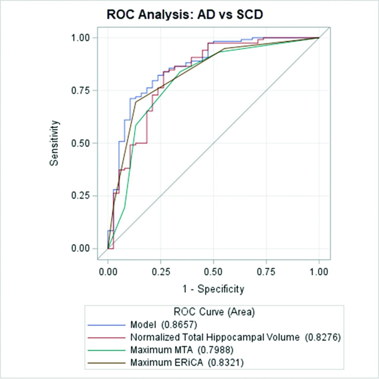
- FIG 4.
ROC curves for AD versus SCD. The overall model includes normalized total hippocampal volume, maximum MTA, maximum ERiCA, and age at diagnosis. Maximum MTA and maximum ERiCA refer to the higher score of the right and left values of the respective parameters.
- FIG 5.
ROC curves for AD versus MCI. The overall model includes normalized total hippocampal volume, maximum MTA, maximum ERiCA, and age at diagnosis. Maximum MTA and maximum ERiCA refer to the higher score of the right and left values of the respective parameters.
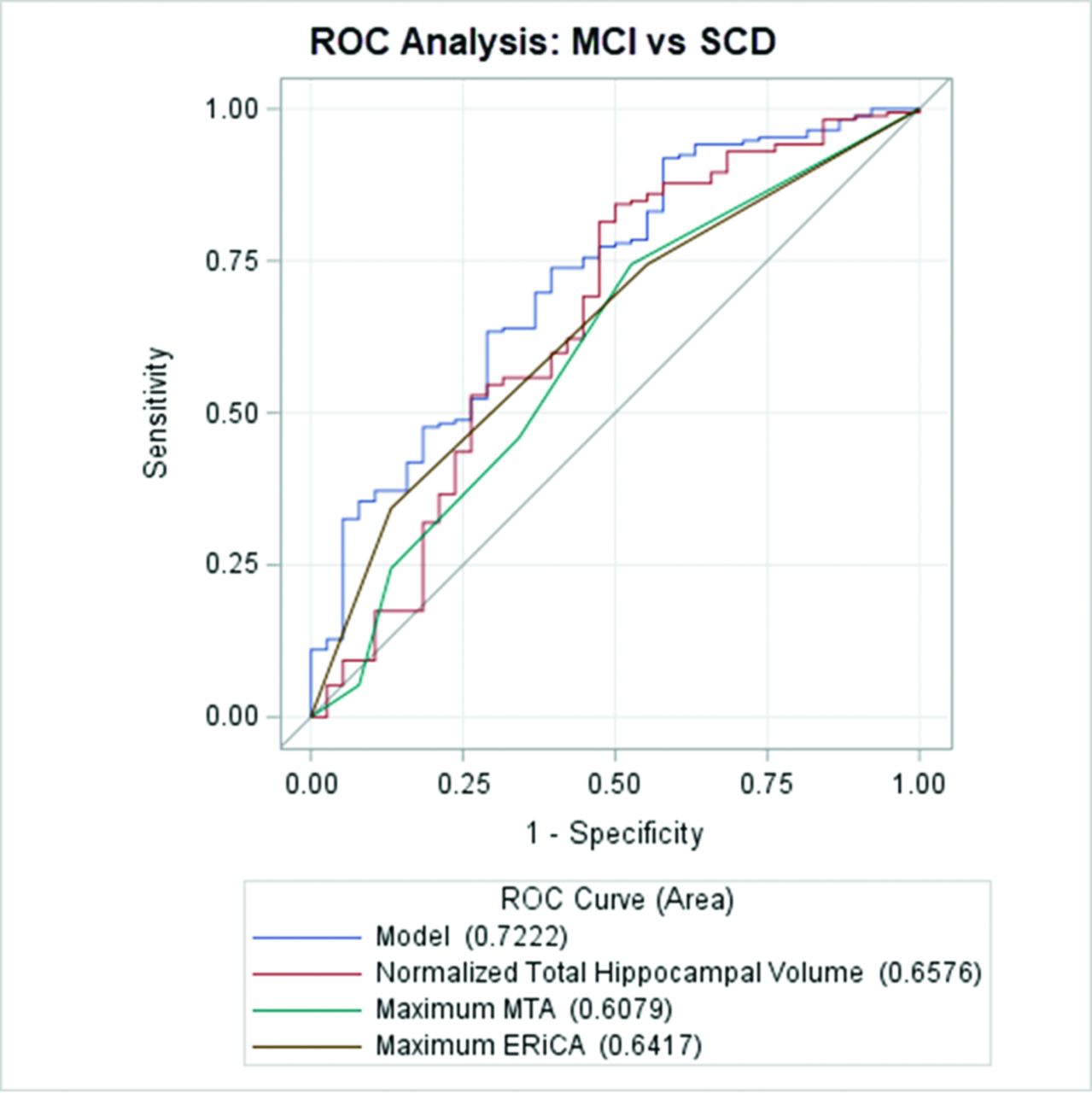
- FIG 6.
ROC curves for MCI versus SCD. The overall model includes NTHV, maximum MTA, maximum ERiCA, and age at diagnosis. Maximum MTA and maximum ERiCA refer to the higher scores of the right and left values of the respective parameters.


Tables
Characteristic AD MCI SCD P Value Sex .93 Male (%) 39.8 41.9 42.1 Female (%) 60.2 58.1 57.9 Race .23 Caucasian or white (%) 58.5 68.0 60.5 Other (%) 41.5 32.0 39.5 Highest education .15 ≤High school (%) 28.8 20.9 15.8 >High school (%) 71.2 79.1 84.2 Arterial hypertension .45 Yes (%) 71.2 66.9 60.5 No (%) 28.8 33.1 39.5 Diabetes mellitus .14 Yes (%) 18.6 23.3 34.2 No (%) 81.4 76.7 65.8 Age at diagnosis (yr) 78 (71–84) 74 (69–78) 67 (54–78) <.001b NTHV (%) 0.35 (0.33–0.37) 0.41 (0.40–0.43) 0.48 (0.42–0.50) <.00b Maximum MTA score 3.0 (2.0–3.0) 1.0 (0.0–2.0) 1.0 (0.0–2.0) <.001b Maximum ERiCA score 2.0 (1.0–2.0) 1.0 (0.0–2.0) 1.0 (0.0–1.0) <.001b a 50% median and interquartile ranges (in parentheses) are shown for age at diagnosis, NTHV, maximum MTA and maximum ERiCA scores. Maximum MTA and maximum ERiCA refer to the higher scores of the right and left values of the respective parameters.
↵b Denotes statistical significance at α = .05.
Parameter Cutoff Value Sensitivity Specificity Accuracy NTHV (%) 0.41 99/118 (84) [76–90] 28/38 (74) [60–88] 127/156 (81) [75–87] Max MTA score: all ages 2.0 99/118 (84) [77–91] 25/38 (66) [51–81] 124/156 (79) [73–86] MTA score: <75 years 2.0 32/44 (73) [60–86] 23/27 (85) [72–99] 55/71 (77) [68–87] MTA score: ≥75 years 3.0 50/74 (68) [57–78] 8/11 (73) [46–99] 58/85 (68) [58–78] Max ERiCA score 2.0 82/118 (69) [61–78] 33/38 (87) [76–98] 115/156 (74) [67–81] Note:—Max indicates maximum.
↵a Patient numbers shown in sensitivity, specificity, and accuracy columns with percentage scores (in parentheses) and 95% confidence intervals [in brackets]. Maximum MTA and maximum ERiCA refer to the higher score of the right and left values of the respective parameters.
Parameter Cutoff Value Sensitivity Specificity Accuracy NTHV (%) 0.38 84/118 (71) [62–79] 115/172 (67) [59–74] 199/290 (69) [63–74] Max MTA score: all ages 2.0 99/118 (84) [77–91] 93/172 (54) [47–62] 192/290 (66) [61–72] MTA score: <75 years 2.0 32/44 (73) [60–86] 63/90 (70) [61–79] 95/134 (71) [63–79] MTA score: ≥75 years 3.0 50/74 (68) [57–78] 51/82 (62) [52–73] 101/156 (65) [57–72] Max ERiCA score 3.0 82/118 (69) [61–78] 113/172 (66) [59–73] 195/290 (67) [62–73] Note:— Max indicates maximum.
↵a Patient numbers shown in sensitivity, specificity, and accuracy columns with percentage scores (in parentheses) and 95% confidence intervals [in brackets].


