Advertisement

Main menu

  • Home
  • Content
    • Current Issue
    • Accepted Manuscripts
    • Article Preview
    • Past Issue Archive
    • Video Articles
    • AJNR Case Collection
    • Case of the Week Archive
    • Case of the Month Archive
    • Classic Case Archive
  • Special Collections
    • AJNR Awards
    • Low-Field MRI
    • Alzheimer Disease
    • ASNR Foundation Special Collection
    • Photon-Counting CT
    • View All
  • Multimedia
    • AJNR Podcasts
    • AJNR SCANtastic
    • Trainee Corner
    • MRI Safety Corner
    • Imaging Protocols
  • For Authors
    • Submit a Manuscript
    • Submit a Video Article
    • Submit an eLetter to the Editor/Response
    • Manuscript Submission Guidelines
    • Statistical Tips
    • Fast Publishing of Accepted Manuscripts
    • Graphical Abstract Preparation
    • Imaging Protocol Submission
    • Author Policies
  • About Us
    • About AJNR
    • Editorial Board
    • Editorial Board Alumni
  • More
    • Become a Reviewer/Academy of Reviewers
    • Subscribers
    • Permissions
    • Alerts
    • Feedback
    • Advertisers
    • ASNR Home

User menu

  • Alerts
  • Log in

Search

  • Advanced search
American Journal of Neuroradiology

American Journal of Neuroradiology

ASHNR American Society of Functional Neuroradiology ASHNR American Society of Pediatric Neuroradiology ASSR
  • Alerts
  • Log in

Advanced Search

  • Home
  • Content
    • Current Issue
    • Accepted Manuscripts
    • Article Preview
    • Past Issue Archive
    • Video Articles
    • AJNR Case Collection
    • Case of the Week Archive
    • Case of the Month Archive
    • Classic Case Archive
  • Special Collections
    • AJNR Awards
    • Low-Field MRI
    • Alzheimer Disease
    • ASNR Foundation Special Collection
    • Photon-Counting CT
    • View All
  • Multimedia
    • AJNR Podcasts
    • AJNR SCANtastic
    • Trainee Corner
    • MRI Safety Corner
    • Imaging Protocols
  • For Authors
    • Submit a Manuscript
    • Submit a Video Article
    • Submit an eLetter to the Editor/Response
    • Manuscript Submission Guidelines
    • Statistical Tips
    • Fast Publishing of Accepted Manuscripts
    • Graphical Abstract Preparation
    • Imaging Protocol Submission
    • Author Policies
  • About Us
    • About AJNR
    • Editorial Board
    • Editorial Board Alumni
  • More
    • Become a Reviewer/Academy of Reviewers
    • Subscribers
    • Permissions
    • Alerts
    • Feedback
    • Advertisers
    • ASNR Home
  • Follow AJNR on Twitter
  • Visit AJNR on Facebook
  • Follow AJNR on Instagram
  • Join AJNR on LinkedIn
  • RSS Feeds

AJNR Awards, New Junior Editors, and more. Read the latest AJNR updates

Research ArticleNeurodegenerative Disorder Imaging

The Value of Quantitative Susceptibility Mapping and Morphometry in the Differential Diagnosis of Parkinsonism

Yi Li, Tingting Yuan, Lulu Gao, Wei Sun, Xiaoxiao Du, Zhihui Sun, Kangli Fan, Ruqing Qiu and Ying Zhang
American Journal of Neuroradiology July 2025, 46 (7) 1429-1438; DOI: https://doi.org/10.3174/ajnr.A8665
Yi Li
aFrom the Department of Neurology (Y.L., W.S., X.D., Z.S., K.F., R.Q., Y.Z.), the First Hospital of Jilin University, Changchun, Jilin Province, China
cDepartment of Neurology (Y.L.), Fujian Medical University Union Hospital, Fuzhou, Fujian Province, China
  • Find this author on Google Scholar
  • Find this author on PubMed
  • Search for this author on this site
Tingting Yuan
bDepartment of Radiology (T.Y.), the First Hospital of Jilin University, Changchun, Jilin Province, China
  • Find this author on Google Scholar
  • Find this author on PubMed
  • Search for this author on this site
Lulu Gao
dDepartment of Neurology (L.G.), the First People’s Hospital of Jining, Jining, Shandong Province, China
  • Find this author on Google Scholar
  • Find this author on PubMed
  • Search for this author on this site
Wei Sun
aFrom the Department of Neurology (Y.L., W.S., X.D., Z.S., K.F., R.Q., Y.Z.), the First Hospital of Jilin University, Changchun, Jilin Province, China
  • Find this author on Google Scholar
  • Find this author on PubMed
  • Search for this author on this site
Xiaoxiao Du
aFrom the Department of Neurology (Y.L., W.S., X.D., Z.S., K.F., R.Q., Y.Z.), the First Hospital of Jilin University, Changchun, Jilin Province, China
  • Find this author on Google Scholar
  • Find this author on PubMed
  • Search for this author on this site
Zhihui Sun
aFrom the Department of Neurology (Y.L., W.S., X.D., Z.S., K.F., R.Q., Y.Z.), the First Hospital of Jilin University, Changchun, Jilin Province, China
  • Find this author on Google Scholar
  • Find this author on PubMed
  • Search for this author on this site
Kangli Fan
aFrom the Department of Neurology (Y.L., W.S., X.D., Z.S., K.F., R.Q., Y.Z.), the First Hospital of Jilin University, Changchun, Jilin Province, China
  • Find this author on Google Scholar
  • Find this author on PubMed
  • Search for this author on this site
Ruqing Qiu
aFrom the Department of Neurology (Y.L., W.S., X.D., Z.S., K.F., R.Q., Y.Z.), the First Hospital of Jilin University, Changchun, Jilin Province, China
  • Find this author on Google Scholar
  • Find this author on PubMed
  • Search for this author on this site
Ying Zhang
aFrom the Department of Neurology (Y.L., W.S., X.D., Z.S., K.F., R.Q., Y.Z.), the First Hospital of Jilin University, Changchun, Jilin Province, China
  • Find this author on Google Scholar
  • Find this author on PubMed
  • Search for this author on this site
  • Article
  • Figures & Data
  • Supplemental
  • Info & Metrics
  • Responses
  • References
  • PDF
Loading

This article requires a subscription to view the full text. If you have a subscription you may use the login form below to view the article. Access to this article can also be purchased.

Graphical Abstract

Figure
  • Download figure
  • Open in new tab
  • Download powerpoint

Abstract

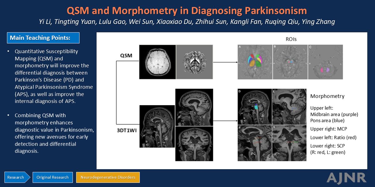
BACKGROUND AND PURPOSE: Differentiating Parkinson disease (PD) from atypical parkinsonism syndrome (APS), including multiple system atrophy (MSA) and progressive supranuclear palsy (PSP), is challenging, and there is no reference standard. Integrating quantitative susceptibility mapping (QSM) and morphometry can help differentiate PD from APS and improve the internal diagnosis of APS.

MATERIALS AND METHODS: In this retrospective study, we enrolled 55 patients with PD, 17 with MSA-parkinsonian type (MSA-P), 15 with MSA-cerebellar type (MSA-C), and 14 with PSP. Thirty-three age-matched healthy subjects served as controls. All subjects underwent QSM imaging and 3D T1WI with manual quantification of ROI and morphometry. ROIs were selected in the basal ganglia and brainstem nuclei, such as the putamen (Pu), globus pallidus (GP), and red nucleus (RN). Morphometry included MR Parkinson disease index (MRPI), the midbrain area-pons area ratio (M/P), and the ratio of the vertical line of the long axis of the midbrain and pons (Ratio). Differential variables between groups were extracted and a binary logistic regression was established to differentiate the differential diagnoses of PD and APS and diseases within APS. The diagnostic value was assessed using the area under the curve (AUC), sensitivity, and specificity.

RESULTS: The combination of Pu and GP performed best when used to distinguish PD from MSA-P, with an AUC of 0.800 (95% CI: 0.664–0.936). The AUC was optimal when MRPI and M/P were combined to distinguish PD from MSA-C at 0.823 (95% CI: 0.686–0.960). Ratio alone performed best in differentiating PD from PSP, with an AUC of 0.848 (95% CI: 0.711–0.985). The AUC for Ratio alone in distinguishing MSA-P from PSP was 0.871 (95% CI: 0.738–1.0). The AUC when using only M/P to distinguish MSA-C from PSP was 0.931 (95% CI: 0.845–1.0). QSM and morphometry each offer distinct advantages in the differential diagnosis among the aforementioned groups. The combination of QSM and morphometry provided the highest diagnostic value in differentiating PD from APS, highlighting the significance of integrating these 2 imaging techniques for enhanced diagnostic precision in clinical practice. The best indicators described above showed equally high differential diagnostic values in patients with a disease duration of ≤3 years.

CONCLUSIONS: QSM and morphometry will improve the differential diagnosis between PD and APS, as well as improve the internal diagnosis of APS.

ABBREVIATIONS:

APS
atypical parkinsonism syndrome
AUC
area under the curve
CN
caudate nucleus
DN
dentate nucleus
GP
globus pallidus
H-Y
Hoehn and Yahr Scale
ICC
intraclass correlation coefficient
M/P
midbrain area-pons area ratio
MRPI
magnetic resonance parkinsonism index
MSA
multiple system atrophy
MSA-C
multiple system atrophy cerebellar subtype
MSA-P
multiple system atrophy parkinsonian subtype
PD
Parkinson disease
PSP
progressive supranuclear palsy
Pu
Putamen
QSM
quantitative susceptibility mapping
Ratio
the ratio of the vertical line of the long axis of the midbrain and pons
RN
red nucleus
ROC
receiver operating characteristic
SN
substantia nigra
SNc
substantia nigra compacta
SNr
substantia nigra reticulate
Th
thalamus
UPDRS-III
Unified Parkinson’s Disease Rating Scale Part III
  • © 2025 by American Journal of Neuroradiology
View Full Text

Log in using your username and password

Forgot your user name or password?

Log in through your institution

You may be able to gain access using your login credentials for your institution. Contact your library if you do not have a username and password.
PreviousNext
Back to top

In this issue

American Journal of Neuroradiology: 46 (7)
American Journal of Neuroradiology
Vol. 46, Issue 7
1 Jul 2025
  • Table of Contents
  • Index by author
  • Complete Issue (PDF)
Advertisement
Print
Download PDF
Email Article

Thank you for your interest in spreading the word on American Journal of Neuroradiology.

NOTE: We only request your email address so that the person you are recommending the page to knows that you wanted them to see it, and that it is not junk mail. We do not capture any email address.

Enter multiple addresses on separate lines or separate them with commas.
The Value of Quantitative Susceptibility Mapping and Morphometry in the Differential Diagnosis of Parkinsonism
(Your Name) has sent you a message from American Journal of Neuroradiology
(Your Name) thought you would like to see the American Journal of Neuroradiology web site.
CAPTCHA
This question is for testing whether or not you are a human visitor and to prevent automated spam submissions.
Cite this article
Yi Li, Tingting Yuan, Lulu Gao, Wei Sun, Xiaoxiao Du, Zhihui Sun, Kangli Fan, Ruqing Qiu, Ying Zhang
The Value of Quantitative Susceptibility Mapping and Morphometry in the Differential Diagnosis of Parkinsonism
American Journal of Neuroradiology Jul 2025, 46 (7) 1429-1438; DOI: 10.3174/ajnr.A8665

Citation Manager Formats

  • BibTeX
  • Bookends
  • EasyBib
  • EndNote (tagged)
  • EndNote 8 (xml)
  • Medlars
  • Mendeley
  • Papers
  • RefWorks Tagged
  • Ref Manager
  • RIS
  • Zotero
0 Responses
Respond to this article
Share
Bookmark this article
QSM and Morphometry in Diagnosing Parkinsonism
Yi Li, Tingting Yuan, Lulu Gao, Wei Sun, Xiaoxiao Du, Zhihui Sun, Kangli Fan, Ruqing Qiu, Ying Zhang
American Journal of Neuroradiology Jul 2025, 46 (7) 1429-1438; DOI: 10.3174/ajnr.A8665
del.icio.us logo Twitter logo Facebook logo Mendeley logo
  • Tweet Widget
  • Facebook Like
  • Google Plus One
Purchase

Jump to section

  • Article
    • Graphical Abstract
    • Abstract
    • ABBREVIATIONS:
    • MATERIALS AND METHODS
    • RESULTS
    • DISCUSSION
    • CONCLUSIONS
    • Acknowledgments
    • Footnotes
    • References
  • Figures & Data
  • Supplemental
  • Info & Metrics
  • Responses
  • References
  • PDF

Related Articles

  • PubMed
  • Google Scholar

Cited By...

  • No citing articles found.
  • Crossref
  • Google Scholar

This article has not yet been cited by articles in journals that are participating in Crossref Cited-by Linking.

More in this TOC Section

  • Role of Brain PET in Alzheimer's Disease
  • Hydrocephalus Impact on FDG PET SPM in Dementia
  • Anti-Amyloid Therapy & CBF as a Response Biomarker
Show more Neurodegenerative Disorder Imaging

Similar Articles

Advertisement

Indexed Content

  • Current Issue
  • Accepted Manuscripts
  • Article Preview
  • Past Issues
  • Editorials
  • Editor's Choice
  • Fellows' Journal Club
  • Letters to the Editor
  • Video Articles

Cases

  • Case Collection
  • Archive - Case of the Week
  • Archive - Case of the Month
  • Archive - Classic Case

More from AJNR

  • Trainee Corner
  • Imaging Protocols
  • MRI Safety Corner
  • Book Reviews

Multimedia

  • AJNR Podcasts
  • AJNR Scantastics

Resources

  • Turnaround Time
  • Submit a Manuscript
  • Submit a Video Article
  • Submit an eLetter to the Editor/Response
  • Manuscript Submission Guidelines
  • Statistical Tips
  • Fast Publishing of Accepted Manuscripts
  • Graphical Abstract Preparation
  • Imaging Protocol Submission
  • Evidence-Based Medicine Level Guide
  • Publishing Checklists
  • Author Policies
  • Become a Reviewer/Academy of Reviewers
  • News and Updates

About Us

  • About AJNR
  • Editorial Board
  • Editorial Board Alumni
  • Alerts
  • Permissions
  • Not an AJNR Subscriber? Join Now
  • Advertise with Us
  • Librarian Resources
  • Feedback
  • Terms and Conditions
  • AJNR Editorial Board Alumni

American Society of Neuroradiology

  • Not an ASNR Member? Join Now

© 2025 by the American Society of Neuroradiology All rights, including for text and data mining, AI training, and similar technologies, are reserved.
Print ISSN: 0195-6108 Online ISSN: 1936-959X

Powered by HighWire