Advertisement

Main menu

  • Home
  • Content
    • Current Issue
    • Accepted Manuscripts
    • Article Preview
    • Past Issue Archive
    • Video Articles
    • AJNR Case Collection
    • Case of the Week Archive
    • Case of the Month Archive
    • Classic Case Archive
  • Special Collections
    • AJNR Awards
    • Low-Field MRI
    • Alzheimer Disease
    • ASNR Foundation Special Collection
    • Photon-Counting CT
    • View All
  • Multimedia
    • AJNR Podcasts
    • AJNR SCANtastic
    • Trainee Corner
    • MRI Safety Corner
    • Imaging Protocols
  • For Authors
    • Submit a Manuscript
    • Submit a Video Article
    • Submit an eLetter to the Editor/Response
    • Manuscript Submission Guidelines
    • Statistical Tips
    • Fast Publishing of Accepted Manuscripts
    • Graphical Abstract Preparation
    • Imaging Protocol Submission
    • Author Policies
  • About Us
    • About AJNR
    • Editorial Board
    • Editorial Board Alumni
  • More
    • Become a Reviewer/Academy of Reviewers
    • Subscribers
    • Permissions
    • Alerts
    • Feedback
    • Advertisers
    • ASNR Home

User menu

  • Alerts
  • Log in

Search

  • Advanced search
American Journal of Neuroradiology

American Journal of Neuroradiology

ASHNR American Society of Functional Neuroradiology ASHNR American Society of Pediatric Neuroradiology ASSR
  • Alerts
  • Log in

Advanced Search

  • Home
  • Content
    • Current Issue
    • Accepted Manuscripts
    • Article Preview
    • Past Issue Archive
    • Video Articles
    • AJNR Case Collection
    • Case of the Week Archive
    • Case of the Month Archive
    • Classic Case Archive
  • Special Collections
    • AJNR Awards
    • Low-Field MRI
    • Alzheimer Disease
    • ASNR Foundation Special Collection
    • Photon-Counting CT
    • View All
  • Multimedia
    • AJNR Podcasts
    • AJNR SCANtastic
    • Trainee Corner
    • MRI Safety Corner
    • Imaging Protocols
  • For Authors
    • Submit a Manuscript
    • Submit a Video Article
    • Submit an eLetter to the Editor/Response
    • Manuscript Submission Guidelines
    • Statistical Tips
    • Fast Publishing of Accepted Manuscripts
    • Graphical Abstract Preparation
    • Imaging Protocol Submission
    • Author Policies
  • About Us
    • About AJNR
    • Editorial Board
    • Editorial Board Alumni
  • More
    • Become a Reviewer/Academy of Reviewers
    • Subscribers
    • Permissions
    • Alerts
    • Feedback
    • Advertisers
    • ASNR Home
  • Follow AJNR on Twitter
  • Visit AJNR on Facebook
  • Follow AJNR on Instagram
  • Join AJNR on LinkedIn
  • RSS Feeds

AJNR Awards, New Junior Editors, and more. Read the latest AJNR updates

Research ArticleADULT BRAIN
Open Access

Prospective, Longitudinal Study of Clinical Outcome and Morphometric Posterior Fossa Changes after Craniocervical Decompression for Symptomatic Chiari I Malformation

Alaaddin Ibrahimy, Tianxia Wu, Jessica Mack, Gretchen C. Scott, Michaela X. Cortes, Fredric K. Cantor, Francis Loth and John D. Heiss
American Journal of Neuroradiology September 2023, DOI: https://doi.org/10.3174/ajnr.A7993
Alaaddin Ibrahimy
aFrom the Surgical Neurology Branch (A.I., J.M., G.C.S., M.X.C., F.K.C., J.D.H.), National Institute of Neurological Disorders and Stroke, National Institutes of Health, Bethesda, Maryland
bDepartment of Biomedical Engineering (A.I.), Yale University, New Haven, Connecticut
  • Find this author on Google Scholar
  • Find this author on PubMed
  • Search for this author on this site
  • ORCID record for Alaaddin Ibrahimy
Tianxia Wu
cClinical Trials Unit (T.W.), National Institute of Neurological Disorders and Stroke, National Institutes of Health, Bethesda, Maryland
  • Find this author on Google Scholar
  • Find this author on PubMed
  • Search for this author on this site
  • ORCID record for Tianxia Wu
Jessica Mack
aFrom the Surgical Neurology Branch (A.I., J.M., G.C.S., M.X.C., F.K.C., J.D.H.), National Institute of Neurological Disorders and Stroke, National Institutes of Health, Bethesda, Maryland
  • Find this author on Google Scholar
  • Find this author on PubMed
  • Search for this author on this site
  • ORCID record for Jessica Mack
Gretchen C. Scott
aFrom the Surgical Neurology Branch (A.I., J.M., G.C.S., M.X.C., F.K.C., J.D.H.), National Institute of Neurological Disorders and Stroke, National Institutes of Health, Bethesda, Maryland
  • Find this author on Google Scholar
  • Find this author on PubMed
  • Search for this author on this site
  • ORCID record for Gretchen C. Scott
Michaela X. Cortes
aFrom the Surgical Neurology Branch (A.I., J.M., G.C.S., M.X.C., F.K.C., J.D.H.), National Institute of Neurological Disorders and Stroke, National Institutes of Health, Bethesda, Maryland
  • Find this author on Google Scholar
  • Find this author on PubMed
  • Search for this author on this site
  • ORCID record for Michaela X. Cortes
Fredric K. Cantor
aFrom the Surgical Neurology Branch (A.I., J.M., G.C.S., M.X.C., F.K.C., J.D.H.), National Institute of Neurological Disorders and Stroke, National Institutes of Health, Bethesda, Maryland
  • Find this author on Google Scholar
  • Find this author on PubMed
  • Search for this author on this site
Francis Loth
dDepartments of Mechanical and Industrial Engineering, and Bioengineering (F.L.), Northeastern University College of Engineering, Boston, Massachusetts
  • Find this author on Google Scholar
  • Find this author on PubMed
  • Search for this author on this site
  • ORCID record for Francis Loth
John D. Heiss
aFrom the Surgical Neurology Branch (A.I., J.M., G.C.S., M.X.C., F.K.C., J.D.H.), National Institute of Neurological Disorders and Stroke, National Institutes of Health, Bethesda, Maryland
  • Find this author on Google Scholar
  • Find this author on PubMed
  • Search for this author on this site
  • ORCID record for John D. Heiss
  • Article
  • Figures & Data
  • Supplemental
  • Info & Metrics
  • Responses
  • References
  • PDF
Loading

Article Figures & Data

Figures

  • Tables
  • FIG 1.
    • Download figure
    • Open in new tab
    • Download powerpoint
    FIG 1.

    Morphometric measurements on a presurgery MR image (A) and a postsurgery MR image (B) for the same patient 1 year later. 1) CSF area posterior to PCF, 2) anterior CSF area, 3) posterior CSF area, 4) cerebellar height, 5a) posterior dorsal width + (5b) anterior dorsal width = total dorsal CSF width, 6) ventral CSF width, 7) cerebellar tonsillar position, 8) occipital bone length, and 9) cerebellum-clivus bone angle. Note that the syrinx in the presurgical MR image resolved after surgery (Supplemental Online Video).

  • FIG 2.
    • Download figure
    • Open in new tab
    • Download powerpoint
    FIG 2.

    Morphometric length, angle, and CSF area measurements (LS-means and 95% CI). A, CSF area posterior to the PCF. B, Posterior CSF area. A and B, RM-ANOVA F-test (df = 6) of TP: P value < .0001. C, Anterior CSF area for CMI-only, and for patients with CMI plus syringomyelia; C, RM-ANCOVA (diagnosis as a covariate), F-test (df = 6) for the interaction (diagnosis × TP): P = .0374. RM-ANOVA F-test (df = 6) for TP: P > .05 for CMI-only, no multiple comparisons: P < .0001 for CMI and syringomyelia (SM). D, Cerebellar height. E, Total dorsal width. F, Ventral CSF width. G, Cerebellar tonsillar position, H, Occipital bone length. I, Cerebellum-clivus bone angle. RM-ANOVA F-test (df = 6) of TP: P value < .0001 except for ventral CSF width (P = .023, F). Hash tag indicates the Dunnett adjusted P value < .05 at the TP compared with TP = –0.5; plus sign, the Dunnett adjusted P value < .05 at the TP compared with TP = +0.5; asterisk, the Dunnett adjusted P value < .01 at the TP compared with TP = –0.5.
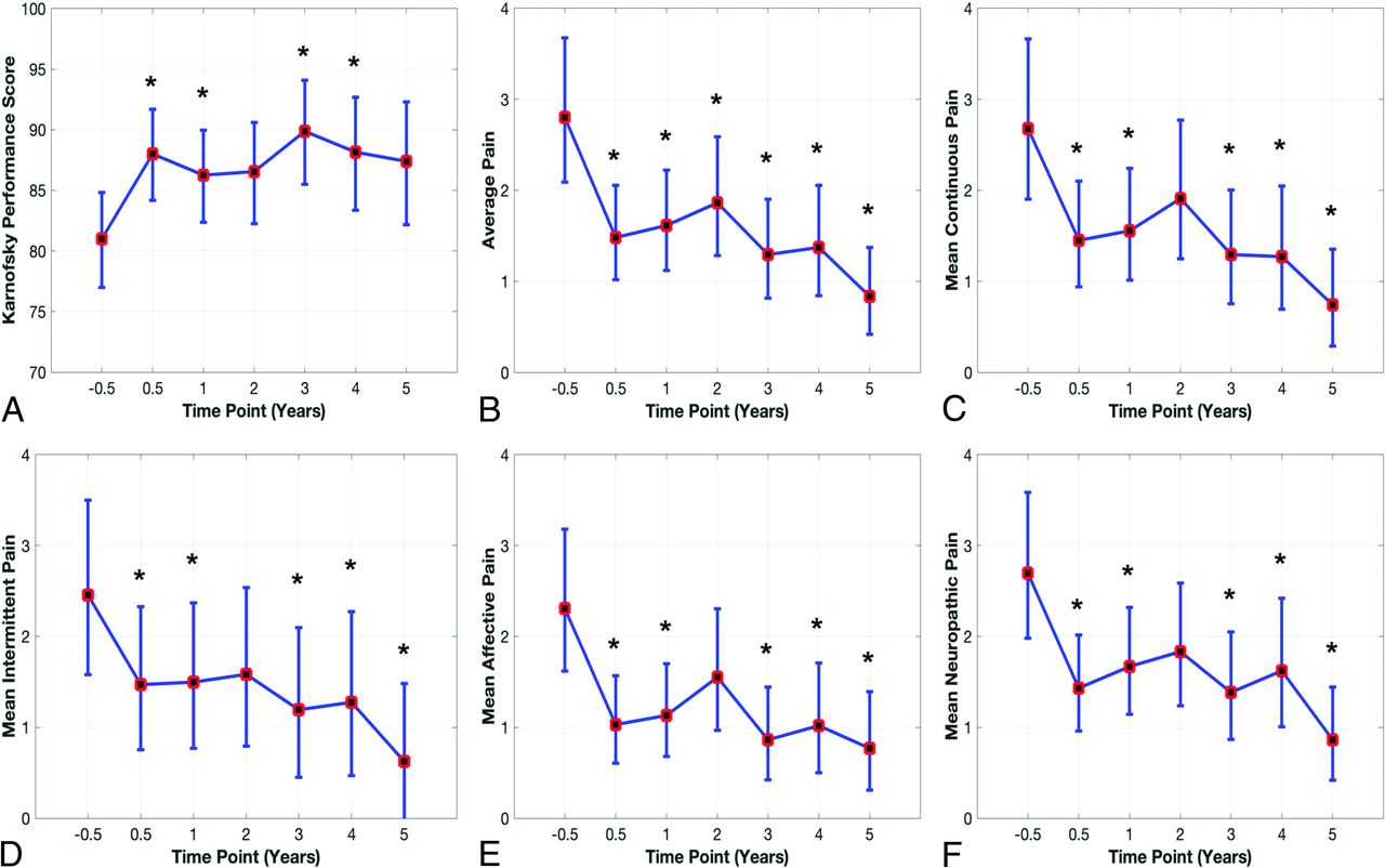
  • FIG 3.
    • Download figure
    • Open in new tab
    • Download powerpoint
    FIG 3.

    Clinical outcomes (back-transformed LS-means and 95% CI): A, KPS score. B, Average pain. C, Mean continuous pain. D, Mean intermittent pain. E, Mean affective pain. F, Mean neuropathic pain. RM-ANOVA F-test (df = 6) of TP: P value < .005. The asterisk indicates the Dunnett adjusted P value < .05 at the TP compared with TP = –0.5.

Tables

  • Figures
    • View popup
    Table 1:

    Number of subjects at each TP category for clinical outcomes and morphometric measurements

    TP categories
     TP (yr)–0.50.512345
     Relative to surgery (mo)–9 to –10–99–1818–3030–4242–54>54
    Number of subjects for each TP
     Morphometric measurements38353123191714
     KPS score36343224221614
     Average pain34333223211414
     Mean of affective pain34333223211414
     Mean of continuous pain33333223211414
     Mean of intermittent pain34333223211414
     Mean of neuropathic pain34333223211414
     ASIA total score35353323241714
     Ambulatory score35353324241714
     Cognitive subtotal35353224221614
     McCormick class score35353224241714
     Motor subtotal35353224221614
     Total FIM35353224221614
    • Note:—ASIA indicates American Spinal Injury Association; FIM, Functional Independence Measure.

    • View popup
    Table 2:

    Description of morphometric measurements

    Morphometric MeasurementDescription
    Area
    1 CSF area posterior to PCF (mm2)CSF area posterior to a line drawn between the IOP and opisthion, and posterior to the cerebellum
    2 Anterior CSF area (mm2)CSF area in the upper cervical spinal canal anterior to the spinal cord between the FM and inferior limit of the C2 vertebra
    3 Posterior CSF area (mm2)CSF area in the upper cervical spinal canal posterior to the spinal cord between the FM and inferior limit of the C2 vertebra
    Length
    4 Cerebellar height (mm)Distance between the most superior point of the superior vermis and the most inferior point of the tonsil
    5 Total dorsal CSF width (mm)Width of the CSF space measured at the level of the FM, anterior to the cerebellum, posterior to the brainstem and posterior the cerebellum, anterior to the subarachnoid space
    6 Ventral CSF width (mm)Width of the CSF space measured at the level of the FM, anterior to the brainstem, posterior to the subarachnoid space
    7 Cerebellar tonsillar position (mm)The perpendicular distance between the most inferior tip of the cerebellar tonsils and the McRae line
    8 Occipital bone length (mm)Length between a point at the midpoint of the occipital bone at the level of the tentorium and IOP and the most inferior tip of the occipital bone
    Angle
    9 Cerebellum-clivus bone angle (degree)The angle subtended by the major axis of the cerebellum with the posterior margin of the clivus bone
    • Note:—IOP indicates internal occipital protuberance; FM, foramen magnum.

PreviousNext
Back to top
Advertisement
Print
Download PDF
Email Article

Thank you for your interest in spreading the word on American Journal of Neuroradiology.

NOTE: We only request your email address so that the person you are recommending the page to knows that you wanted them to see it, and that it is not junk mail. We do not capture any email address.

Enter multiple addresses on separate lines or separate them with commas.
Prospective, Longitudinal Study of Clinical Outcome and Morphometric Posterior Fossa Changes after Craniocervical Decompression for Symptomatic Chiari I Malformation
(Your Name) has sent you a message from American Journal of Neuroradiology
(Your Name) thought you would like to see the American Journal of Neuroradiology web site.
CAPTCHA
This question is for testing whether or not you are a human visitor and to prevent automated spam submissions.
Cite this article
Alaaddin Ibrahimy, Tianxia Wu, Jessica Mack, Gretchen C. Scott, Michaela X. Cortes, Fredric K. Cantor, Francis Loth, John D. Heiss
Prospective, Longitudinal Study of Clinical Outcome and Morphometric Posterior Fossa Changes after Craniocervical Decompression for Symptomatic Chiari I Malformation
American Journal of Neuroradiology Sep 2023, DOI: 10.3174/ajnr.A7993

Citation Manager Formats

  • BibTeX
  • Bookends
  • EasyBib
  • EndNote (tagged)
  • EndNote 8 (xml)
  • Medlars
  • Mendeley
  • Papers
  • RefWorks Tagged
  • Ref Manager
  • RIS
  • Zotero
0 Responses
Respond to this article
Share
Bookmark this article
Prospective, Longitudinal Study of Clinical Outcome and Morphometric Posterior Fossa Changes after Craniocervical Decompression for Symptomatic Chiari I Malformation
Alaaddin Ibrahimy, Tianxia Wu, Jessica Mack, Gretchen C. Scott, Michaela X. Cortes, Fredric K. Cantor, Francis Loth, John D. Heiss
American Journal of Neuroradiology Sep 2023, DOI: 10.3174/ajnr.A7993
del.icio.us logo Twitter logo Facebook logo Mendeley logo
  • Tweet Widget
  • Facebook Like
  • Google Plus One
Purchase

Jump to section

  • Article
    • Abstract
    • ABBREVIATIONS:
    • MATERIALS AND METHODS
    • RESULTS
    • DISCUSSION
    • CONCLUSIONS
    • Footnotes
    • References
  • Figures & Data
  • Supplemental
  • Info & Metrics
  • Responses
  • References
  • PDF

Related Articles

  • PubMed
  • Google Scholar

Cited By...

  • No citing articles found.
  • Crossref (2)
  • Google Scholar

This article has been cited by the following articles in journals that are participating in Crossref Cited-by Linking.

  • ChatGPT as a Decision Support Tool in the Management of Chiari I Malformation: A Comparison to 2023 CNS Guidelines
    Ethan D.L. Brown, Apratim Maity, Max Ward, Daniel Toscano, Griffin R. Baum, Mark A. Mittler, Sheng-Fu Larry Lo, Randy S. D'Amico
    World Neurosurgery 2024 191
  • Radiopaedia.org
    Frank Gaillard, Rohit Sharma, Tariq Walizai
    2008

More in this TOC Section

  • Diagnostic Neuroradiology of Monoclonal Antibodies
  • Segmentation of Brain Metastases with BLAST
Show more ADULT BRAIN

Similar Articles

Advertisement

Indexed Content

  • Current Issue
  • Accepted Manuscripts
  • Article Preview
  • Past Issues
  • Editorials
  • Editor's Choice
  • Fellows' Journal Club
  • Letters to the Editor
  • Video Articles

Cases

  • Case Collection
  • Archive - Case of the Week
  • Archive - Case of the Month
  • Archive - Classic Case

More from AJNR

  • Trainee Corner
  • Imaging Protocols
  • MRI Safety Corner
  • Book Reviews

Multimedia

  • AJNR Podcasts
  • AJNR Scantastics

Resources

  • Turnaround Time
  • Submit a Manuscript
  • Submit a Video Article
  • Submit an eLetter to the Editor/Response
  • Manuscript Submission Guidelines
  • Statistical Tips
  • Fast Publishing of Accepted Manuscripts
  • Graphical Abstract Preparation
  • Imaging Protocol Submission
  • Evidence-Based Medicine Level Guide
  • Publishing Checklists
  • Author Policies
  • Become a Reviewer/Academy of Reviewers
  • News and Updates

About Us

  • About AJNR
  • Editorial Board
  • Editorial Board Alumni
  • Alerts
  • Permissions
  • Not an AJNR Subscriber? Join Now
  • Advertise with Us
  • Librarian Resources
  • Feedback
  • Terms and Conditions
  • AJNR Editorial Board Alumni

American Society of Neuroradiology

  • Not an ASNR Member? Join Now

© 2025 by the American Society of Neuroradiology All rights, including for text and data mining, AI training, and similar technologies, are reserved.
Print ISSN: 0195-6108 Online ISSN: 1936-959X

Powered by HighWire