
Article Figures & Data
Figures
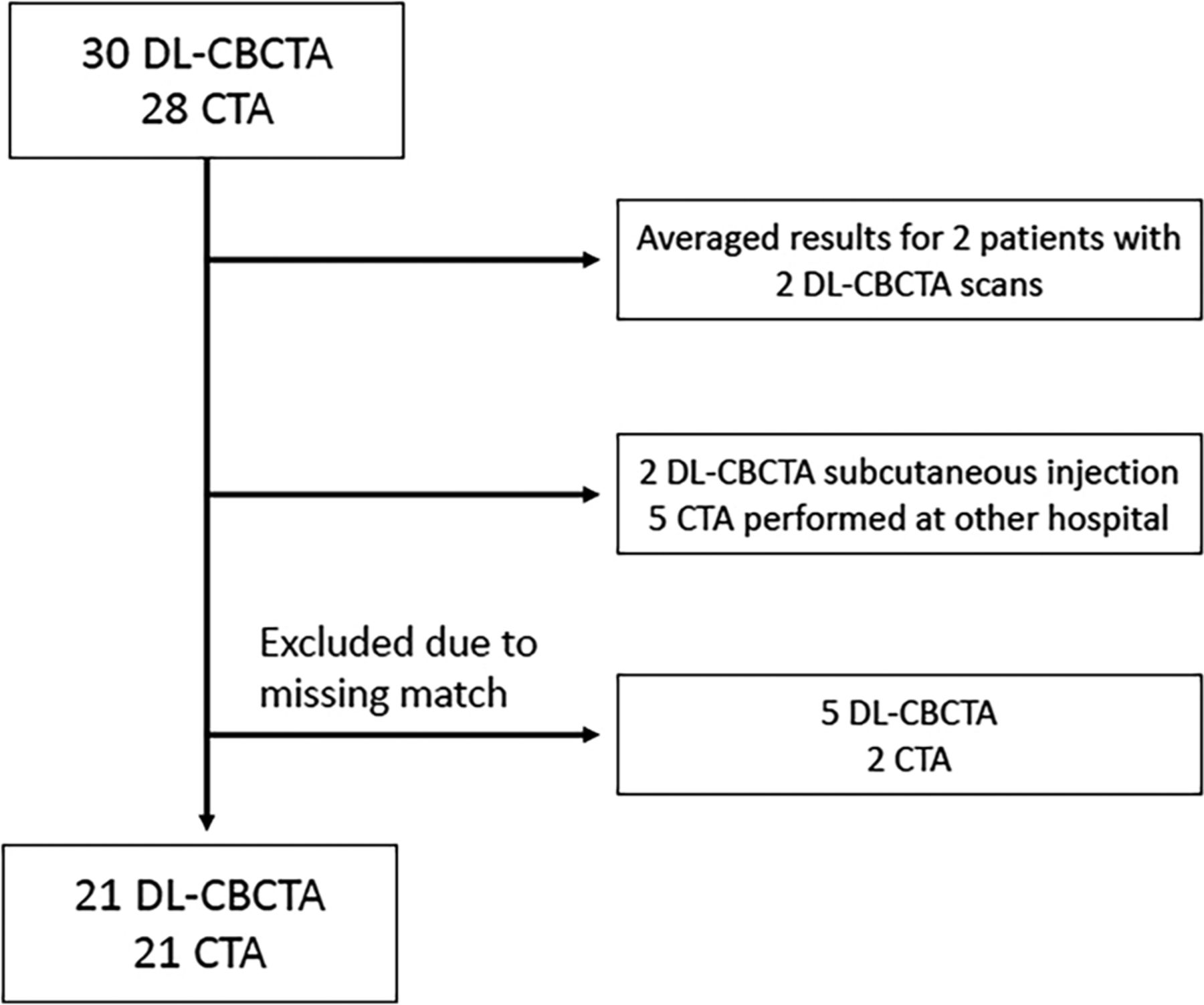
- FIG 1.
Of 28 consecutively enrolled patients, 5 had no in-house CTA and 2 had subcutaneous IV contrast media injection during the DL-CBCTA scan. Two patients were imaged twice with DL-CBCTA, and for those, the results from both scans were averaged. Consequently, 21 complete and matched DL-CBCTA and CTA image sets from 21 patients were included.
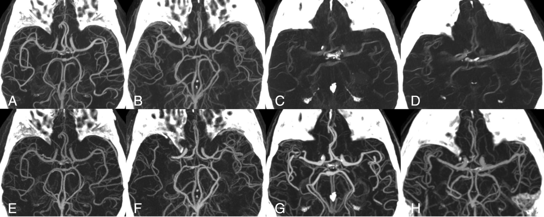
- FIG 2.
DL-CBCTA 70-keV images (upper row) and CTA (lower row) with MIP 35-mm section thickness. A and B, Acceptable-quality DL-CBCTA scans. C and D, Typical scans in the data set affected by motion artifacts. Lower row (E–H) shows the corresponding CTA. Note that images F, G, and H show a right-sided MCA occlusion, which have been resolved at the time for DL-CBCTA imaging. Only the arterial anatomy in the unaffected hemisphere was evaluated in this study.


Tables
Canon Aquilion ONE Philips IQon Prototype DL-CBCT Tube (kV) 100 120 120 Tube current (mAs/mA) (average) 196 (Auto-modulation) 114 (Auto-modulation) 310 Scan time/rotation time (sec) 3.3/0.5 (Full rotation) 2.5/0.3 (Full rotation) 20.0/20.0 (200° rotation) Nominal beam width (mm) 80 × 0.500 64 × 0.625 194.700 Pitch factor 0.813 0.671 NA Display FOV coronal × sagittal × axial (mm3) 210.9 × 210.9 × 160.0 210.0 × 210.0 × 160.0 251.8 × 251.8 × 194.7 Section thickness (mm) 0.50 0.67 0.66 Matrix size 512 × 512 512 × 512 384 × 384 Reconstruction kernel FC43 Filter UA Ståhl et al15a Reconstruction algorithm AIDR 3D eStandard iDose4 level 4 Ståhl et al15a Avg CTDIvol (16-cm phantom) 20.0 mGy 21.3 mGy NA Air kerma (in an 18-cm water phantom)b NA NA 57.6 mGy MTFc 50%: 3.78 50%: 3.46 50%: 3.57 10%: 6.57 10%: 6.65 10%: 6.04 Note:—CTDIvol indicates volume CT dose index; MTF, modulation transfer function; NA, not applicable.
↵a Details of the prototype algorithm are described in Ståhl et al.15
↵b Air kerma in an 18-cm diameter plastic water phantom at the center of the scan length, measured in accordance with American Association of Physicists in Medicine Task Group Report 111.26
↵c Generated from consecutive scans of the upper bead of a Catphan CTP528 module (The Phantom Laboratory).
16 Segments 11 Segments (Powered) 11 Segments (Thrombectomy) ICA ICA ICA M1 M1 M1 M2 M2 M2 M3 M3-M4 M3 M4 M4 A1 A1-A2 A1 A2 A2 Lenticulostriate Lenticulostriate Vertebral Vertebral Vertebral Basilar Basilar Basilar AICA AICA-PICA-SCA PICA SCA Basilar perforators Basilar perforators P1 P1-P2 P1 P2 P2 Note:—Lenticulostriate indicates lenticulostriate artery perforators; Vertebral, intracranial vertebral artery; Basilar perforators, basilar artery perforating branches; SCA, superior cerebellar artery.
Powered Data Set Powered Subset Thrombectomy Data Set Thrombectomy Subset Patients 21 12 21 12 Segments rated 231 132 231 132 Majority 0.77 (0.70) 0.98 (0.93)b 0.77 (0.71) 0.98 (0.93)b Reader 1 0.65 (0.58) 0.88 (0.80)b 0.67 (0.60) 0.91 (0.84)b Reader 2 0.90 (0.84)b 0.98 (0.93)b 0.89 (0.83)b 0.98 (0.93)b Reader 3 0.60 (0.53) 0.78 (0.69) 0.68 (0.61) 0.88 (0.80)b ↵a Proportion of DL-CBCTA arterial segment visibility rated equal or superior to CTA. The data set (21 patients) includes all scans; the subset (12 patients) excluded inferior scans. The 98.75% CI of the 1-sided lower performance boundary is in parentheses (lower boundary is defined as 80% rated equal or superior).
↵b Statistically significant result.
Powered Data Set Powered Subset Thrombectomy Data Set Thrombectomy Subset Patients 21 12 21 12 Segments rated 231 132 231 132 Majority 0.41 (0.34) 0.63 (0.53) 0.55 (0.48) 0.81 (0.72) Reader 1 0.42 (0.35) 0.68 (0.58) 0.54 (0.46) 0.85 (0.77) Reader 2 0.58 (0.50) 0.66 (0.56) 0.65 (0.58) 0.74 (0.64) Reader 3 0.36 (0.29) 0.55 (0.44) 0.52 (0.44) 0.73 (0.63) ↵a Proportion of DL-CBCTA arterial segment artifacts rated equal or superior to CTA. Data set (21 patients) includes all scans; subset (12 patients) excluded inferior scans. The 98.75% CI of the 1-sided lower performance boundary is in parentheses (lower boundary is defined as 80% rated equal or superior).


Jump to section
Related Articles
Cited By...
- No citing articles found.