
Article Figures & Data
Figures
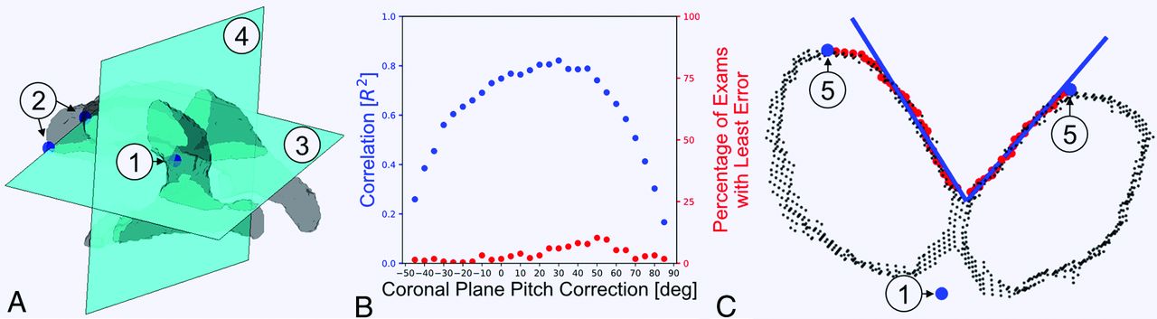
- FIG 1.
CA algorithm. CA measurements are automatically calculated with the following approach. A, Ventricles are segmented in FreeSurfer, and 3 reference points are calculated: the centroid of the extracted ventricles (1) and the most anterior points to the left and right of the centroid (2). These 3 points are used to calculate the axial reference plane (3) and the coronal reference plane (4), shown here without pitch correction. B, The coronal plane pitch correction is optimized by finding the angle that maximizes the correlation of manual and automated CA measurements (30°). For each pitch correction, the percentage of examinations with the least error is also shown. C, The pitch-corrected coronal section is analyzed by finding the most superior point to the left and right of the centroid (5). A greedy pathfinding algorithm connects the 2 superior reference points (5) to identify the medial walls of the lateral ventricles (red points). The inferior 20% and superior 20% of red points are excluded, selecting the middle 60% for angle calculation; this was found empirically to exclude the portions of the ventricle walls with higher curvature. A first-order polynomial fit produces the fit lines (blue lines), and the angle between them is calculated.
- FIG 2.
Measurement variability. A, We found a slight bias (−3.78°) with 95% limits of agreement of −14.95° to +7.39° between 2 radiologists. This translates to an intraclass correlation coefficient of 0.97, demonstrating good reproducibility of the callosal angle biomarker between 2 readers. B, Comparison of manual and linearly corrected automatic CA measurements for n = 281 images had 95% limits of agreement of about ±22° and an ICC of 0.90. We observed lower agreement between manual and automatic measurements than between the 2 neuroradiologists, which highlights the impact of differences in measurement methods.
- FIG 3.
Histogram of coefficients of variation for 906 patients. The variation of automatic CA measurements is calculated for patients with 3–8 separate MR imaging acquisitions. The median coefficient of variation is 4.2%. This is noteworthy because it means that the CA measurement is highly reproducible in a large, real-world sample of MR imaging examinations.
- FIG 4.
Histogram of automatically measured CAs. Angles are measured from 5264 MR images from 1856 patients. The median CA is 113° (Q1 = 101°, Q3 = 123°). The distribution has an acute skewness of −1.048 and excess kurtosis of 0.456. Suggested thresholds for suspected normal pressure hydrocephalus of 90°, 90.8°, and 100° are shown, and we note that 12.4%, 13.0%, and 23.5% of images have CAs narrower than the corresponding cutoffs.


Jump to section
Related Articles
Cited By...
- Automated Detection of Normal Pressure Hydrocephalus Using CT Imaging for Calculating the Ventricle-to-Subarachnoid Volume Ratio
- Automated Idiopathic Normal Pressure Hydrocephalus Diagnosis via Artificial Intelligence-Based 3D T1 MRI Volumetric Analysis
- Prediction of Surgical Outcomes in Normal Pressure Hydrocephalus by MR Elastography
- Callosal Angle Narrowing in Research Data Bases of the Cognitively Impaired