Advertisement

Main menu

  • Home
  • Content
    • Current Issue
    • Accepted Manuscripts
    • Article Preview
    • Past Issue Archive
    • Video Articles
    • AJNR Case Collection
    • Case of the Week Archive
    • Case of the Month Archive
    • Classic Case Archive
  • Special Collections
    • AJNR Awards
    • Low-Field MRI
    • Alzheimer Disease
    • ASNR Foundation Special Collection
    • Photon-Counting CT
    • View All
  • Multimedia
    • AJNR Podcasts
    • AJNR SCANtastic
    • Trainee Corner
    • MRI Safety Corner
    • Imaging Protocols
  • For Authors
    • Submit a Manuscript
    • Submit a Video Article
    • Submit an eLetter to the Editor/Response
    • Manuscript Submission Guidelines
    • Statistical Tips
    • Fast Publishing of Accepted Manuscripts
    • Graphical Abstract Preparation
    • Imaging Protocol Submission
    • Author Policies
  • About Us
    • About AJNR
    • Editorial Board
    • Editorial Board Alumni
  • More
    • Become a Reviewer/Academy of Reviewers
    • Subscribers
    • Permissions
    • Alerts
    • Feedback
    • Advertisers
    • ASNR Home

User menu

  • Alerts
  • Log in

Search

  • Advanced search
American Journal of Neuroradiology

American Journal of Neuroradiology

ASHNR American Society of Functional Neuroradiology ASHNR American Society of Pediatric Neuroradiology ASSR
  • Alerts
  • Log in

Advanced Search

  • Home
  • Content
    • Current Issue
    • Accepted Manuscripts
    • Article Preview
    • Past Issue Archive
    • Video Articles
    • AJNR Case Collection
    • Case of the Week Archive
    • Case of the Month Archive
    • Classic Case Archive
  • Special Collections
    • AJNR Awards
    • Low-Field MRI
    • Alzheimer Disease
    • ASNR Foundation Special Collection
    • Photon-Counting CT
    • View All
  • Multimedia
    • AJNR Podcasts
    • AJNR SCANtastic
    • Trainee Corner
    • MRI Safety Corner
    • Imaging Protocols
  • For Authors
    • Submit a Manuscript
    • Submit a Video Article
    • Submit an eLetter to the Editor/Response
    • Manuscript Submission Guidelines
    • Statistical Tips
    • Fast Publishing of Accepted Manuscripts
    • Graphical Abstract Preparation
    • Imaging Protocol Submission
    • Author Policies
  • About Us
    • About AJNR
    • Editorial Board
    • Editorial Board Alumni
  • More
    • Become a Reviewer/Academy of Reviewers
    • Subscribers
    • Permissions
    • Alerts
    • Feedback
    • Advertisers
    • ASNR Home
  • Follow AJNR on Twitter
  • Visit AJNR on Facebook
  • Follow AJNR on Instagram
  • Join AJNR on LinkedIn
  • RSS Feeds

AJNR Awards, New Junior Editors, and more. Read the latest AJNR updates

Research ArticleADULT BRAIN
Open Access

Comparison of Hippocampal Subfield Segmentation Agreement between 2 Automated Protocols across the Adult Life Span

A. Samara, C.A. Raji, Z. Li and T. Hershey
American Journal of Neuroradiology August 2021, DOI: https://doi.org/10.3174/ajnr.A7244
A. Samara
aFrom the Department of Psychiatry (A.S., Z.L., T.H.)
  • Find this author on Google Scholar
  • Find this author on PubMed
  • Search for this author on this site
  • ORCID record for A. Samara
C.A. Raji
bMallinckrodt Institute of Radiology (C.A.R., T.H.)
cDepartments of Neurology (C.A.R., T.H.)
  • Find this author on Google Scholar
  • Find this author on PubMed
  • Search for this author on this site
  • ORCID record for C.A. Raji
Z. Li
aFrom the Department of Psychiatry (A.S., Z.L., T.H.)
dPsychological and Brain Sciences (Z.L.), Washington University School of Medicine, St. Louis, Missouri
  • Find this author on Google Scholar
  • Find this author on PubMed
  • Search for this author on this site
  • ORCID record for Z. Li
T. Hershey
aFrom the Department of Psychiatry (A.S., Z.L., T.H.)
bMallinckrodt Institute of Radiology (C.A.R., T.H.)
cDepartments of Neurology (C.A.R., T.H.)
  • Find this author on Google Scholar
  • Find this author on PubMed
  • Search for this author on this site
  • ORCID record for T. Hershey
  • Article
  • Figures & Data
  • Supplemental
  • Info & Metrics
  • Responses
  • References
  • PDF
Loading

Article Figures & Data

Figures

  • Tables
  • FIG 1.
    • Download figure
    • Open in new tab
    • Download powerpoint
    FIG 1.

    An example of hippocampal subfield segmentation by FreeSurfer (upper row) and volBrain (lower row) shown in axial, coronal, and sagittal sections. GC-ML-DG indicates granule cell and molecular layers of the dentate gyrus; HATA, hippocampus-amygdala transition area.
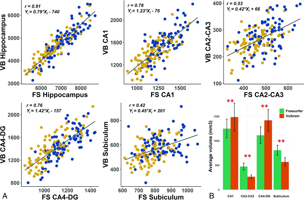
  • FIG 2.
    • Download figure
    • Open in new tab
    • Download powerpoint
    FIG 2.

    A, Comparison of uncorrected total hippocampus, CA1, CA2-CA3, CA4-DG, and subiculum volumes (cubic millimeters) between FreeSurfer and volBrain (yellow, women; blue, men). Regression lines relating volBrain to FreeSurfer volumes are shown for each subfield. The average values of subfield volumes reported here are the sum of right and left hemisphere volumes combined. All Pearson r correlations are significant (P < .001). B, Bar graphs show means (SDs). Double asterisks indicate statistical significance (P < .001); FS, FreeSurfer; VB, volBrain.

  • FIG 3.
    • Download figure
    • Open in new tab
    • Download powerpoint
    FIG 3.

    Bland-Altman plots for uncorrected subfield volumes (CA1, CA2-CA3, CA4-DG, and subiculum volumes [cubic millimeters]) generated by FreeSurfer and volBrain (yellow, women; blue, men). Intrasubject volume difference (y-axis) is defined as (FreeSurfer volume) – (volBrain volume). Mean volume is represented on the x-axis. The mean (SD, 1.96) volume difference and 95% confidence intervals are plotted as dashed horizontal lines. Except for the subiculum, all Pearson r correlations are significant (P < .001).

Tables

  • Figures
  • Uncorrected hippocampal subfield, total hippocampal, and intracranial volumes measured by FreeSurfer and volBrain protocolsa

    Total (n = 172)Male (n = 83)Female (n = 89)Cohen’s D
    MeanSDMeanSDMeanSD
    FreeSurfer v6.0
     CA11251193132116911841590.83
     CA2-CA34727149751449540.91
     CA4-DG1113171117314110571350.84
     Subiculum807102841101775870.7
     Parasubiculum1212112616117210.48
     Presubiculum5537258175527590.8
     Molecular layer1205168124113611721500.48
     HATA1363114219130230.57
     Fimbria1786718862168480.36
     Tail99916010491559521180.7
     Fissureb2697127649262700.23
     THV6839957716384665377580.78
     eICV1,563,261146,9831,677,235158,9671,456,885231,3101.11
    volBrain, HIPS segmentation protocol
     CA11486286157426714052340.67
     CA2-CA32643727643253320.61
     CA4-DG1420221146220713822600.34
     SR-SL-SM9381589881738921450.6
     Subiculum5668759386541930.58
     THV4689771490877644846650.59
     eICV1,374,768133,4671,462,729139,9051,292,671126,1291.28
    • Note:—HATA indicates hippocampus-amygdala transition area; eICV, estimated intracranial volume; HIPS, hippocampal subfield segmentation protocol.

    • ↵a Uuits are cubic millimeters.

    • ↵b Not significantly different. Men had significantly larger values than females in all volumes, except hippocampus fissure (P < .05, Student t test). The average values of total and subfield volumes reported here are the sum of right and left hemispheres combined.

PreviousNext
Back to top
Advertisement
Print
Download PDF
Email Article

Thank you for your interest in spreading the word on American Journal of Neuroradiology.

NOTE: We only request your email address so that the person you are recommending the page to knows that you wanted them to see it, and that it is not junk mail. We do not capture any email address.

Enter multiple addresses on separate lines or separate them with commas.
Comparison of Hippocampal Subfield Segmentation Agreement between 2 Automated Protocols across the Adult Life Span
(Your Name) has sent you a message from American Journal of Neuroradiology
(Your Name) thought you would like to see the American Journal of Neuroradiology web site.
CAPTCHA
This question is for testing whether or not you are a human visitor and to prevent automated spam submissions.
Cite this article
A. Samara, C.A. Raji, Z. Li, T. Hershey
Comparison of Hippocampal Subfield Segmentation Agreement between 2 Automated Protocols across the Adult Life Span
American Journal of Neuroradiology Aug 2021, DOI: 10.3174/ajnr.A7244

Citation Manager Formats

  • BibTeX
  • Bookends
  • EasyBib
  • EndNote (tagged)
  • EndNote 8 (xml)
  • Medlars
  • Mendeley
  • Papers
  • RefWorks Tagged
  • Ref Manager
  • RIS
  • Zotero
0 Responses
Respond to this article
Share
Bookmark this article
Comparison of Hippocampal Subfield Segmentation Agreement between 2 Automated Protocols across the Adult Life Span
A. Samara, C.A. Raji, Z. Li, T. Hershey
American Journal of Neuroradiology Aug 2021, DOI: 10.3174/ajnr.A7244
del.icio.us logo Twitter logo Facebook logo Mendeley logo
  • Tweet Widget
  • Facebook Like
  • Google Plus One
Purchase

Jump to section

  • Article
    • Abstract
    • ABBREVIATIONS:
    • MATERIALS AND METHODS
    • RESULTS
    • DISCUSSION
    • CONCLUSIONS
    • Footnotes
    • References
  • Figures & Data
  • Supplemental
  • Info & Metrics
  • Responses
  • References
  • PDF

Related Articles

  • PubMed
  • Google Scholar

Cited By...

  • No citing articles found.
  • Crossref
  • Google Scholar

This article has not yet been cited by articles in journals that are participating in Crossref Cited-by Linking.

More in this TOC Section

ADULT BRAIN

  • Diagnostic Neuroradiology of Monoclonal Antibodies
  • Cerebral ADC Changes in Fabry Disease
  • ML for Glioma Molecular Subtype Prediction
Show more ADULT BRAIN

FUNCTIONAL

  • Kurtosis and Epileptogenic Tubers: A Pilot Study
  • Glutaric Aciduria Type 1: DK vs. Conventional MRI
  • Multiparametric MRI in PEDS Pontine Glioma
Show more FUNCTIONAL

Similar Articles

Advertisement

Indexed Content

  • Current Issue
  • Accepted Manuscripts
  • Article Preview
  • Past Issues
  • Editorials
  • Editor's Choice
  • Fellows' Journal Club
  • Letters to the Editor
  • Video Articles

Cases

  • Case Collection
  • Archive - Case of the Week
  • Archive - Case of the Month
  • Archive - Classic Case

More from AJNR

  • Trainee Corner
  • Imaging Protocols
  • MRI Safety Corner
  • Book Reviews

Multimedia

  • AJNR Podcasts
  • AJNR Scantastics

Resources

  • Turnaround Time
  • Submit a Manuscript
  • Submit a Video Article
  • Submit an eLetter to the Editor/Response
  • Manuscript Submission Guidelines
  • Statistical Tips
  • Fast Publishing of Accepted Manuscripts
  • Graphical Abstract Preparation
  • Imaging Protocol Submission
  • Evidence-Based Medicine Level Guide
  • Publishing Checklists
  • Author Policies
  • Become a Reviewer/Academy of Reviewers
  • News and Updates

About Us

  • About AJNR
  • Editorial Board
  • Editorial Board Alumni
  • Alerts
  • Permissions
  • Not an AJNR Subscriber? Join Now
  • Advertise with Us
  • Librarian Resources
  • Feedback
  • Terms and Conditions
  • AJNR Editorial Board Alumni

American Society of Neuroradiology

  • Not an ASNR Member? Join Now

© 2025 by the American Society of Neuroradiology All rights, including for text and data mining, AI training, and similar technologies, are reserved.
Print ISSN: 0195-6108 Online ISSN: 1936-959X

Powered by HighWire