
Article Figures & Data
Figures
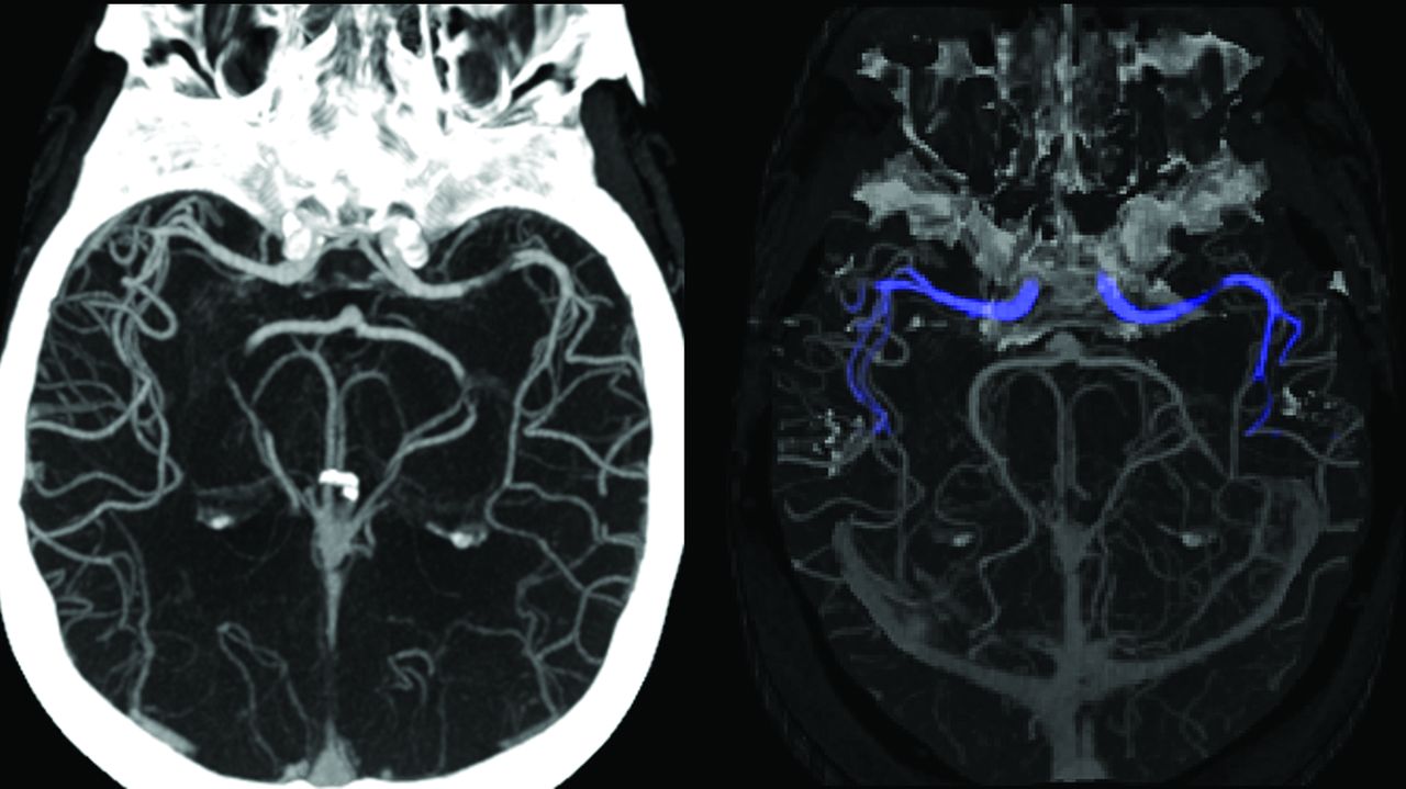
- FIG 1.
Division of the M2 segment of the MCA into proximal and distal segments at the curve of the artery into the Sylvian fissure (marked bilaterally by the dashed lines).
- FIG 2.
Alerts as they appear on the user end of the mobile application, showing the overview screen of examinations with (A) and without (B) a suspected LVO. An overview screen of failed processing is shown in C, in this case, due to metallic artifacts.
- FIG 3.
Flow diagram delineating the various steps of the algorithm. App indicates mobile application.
- FIG 4.
Overview of the algorithm steps. A, Identification of an applicable scan based on metadata. B, Cropping the head region. Registration (C) and segmentation (D) of ICA-T/M1 regions. E, Additional segmentation of all vessels. Refinement of the segmentations to include only the MCA branches (F) and detection of suspected LVO based on vessel length (G).
- FIG 5.
Algorithm processing of a partial occlusion. The cropped scan on the left visualizes a left M1 partial occlusion. The segmentation (on the right) extends through the partial occlusion. However, the average Hounsfield unit value decreases and then increases and a notification is triggered, even though the length of the segmentation exceeds the threshold.
- FIG 6.
System identification illustration demonstrates stenosis of the M1 segment of the left MCA (A), occlusion of the M1 segment of the left MCA (C), and occlusion of the proximal M2 segment of the right MCA (E), as they appear as preliminary convolutional neural network outcomes (green boxes represent original annotations by the Viz LVO system during identification). The images on the lower row (B, D, and F, respectively) match processed images sent by the system via the application and received by the viewer during an alert.
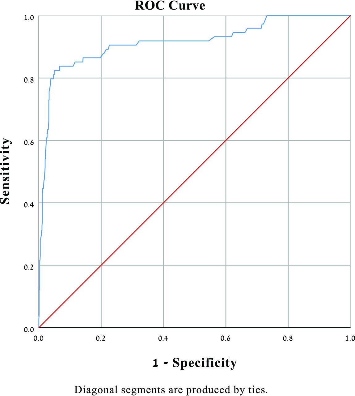
- FIG 7.
Prediction of LVO logistic regression (adjusted for age and sex). The area under the curve is shown to be 0.91. ROC indicates receiver operating characteristic.


Tables
Patients (n = 1167) Age (mean) [SD] 62.2 19.6 Male 689 59 Stenosis (50%>) 66 5.7 Extracranial ICA 43 3.7 Intracranial 23 2.0 Stroke protocol 404 34.6 Hemorrhage 80 6.8 Tumor 12 1.0 LVO 75 6.4 LVO location (n = 75) Carotid terminus 28 37.3 M1 47 62.6 Distal occlusion (non-LVO) (n = 44) Proximal M2 21 47.7 Distal M2–3 23 52.3 ↵a Data are number and percentage unless otherwise indicated.
Pathology No. % Stenosis (>50%) 9 16.1 Distal occlusions 12 21.4 Proximal M2 8 14.3 Distal M2/M3 4 7.14 Hemorrhage 12 21.4 Tumor 4 7.14 No revealed pathology 19 33.9 Overall 56 100 - Table 3:
Prediction of LVO by the Viz LVO system—logistic regression (adjusted for age and sex)
Variable OR SE Sig 95% CI Lower Upper Suspected LVO 51.75 0.298 .000 28.84 92.84 Age 1.030 0.009 .001 1.013 1.048 Sex 1.474 0.295 .188 0.828 2.626 Note:—SE indicates standard error; Sig, significance.
System LVO Detection Sensitivity 95% CI Specificity 95% CI NPV 95% CI PPV 95% CI Accuracy 95% CI Entire cohort (n = 1167) 0.81 0.74–0.91 0.96 0.95–0.97 0.99 0.98–0.99 0.65 0.55–0.74 0.94 0.92–0.96 Stroke protocol subgroup (n = 404) 0.82 0.71–0.89 0.90 0.86–0.93 0.96 0.93–0.98 0.64 0.53–0.73 0.89 0.86–0.94 Note:—NPV indicates negative predictive value.


Jump to section
Related Articles
Cited By...
- Correspondence on: 'Viz LVO versus Rapid LVO in detection of large vessel occlusion on CT angiography for acute stroke by Delora et al
- Workflow improvements from automated large vessel occlusion detection algorithms are dependent on care team engagement
- Automated detection of large vessel occlusion using deep learning: a pivotal multicenter study and reader performance study
- Viz LVO versus Rapid LVO in detection of large vessel occlusion on CT angiography for acute stroke
- Automated detection of large vessel occlusion using deep learning: a pivotal multicenter clinical trial and reader assessment study
- The Impact of Artificial Intelligence on Large Vessel Occlusion Stroke Detection and Management: A Systematic Review Meta-analysis
- Excellence is a habit: Enhancing predictions of language impairment by identifying stable features in clinical perfusion scans
- Machine learning and acute stroke imaging
- JNIS spotlight: commissioned reviews
- AI software detection of large vessel occlusion stroke on CT angiography: a real-world prospective diagnostic test accuracy study
- Diagnostic performance of an algorithm for automated large vessel occlusion detection on CT angiography
- Automated emergent large vessel occlusion detection by artificial intelligence improves stroke workflow in a hub and spoke stroke system of care
- The True Potential of Artificial Intelligence for Detection of Large-Vessel Occlusion: The Role of M2 Occlusions