Advertisement

Main menu

  • Home
  • Content
    • Current Issue
    • Accepted Manuscripts
    • Article Preview
    • Past Issue Archive
    • Video Articles
    • AJNR Case Collection
    • Case of the Week Archive
    • Case of the Month Archive
    • Classic Case Archive
  • Special Collections
    • Advancing NeuroMRI with High-Relaxivity Contrast Agents
    • AJNR Awards
    • Low-Field MRI
    • Alzheimer Disease
    • ASNR Foundation Special Collection
    • Photon-Counting CT
    • View All
  • Multimedia
    • AJNR Podcasts
    • AJNR SCANtastic
    • Trainee Corner
    • MRI Safety Corner
    • Imaging Protocols
  • For Authors
    • Submit a Manuscript
    • Submit a Video Article
    • Submit an eLetter to the Editor/Response
    • Manuscript Submission Guidelines
    • Statistical Tips
    • Fast Publishing of Accepted Manuscripts
    • Graphical Abstract Preparation
    • Imaging Protocol Submission
    • Author Policies
  • About Us
    • About AJNR
    • Editorial Board
    • Editorial Board Alumni
  • More
    • Become a Reviewer/Academy of Reviewers
    • Subscribers
    • Permissions
    • Alerts
    • Feedback
    • Advertisers
    • ASNR Home

User menu

  • Alerts
  • Log in

Search

  • Advanced search
American Journal of Neuroradiology

American Journal of Neuroradiology

ASHNR American Society of Functional Neuroradiology ASHNR American Society of Pediatric Neuroradiology ASSR
  • Alerts
  • Log in

Advanced Search

  • Home
  • Content
    • Current Issue
    • Accepted Manuscripts
    • Article Preview
    • Past Issue Archive
    • Video Articles
    • AJNR Case Collection
    • Case of the Week Archive
    • Case of the Month Archive
    • Classic Case Archive
  • Special Collections
    • Advancing NeuroMRI with High-Relaxivity Contrast Agents
    • AJNR Awards
    • Low-Field MRI
    • Alzheimer Disease
    • ASNR Foundation Special Collection
    • Photon-Counting CT
    • View All
  • Multimedia
    • AJNR Podcasts
    • AJNR SCANtastic
    • Trainee Corner
    • MRI Safety Corner
    • Imaging Protocols
  • For Authors
    • Submit a Manuscript
    • Submit a Video Article
    • Submit an eLetter to the Editor/Response
    • Manuscript Submission Guidelines
    • Statistical Tips
    • Fast Publishing of Accepted Manuscripts
    • Graphical Abstract Preparation
    • Imaging Protocol Submission
    • Author Policies
  • About Us
    • About AJNR
    • Editorial Board
    • Editorial Board Alumni
  • More
    • Become a Reviewer/Academy of Reviewers
    • Subscribers
    • Permissions
    • Alerts
    • Feedback
    • Advertisers
    • ASNR Home
  • Follow AJNR on Twitter
  • Visit AJNR on Facebook
  • Follow AJNR on Instagram
  • Join AJNR on LinkedIn
  • RSS Feeds

AJNR Awards, New Junior Editors, and more. Read the latest AJNR updates


Improved Turnaround Times | Median time to first decision: 12 days

Research ArticleRADIOLOGY-PATHOLOGY CORRELATION

Optic Nerve Choristoma Mimicking a Neurenteric Cyst

J.C. Benson, C. Giannini, S. Cohen Cohen, J. Van Gompel, D.K. Kim, J. Port, F. Diehn, L. Eckel and C. Carr
American Journal of Neuroradiology December 2020, DOI: https://doi.org/10.3174/ajnr.A6892
J.C. Benson
aFrom the Department of Radiology (J.C.B., D.K.K., J.P., F.D., L.E., C.C.)
  • Find this author on Google Scholar
  • Find this author on PubMed
  • Search for this author on this site
  • ORCID record for J.C. Benson
C. Giannini
bPathology (C.G.), Mayo Clinic, Rochester, Minnesota
  • Find this author on Google Scholar
  • Find this author on PubMed
  • Search for this author on this site
  • ORCID record for C. Giannini
S. Cohen Cohen
cNeurosurgery (S.C.C., J.V.G.), Mayo Clinic, Rochester, Minnesota
  • Find this author on Google Scholar
  • Find this author on PubMed
  • Search for this author on this site
  • ORCID record for S. Cohen Cohen
J. Van Gompel
cNeurosurgery (S.C.C., J.V.G.), Mayo Clinic, Rochester, Minnesota
  • Find this author on Google Scholar
  • Find this author on PubMed
  • Search for this author on this site
  • ORCID record for J. Van Gompel
D.K. Kim
aFrom the Department of Radiology (J.C.B., D.K.K., J.P., F.D., L.E., C.C.)
  • Find this author on Google Scholar
  • Find this author on PubMed
  • Search for this author on this site
  • ORCID record for D.K. Kim
J. Port
aFrom the Department of Radiology (J.C.B., D.K.K., J.P., F.D., L.E., C.C.)
  • Find this author on Google Scholar
  • Find this author on PubMed
  • Search for this author on this site
  • ORCID record for J. Port
F. Diehn
aFrom the Department of Radiology (J.C.B., D.K.K., J.P., F.D., L.E., C.C.)
  • Find this author on Google Scholar
  • Find this author on PubMed
  • Search for this author on this site
  • ORCID record for F. Diehn
L. Eckel
aFrom the Department of Radiology (J.C.B., D.K.K., J.P., F.D., L.E., C.C.)
  • Find this author on Google Scholar
  • Find this author on PubMed
  • Search for this author on this site
  • ORCID record for L. Eckel
C. Carr
aFrom the Department of Radiology (J.C.B., D.K.K., J.P., F.D., L.E., C.C.)
  • Find this author on Google Scholar
  • Find this author on PubMed
  • Search for this author on this site
  • ORCID record for C. Carr
  • Article
  • Figures & Data
  • Info & Metrics
  • Responses
  • References
  • PDF
Loading

Article Figures & Data

Figures

  • FIG 1.
    • Download figure
    • Open in new tab
    • Download powerpoint
    FIG 1.

    Preoperative CT and MR images of the lesion. Axial CT (A), axial (B) and sagittal (C) MPRAGE, and axial T2 Cube (GE Healthcare) (D), T2-FLAIR (E), and DWI (F) demonstrate an extra-axial cystic lesion in the anterior cranial fossa (between arrows, A). The cyst drapes around the anterior falx and causes substantial mass effect on the adjacent bifrontal gyri. The optic chiasm (curved arrow, C) and pituitary gland (dashed arrow, C) are deviated posteriorly. The intracystic fluid signal is slightly hyperintense to CSF on T2 Cube, does not suppress on FLAIR, and does not restrict on DWI.
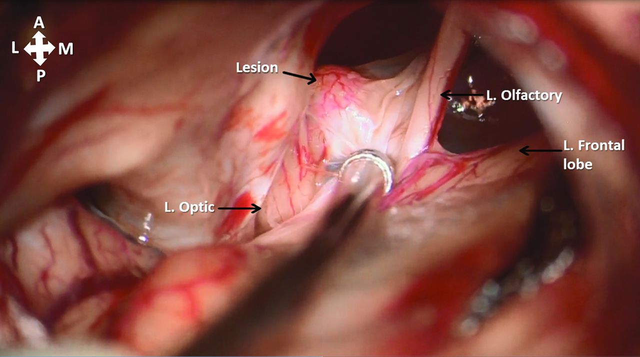
  • FIG 2.
    • Download figure
    • Open in new tab
    • Download powerpoint
    FIG 2.

    Intraoperative view of a left frontal craniotomy with a subfrontal approach for resection of an anterior cranial fossa cyst. A granular lesion adhering to the medial surface of the left optic nerve is shown, with adjacent structures labeled. L. indicates left.

  • FIG 3.
    • Download figure
    • Open in new tab
    • Download powerpoint
    FIG 3.

    Re-review of preoperative coronal MPRAGE (A) and postcontrast (B) images demonstrates a T1-hyperintense focus along the left optic nerve (arrows), correlating with the surgical findings. Intrinsic T1 signal was presumably related to the intralesional fat tissue noted on subsequent histologic analysis. Although faint enhancement was seen in the adjacent tissue, it was not certain whether a component of this solid focus demonstrated definite enhancement (B).

  • FIG 4.
    • Download figure
    • Open in new tab
    • Download powerpoint
    FIG 4.

    Optic nerve choristoma composed of adipose tissue, glandular lobules, and ducts (A), with predominant mucinous (B) and scant serous (C) glands consistent with salivary gland tissue. The duct (D) shows a typical basal cell layer (black arrow) and columnar lining.

  • FIG 5.
    • Download figure
    • Open in new tab
    • Download powerpoint
    FIG 5.

    The collapsed cyst wall shows largely a flattened lining (A and B) bistratified positive for CAM5.2 (C). p63-positivity is limited to the basal cells of the bistratified epithelium lining the cyst wall as typically seen in duct structures (D). BRAF V600E, a marker of papillary craniopharyngioma, is negative (E); the stain is also typically positive in normal cilia, which were not present in the cyst lining. The cyst lining shows normal cytoplasmic beta-catenin expression (F) and also of epithelial membrane antigen (EMA) (G), while progesterone receptor (PR) stain is present in a subset of the arachnoidal cells observed in the fibrous stroma associated with the cyst wall (J).

PreviousNext
Back to top
Advertisement
Print
Download PDF
Email Article

Thank you for your interest in spreading the word on American Journal of Neuroradiology.

NOTE: We only request your email address so that the person you are recommending the page to knows that you wanted them to see it, and that it is not junk mail. We do not capture any email address.

Enter multiple addresses on separate lines or separate them with commas.
Optic Nerve Choristoma Mimicking a Neurenteric Cyst
(Your Name) has sent you a message from American Journal of Neuroradiology
(Your Name) thought you would like to see the American Journal of Neuroradiology web site.
CAPTCHA
This question is for testing whether or not you are a human visitor and to prevent automated spam submissions.
Cite this article
J.C. Benson, C. Giannini, S. Cohen Cohen, J. Van Gompel, D.K. Kim, J. Port, F. Diehn, L. Eckel, C. Carr
Optic Nerve Choristoma Mimicking a Neurenteric Cyst
American Journal of Neuroradiology Dec 2020, DOI: 10.3174/ajnr.A6892

Citation Manager Formats

  • BibTeX
  • Bookends
  • EasyBib
  • EndNote (tagged)
  • EndNote 8 (xml)
  • Medlars
  • Mendeley
  • Papers
  • RefWorks Tagged
  • Ref Manager
  • RIS
  • Zotero
0 Responses
Respond to this article
Share
Bookmark this article
Optic Nerve Choristoma Mimicking a Neurenteric Cyst
J.C. Benson, C. Giannini, S. Cohen Cohen, J. Van Gompel, D.K. Kim, J. Port, F. Diehn, L. Eckel, C. Carr
American Journal of Neuroradiology Dec 2020, DOI: 10.3174/ajnr.A6892
del.icio.us logo Twitter logo Facebook logo Mendeley logo
  • Tweet Widget
  • Facebook Like
  • Google Plus One
Purchase

Jump to section

  • Article
    • Abstract
    • DISCUSSION
    • Footnotes
    • References
  • Figures & Data
  • Info & Metrics
  • Responses
  • References
  • PDF

Related Articles

  • No related articles found.
  • PubMed
  • Google Scholar

Cited By...

  • No citing articles found.
  • Crossref
  • Google Scholar

This article has not yet been cited by articles in journals that are participating in Crossref Cited-by Linking.

More in this TOC Section

RADIOLOGY-PATHOLOGY CORRELATION

  • Histopathological Analysis of Five Patients with CSF-Venous Fistulas After Surgical Nerve Root Ligation and Resection
  • Calcified Pseudoneoplasm of the Neuraxis
  • Hypothalamic Pilomyxoid Astrocytoma in a Child with Lipodystrophy
Show more RADIOLOGY-PATHOLOGY CORRELATION

Adult Brain

  • Diagnostic Neuroradiology of Monoclonal Antibodies
  • Clinical Outcomes After Chiari I Decompression
  • Segmentation of Brain Metastases with BLAST
Show more Adult Brain

Similar Articles

Advertisement

Indexed Content

  • Current Issue
  • Accepted Manuscripts
  • Article Preview
  • Past Issues
  • Editorials
  • Editor's Choice
  • Fellows' Journal Club
  • Letters to the Editor
  • Video Articles

Cases

  • Case Collection
  • Archive - Case of the Week
  • Archive - Case of the Month
  • Archive - Classic Case

More from AJNR

  • Trainee Corner
  • Imaging Protocols
  • MRI Safety Corner
  • Book Reviews

Multimedia

  • AJNR Podcasts
  • AJNR Scantastics

Resources

  • Turnaround Time
  • Submit a Manuscript
  • Submit a Video Article
  • Submit an eLetter to the Editor/Response
  • Manuscript Submission Guidelines
  • Statistical Tips
  • Fast Publishing of Accepted Manuscripts
  • Graphical Abstract Preparation
  • Imaging Protocol Submission
  • Evidence-Based Medicine Level Guide
  • Publishing Checklists
  • Author Policies
  • Become a Reviewer/Academy of Reviewers
  • News and Updates

About Us

  • About AJNR
  • Editorial Board
  • Editorial Board Alumni
  • Alerts
  • Permissions
  • Not an AJNR Subscriber? Join Now
  • Advertise with Us
  • Librarian Resources
  • Feedback
  • Terms and Conditions
  • AJNR Editorial Board Alumni

American Society of Neuroradiology

  • Not an ASNR Member? Join Now

© 2025 by the American Society of Neuroradiology All rights, including for text and data mining, AI training, and similar technologies, are reserved.
Print ISSN: 0195-6108 Online ISSN: 1936-959X

Powered by HighWire