
Article Figures & Data
Figures
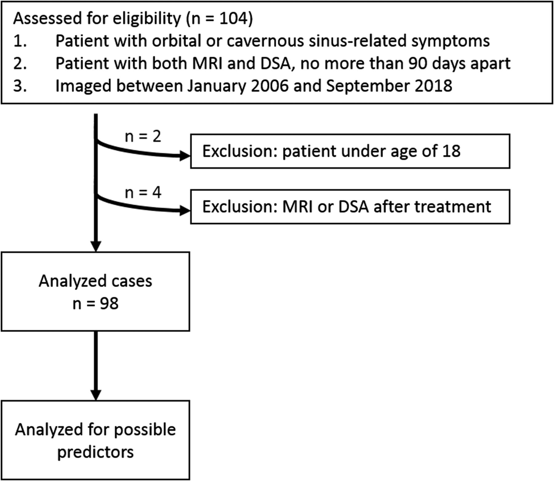
- FIG 1.
Flow diagram of the case selection procedure and case numbers in each subgroup.
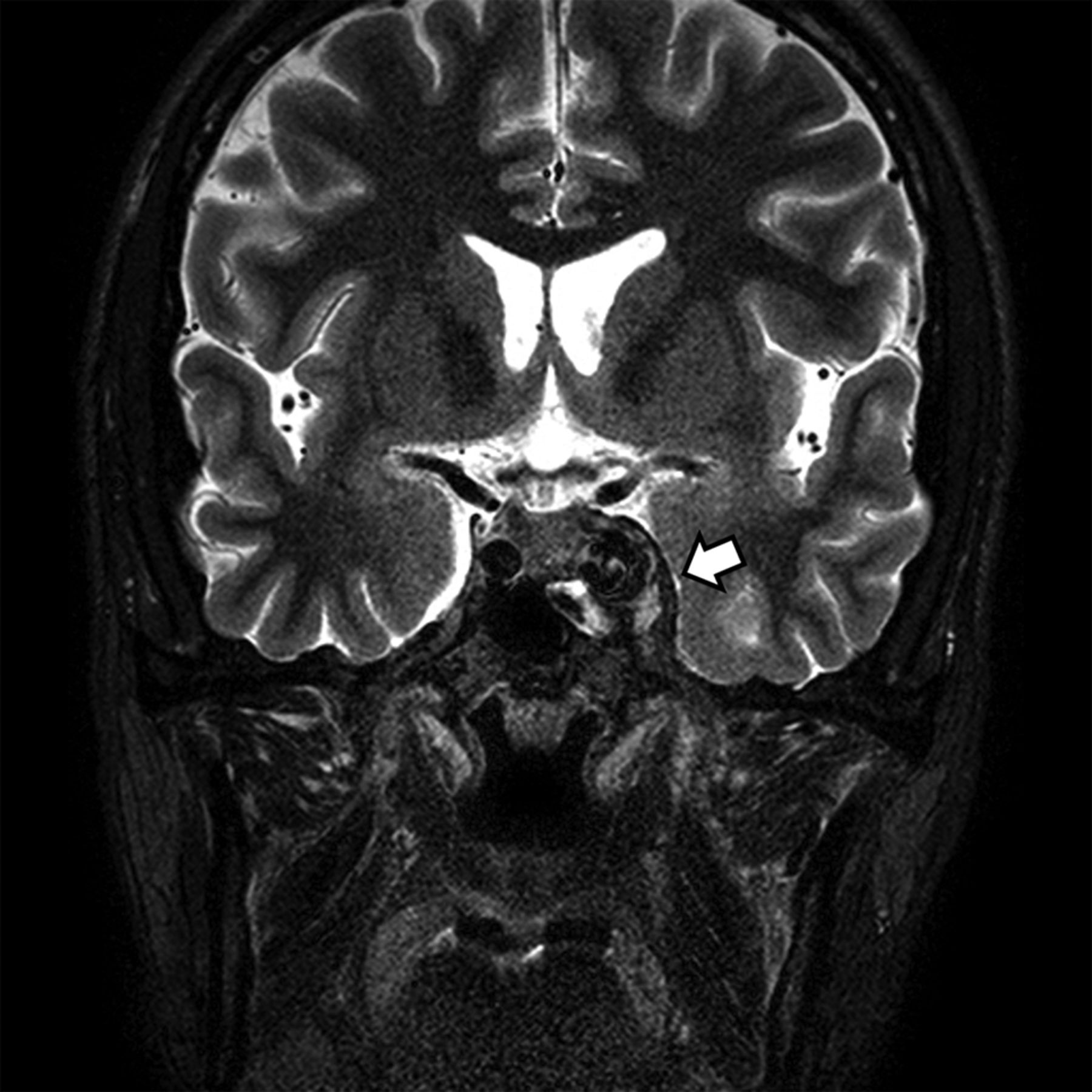
- FIG 2.
A CCF case with abnormal contour of the cavernous sinus. Coronal T2-weighted image of a patient with diplopia, confirmed to be left sixth cranial nerve palsy on neurologic examination. Note the abnormal contour bulging of the left cavernous sinus (arrow). An internal signal void was also noted on both T2-weighted (arrow) and T1-weighted imaging (not shown). The patient was confirmed as having a direct CCF on digital subtraction angiography.
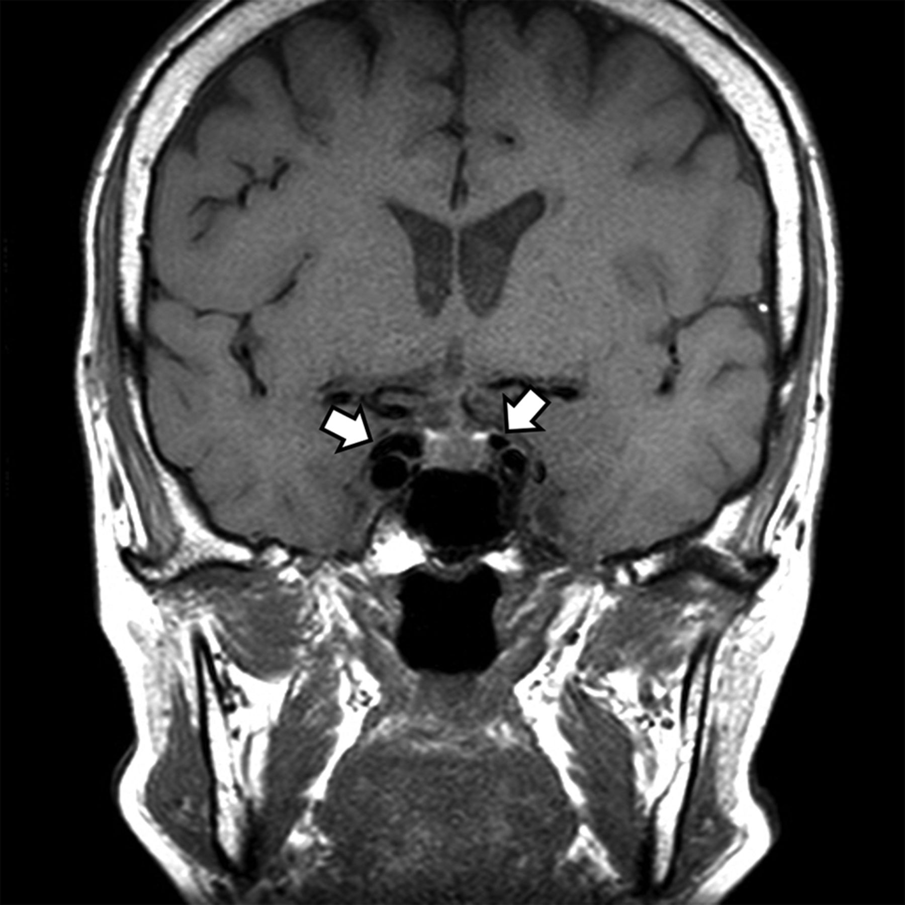
- FIG 3.
A CCF case with internal signal void of the cavernous sinuses. Coronal T1-weighted image of a patient with diplopia, confirmed to be right third cranial nerve palsy on neurologic examination. Note the internal signal void in both cavernous sinuses visible on T1-weighted image (arrows). The patient was confirmed to have an indirect CCF on digital subtraction angiography.
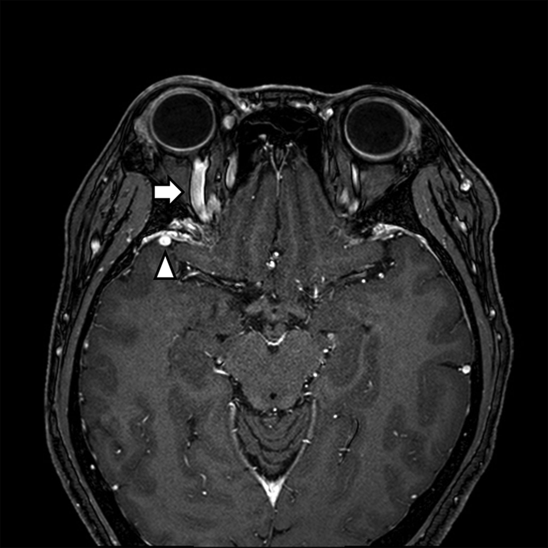
- FIG 4.
A CCF case with prominent venous drainage flow in the anterior and lateral venous structures. Axial contrast-enhanced T1-weighted image of a patient with right ocular pain and conjunctival injection. Note the enlarged right superior ophthalmic vein (anterior; arrow) and right sphenoparietal sinus (lateral; arrowhead). The patient was confirmed as having an indirect CCF on digital subtraction angiography.
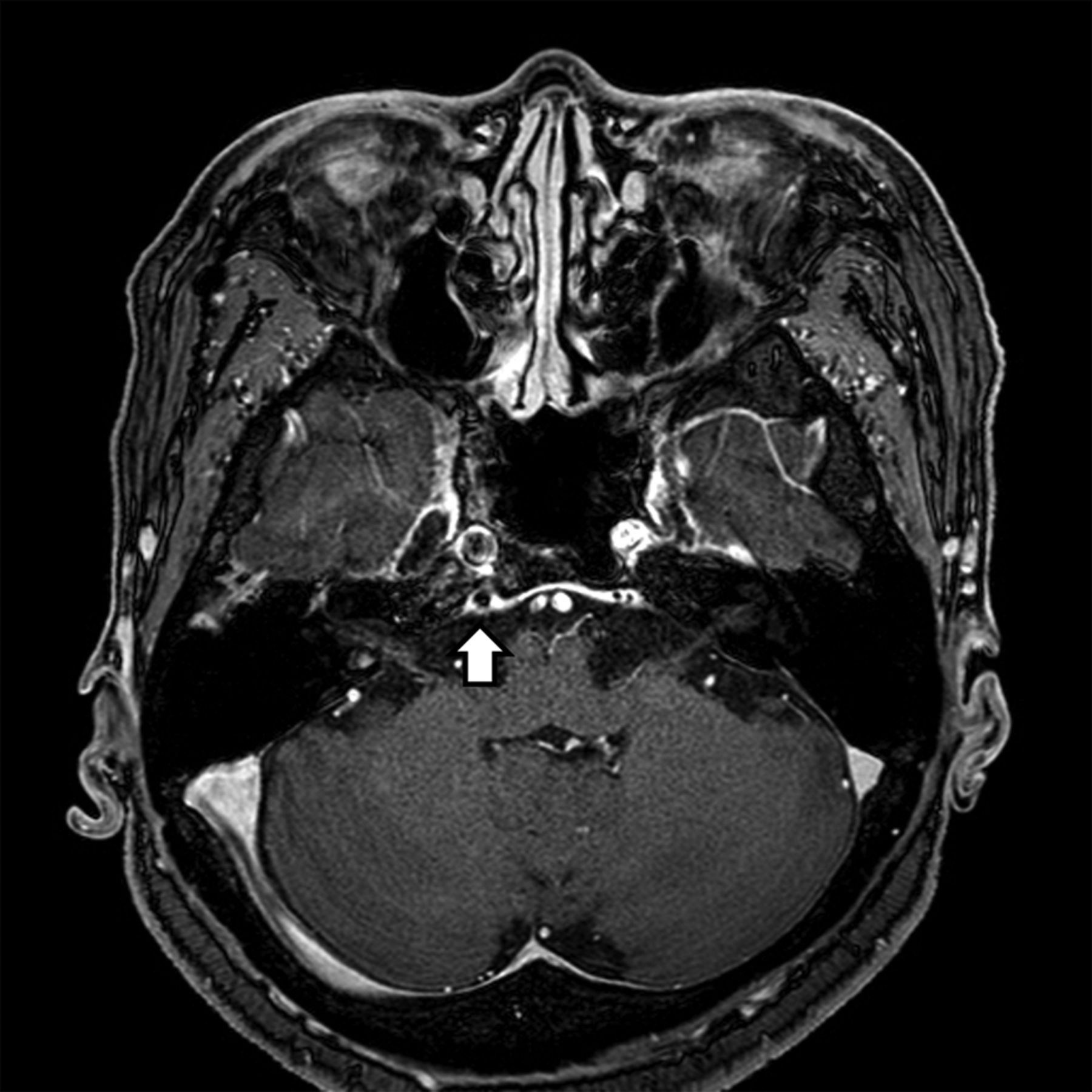
- FIG 5.
A CCF case with prominent venous drainage flow in the posterior venous structure. Axial contrast-enhanced T1-weighted image of a patient with diplopia, confirmed to be right sixth cranial nerve palsy on neurologic examination. Note the enlarged right inferior petrosal sinus (posterior) with an internal signal void (arrow) indicating increased flow rate. The patient was confirmed as having an indirect CCF on digital subtraction angiography.
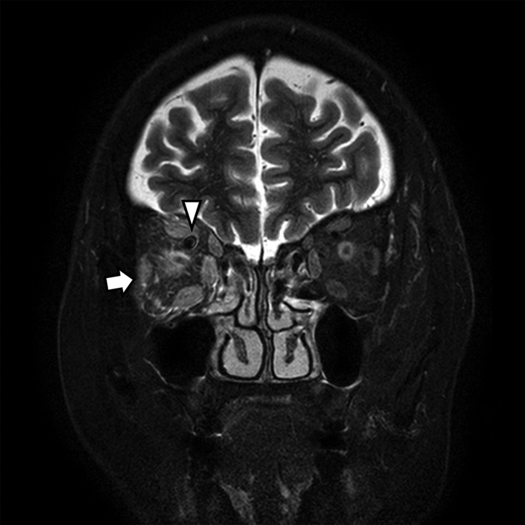
- FIG 6.
A CCF case with high signal change and orbital soft tissue thickening. Coronal T2-weighted image of a patient with periorbital swelling, conjunctival injection, ocular pain, and diplopia. Fat stranding and swelling of extraocular muscles (arrow) are noted. Prominent venous drainage flow in the superior ophthalmic vein is also noted (arrowhead). The patient was confirmed as having an indirect CCF on digital subtraction angiography.


Tables
All Patients(n = 98) CCF Positive Patients (n = 38) CCF Negative Patients (n = 60) Sex, n Male/Female 32/66 11/27 21/39 Age, years Mean (range) 54.6 (20–85) 66.0 (24–85) 51.4 (20–81) Symptoms, n (%) Diplopia 54 (55.1) 20 (52.6) 34 (56.7) Eyeball pain 27 (27.6) 14 (36.8) 13 (21.7) Facial pain 2 (2.0) 1 (2.6) 1 (1.7) Ptosis 30 (30.6) 8 (21.1) 22 (36.7) Proptosis 14 (14.3) 9 (23.7) 5 (8.3) Periorbital swelling 19 (19.4) 16 (42.1) 3 (5) Conjunctival injection 23 (23.5) 16 (42.1) 7 (11.7) Visual disturbance 18 (18.4) 7 (18.4) 11 (18.3) Headache 40 (40.8) 14 (36.8) 26 (43.3) Dizziness 13 (13.3) 5 (13.2) 8 (13.3) Neurologic signs, n (%)Laterality Right 30 (30.6) 8 (21.1) 22 (36.7) Left 22 (22.4) 6 (15.8) 16 (26.7) 3rd cranial neve palsy 26 (26.5) 4 (10.5) 22 (36.7) 4th cranial nerve palsy 5 (5.1) 2 (5.3) 3 (5) 6th cranial nerve palsy 22 (22.4) 8 (21.1) 14 (23.3) Trauma history, n (%) 5 (5.1) 3 (7.9) 2 (3.3) Feature Total CCF Cases Univariable Logistic Regression OR (95% CI) P Value Abnormal contour of cavernous sinus 57.1% (56/98) 92.1% (35/38) 21.7 (6.0–78.9) <.001 Signal void of cavernous sinus 73.5% (72/98) 92.1% (35/38) 15.3 (4.2–55.1) <.001 Prominent venous drainage flowa 52.0% (51/98) 94.7% (36/38) 54.0 (11.6–251.7) <.001 Anterior 36.7% (36/98) 78.9% (30/38) 33.8 (10.7–106.5) <.001 Lateral 22.4% (22/98) 34.2% (13/38) 3.0 (1.1–7.8) .030 Posterior 23.5% (23/98) 47.4% (18/38) 9.9 (3.3–30.2) <.001 Orbital/periorbital soft tissue swelling 39.8% (39/98) 84.2% (32/38) 40.4 (12.5–130.8) <.001 ↵a Prominent venous drainage flow indicates the presence of prominent venous drainage flow in at least 1 of the anterior, lateral, and posterior prominent venous drainage flows.
- Table 3:
Diagnostic performance of each thin-section MR imaging feature and combination of imaging features for CCFa
Accuracy Sensitivity Specificity PLR NLR Abnormal contour of cavernous sinus 75.5% (65.8–83.6%) 92.1% (78.6–98.3) 65% (51.6–76.9) 2.6 (1.8–3.8) 0.1 (0.0–0.4) Signal void of cavernous sinus 70.4% (60.7–78.5%) 92.1% (79.2–97.3) 56.7% (44.1–68.4) 2.1 (1.6–2.9) 0.1 (0.0–0.4) Any prominent venous drainage flow 82.7% (74.0–88.9) 94.7% (82.7–98.5) 75% (62.8–84.2) 3.8 (2.4–5.9) 0.1 (0.0–0.3) Prominent anterior venous drainage flow 85.7% (77.4–91.3) 79.0% (63.7–88.9) 90% (79.9–95.3) 7.9 (3.6–7.2) 0.2 (0.1–0.4) Prominent lateral venous drainage flow 65.3% (55.0–74.6) 34.2% (19.6–51.2) 85% (73.4–92.9) 2.3 (1.1–4.8) 0.8 (0.6–1.0) Prominent posterior venous drainage flow 74.5% (64.7–82.8) 47.4% (31.0–64.2) 91.7% (81.6–97.2) 5.7 (2.3–14.0) 0.6 (0.4–0.8) Orbital/periorbital soft tissue swelling 86.7% (78.6–92.1) 84.2% (69.6–92.6) 88.3% (77.8–94.2) 7.2 (3.6–14.7) 0.2 (0.1–0.4) Combination 1 (any prominent venous drainage flow AND internal signal void of cavernous sinus) 91.8% (84.7–95.8) 89.5% (75.9–95.8) 93.3% (84.1–97.4) 13.4 (5.2–34.8) 0.1 (0.0–0.3) Combination 2 (any prominent venous drainage flow AND orbital/periorbital soft tissue swelling) 89.8% (82.2–94.4) 79.0% (63.7–88.9) 96.7% (88.6–99.1) 23.7 (6.0–93.4) 0.2 (0.1–0.4) Combination 3 (prominent anterior venous drainage flow OR orbital/periorbital soft tissue swelling) 85.7% (77.4–91.3) 92.1% (79.2–97.3) 81.7% (70.1–89.4) 5.0 (2.9–8.6) 0.1 (0.0–0.3) Note:—PLR indicates positive likelihood ratio; NLR, negative likelihood ratio.
↵a Data in parentheses are 95% confidence intervals.


Jump to section
Related Articles
Cited By...
- No citing articles found.