Advertisement

Main menu

  • Home
  • Content
    • Current Issue
    • Accepted Manuscripts
    • Article Preview
    • Past Issue Archive
    • Video Articles
    • AJNR Case Collection
    • Case of the Week Archive
    • Case of the Month Archive
    • Classic Case Archive
  • Special Collections
    • AJNR Awards
    • Low-Field MRI
    • Alzheimer Disease
    • ASNR Foundation Special Collection
    • Photon-Counting CT
    • View All
  • Multimedia
    • AJNR Podcasts
    • AJNR SCANtastic
    • Trainee Corner
    • MRI Safety Corner
    • Imaging Protocols
  • For Authors
    • Submit a Manuscript
    • Submit a Video Article
    • Submit an eLetter to the Editor/Response
    • Manuscript Submission Guidelines
    • Statistical Tips
    • Fast Publishing of Accepted Manuscripts
    • Graphical Abstract Preparation
    • Imaging Protocol Submission
    • Author Policies
  • About Us
    • About AJNR
    • Editorial Board
    • Editorial Board Alumni
  • More
    • Become a Reviewer/Academy of Reviewers
    • Subscribers
    • Permissions
    • Alerts
    • Feedback
    • Advertisers
    • ASNR Home

User menu

  • Alerts
  • Log in

Search

  • Advanced search
American Journal of Neuroradiology

American Journal of Neuroradiology

ASHNR American Society of Functional Neuroradiology ASHNR American Society of Pediatric Neuroradiology ASSR
  • Alerts
  • Log in

Advanced Search

  • Home
  • Content
    • Current Issue
    • Accepted Manuscripts
    • Article Preview
    • Past Issue Archive
    • Video Articles
    • AJNR Case Collection
    • Case of the Week Archive
    • Case of the Month Archive
    • Classic Case Archive
  • Special Collections
    • AJNR Awards
    • Low-Field MRI
    • Alzheimer Disease
    • ASNR Foundation Special Collection
    • Photon-Counting CT
    • View All
  • Multimedia
    • AJNR Podcasts
    • AJNR SCANtastic
    • Trainee Corner
    • MRI Safety Corner
    • Imaging Protocols
  • For Authors
    • Submit a Manuscript
    • Submit a Video Article
    • Submit an eLetter to the Editor/Response
    • Manuscript Submission Guidelines
    • Statistical Tips
    • Fast Publishing of Accepted Manuscripts
    • Graphical Abstract Preparation
    • Imaging Protocol Submission
    • Author Policies
  • About Us
    • About AJNR
    • Editorial Board
    • Editorial Board Alumni
  • More
    • Become a Reviewer/Academy of Reviewers
    • Subscribers
    • Permissions
    • Alerts
    • Feedback
    • Advertisers
    • ASNR Home
  • Follow AJNR on Twitter
  • Visit AJNR on Facebook
  • Follow AJNR on Instagram
  • Join AJNR on LinkedIn
  • RSS Feeds

AJNR Awards, New Junior Editors, and more. Read the latest AJNR updates

Research ArticleArtificial Intelligence

Synthesizing Contrast-Enhanced MR Images from Noncontrast MR Images Using Deep Learning

Gowtham Murugesan, Fang F. Yu, Michael Achilleos, John DeBevits, Sahil Nalawade, Chandan Ganesh, Ben Wagner, Ananth J Madhuranthakam and Joseph A. Maldjian
American Journal of Neuroradiology March 2024, 45 (3) 312-319; DOI: https://doi.org/10.3174/ajnr.A8107
Gowtham Murugesan
aDepartment of Radiology, University of Texas Southwestern Medical Center, Dallas, Texas
  • Find this author on Google Scholar
  • Find this author on PubMed
  • Search for this author on this site
Fang F. Yu
aDepartment of Radiology, University of Texas Southwestern Medical Center, Dallas, Texas
  • Find this author on Google Scholar
  • Find this author on PubMed
  • Search for this author on this site
  • ORCID record for Fang F. Yu
Michael Achilleos
aDepartment of Radiology, University of Texas Southwestern Medical Center, Dallas, Texas
  • Find this author on Google Scholar
  • Find this author on PubMed
  • Search for this author on this site
  • ORCID record for Michael Achilleos
John DeBevits
aDepartment of Radiology, University of Texas Southwestern Medical Center, Dallas, Texas
  • Find this author on Google Scholar
  • Find this author on PubMed
  • Search for this author on this site
  • ORCID record for John DeBevits
Sahil Nalawade
aDepartment of Radiology, University of Texas Southwestern Medical Center, Dallas, Texas
  • Find this author on Google Scholar
  • Find this author on PubMed
  • Search for this author on this site
  • ORCID record for Sahil Nalawade
Chandan Ganesh
aDepartment of Radiology, University of Texas Southwestern Medical Center, Dallas, Texas
  • Find this author on Google Scholar
  • Find this author on PubMed
  • Search for this author on this site
  • ORCID record for Chandan Ganesh
Ben Wagner
aDepartment of Radiology, University of Texas Southwestern Medical Center, Dallas, Texas
  • Find this author on Google Scholar
  • Find this author on PubMed
  • Search for this author on this site
  • ORCID record for Ben Wagner
Ananth J Madhuranthakam
aDepartment of Radiology, University of Texas Southwestern Medical Center, Dallas, Texas
  • Find this author on Google Scholar
  • Find this author on PubMed
  • Search for this author on this site
  • ORCID record for Ananth J Madhuranthakam
Joseph A. Maldjian
aDepartment of Radiology, University of Texas Southwestern Medical Center, Dallas, Texas
  • Find this author on Google Scholar
  • Find this author on PubMed
  • Search for this author on this site
  • ORCID record for Joseph A. Maldjian
  • Article
  • Figures & Data
  • Info & Metrics
  • Responses
  • References
  • PDF
Loading

Article Figures & Data

Figures

  • Tables
  • FIG 1.
    • Download figure
    • Open in new tab
    • Download powerpoint
    FIG 1.

    Residual inception DenseNet (RID). A, RID model for virtual contrast enhancement (vT1c prediction) and enhancing tumor (ET) segmentation. B, RID model for whole tumor (WT) segmentation.

  • FIG 2.
    • Download figure
    • Open in new tab
    • Download powerpoint
    FIG 2.

    Residual inception DenseNet (RID). A, RID model for whole tumor segmentation. B, RID model for virtual contrast enhancement and enhancing tumor segmentation.

  • FIG 3.
    • Download figure
    • Open in new tab
    • Download powerpoint
    FIG 3.

    Building blocks of residual inception network. From left to right, dense block, convolution block, transition block, and projection block.

  • FIG 4.
    • Download figure
    • Open in new tab
    • Download powerpoint
    FIG 4.

    Synthesized virtual contrast enhanced T1w (vT1c) images in 3 different subjects. Ground truth (left column) and synthesized vT1c (right column) image pairs for 9 subjects.
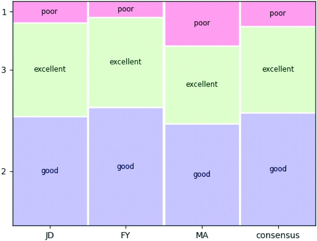
  • FIG 5.
    • Download figure
    • Open in new tab
    • Download powerpoint
    FIG 5.

    Mosaic plot illustrating the distribution of 3 expert radiologists and their consensus along a 3-point Likert scale.
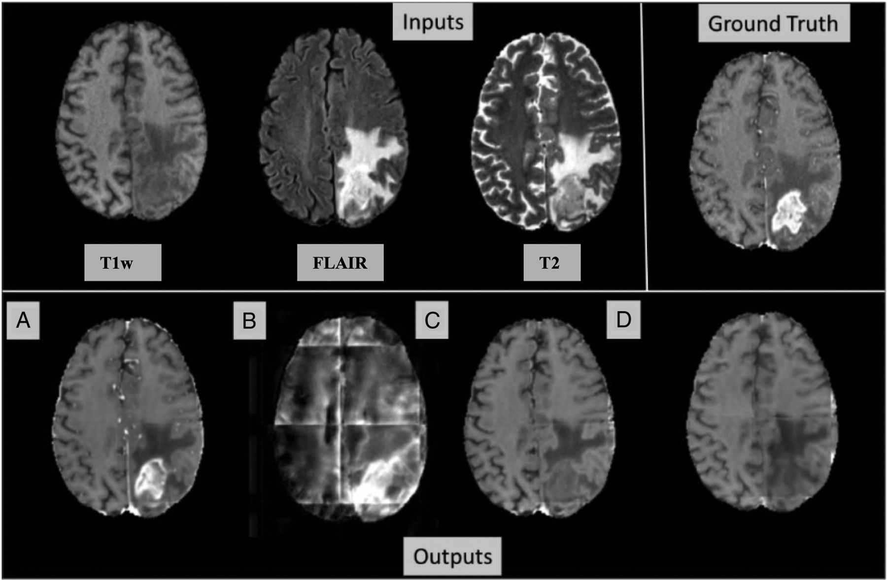
  • FIG 6.
    • Download figure
    • Open in new tab
    • Download powerpoint
    FIG 6.

    Importance of input sequences example. Top row, input images: T1w, FLAIR, T2, and the ground truth T1c. Bottom row, output images with (A) all inputs (T1w, FLAIR, and T2w) given to the model, (B) T1w replaced with zeros in the input, (C) FLAIR replaced with zeros in the input, and (D) T2 replaced with zeros in the input. The T2 and FLAIR inputs together provide contrast enhancement prediction, whereas T1w input provides primarily anatomic detail.

Tables

  • Figures
    • View popup
    Table 1:

    Quantitative evaluation. Analysis of virtual enhancement prediction by using various masks generated by an external model

    MaskSSIMNMSEDicePSNR
    Whole brain0.910.030.3264.35
    Whole tumor0.900.010.3548.99
    Enhancing tumor0.900.010.6249.93
    • View popup
    Table 2:

    Quantitative presence and location of the under/overestimation of synthetic contrast enhancement, the introduction of artifacts, and the image quality improvement on vT1c

     Reviewer 1 (FY)Reviewer 2 (MA)Reviewer 3 (JD)
    Overestimate (O)262720
    Underestimate (U)716958
    Both (O and U)11834
    Central543564
    Peripheral947197
    False distant enhancement10915
    Missed distant enhancement542
    Artifact172225
    Image quality improved10NA3
    • Note:—NA indicates not applicable.

PreviousNext
Back to top

In this issue

American Journal of Neuroradiology: 45 (3)
American Journal of Neuroradiology
Vol. 45, Issue 3
1 Mar 2024
  • Table of Contents
  • Index by author
  • Complete Issue (PDF)
Advertisement
Print
Download PDF
Email Article

Thank you for your interest in spreading the word on American Journal of Neuroradiology.

NOTE: We only request your email address so that the person you are recommending the page to knows that you wanted them to see it, and that it is not junk mail. We do not capture any email address.

Enter multiple addresses on separate lines or separate them with commas.
Synthesizing Contrast-Enhanced MR Images from Noncontrast MR Images Using Deep Learning
(Your Name) has sent you a message from American Journal of Neuroradiology
(Your Name) thought you would like to see the American Journal of Neuroradiology web site.
CAPTCHA
This question is for testing whether or not you are a human visitor and to prevent automated spam submissions.
Cite this article
Gowtham Murugesan, Fang F. Yu, Michael Achilleos, John DeBevits, Sahil Nalawade, Chandan Ganesh, Ben Wagner, Ananth J Madhuranthakam, Joseph A. Maldjian
Synthesizing Contrast-Enhanced MR Images from Noncontrast MR Images Using Deep Learning
American Journal of Neuroradiology Mar 2024, 45 (3) 312-319; DOI: 10.3174/ajnr.A8107

Citation Manager Formats

  • BibTeX
  • Bookends
  • EasyBib
  • EndNote (tagged)
  • EndNote 8 (xml)
  • Medlars
  • Mendeley
  • Papers
  • RefWorks Tagged
  • Ref Manager
  • RIS
  • Zotero
0 Responses
Respond to this article
Share
Bookmark this article
Synthesizing Contrast-Enhanced MR Images via DL
Gowtham Murugesan, Fang F. Yu, Michael Achilleos, John DeBevits, Sahil Nalawade, Chandan Ganesh, Ben Wagner, Ananth J Madhuranthakam, Joseph A. Maldjian
American Journal of Neuroradiology Mar 2024, 45 (3) 312-319; DOI: 10.3174/ajnr.A8107
del.icio.us logo Twitter logo Facebook logo Mendeley logo
  • Tweet Widget
  • Facebook Like
  • Google Plus One
Purchase

Jump to section

  • Article
    • Abstract
    • ABBREVIATIONS:
    • MATERIALS AND METHODS
    • RESULTS
    • DISCUSSION
    • CONCLUSIONS
    • Footnotes
    • References
  • Figures & Data
  • Info & Metrics
  • Responses
  • References
  • PDF

Related Articles

  • No related articles found.
  • PubMed
  • Google Scholar

Cited By...

  • No citing articles found.
  • Crossref
  • Google Scholar

This article has not yet been cited by articles in journals that are participating in Crossref Cited-by Linking.

More in this TOC Section

  • Large Language Models in Radiology
  • Improving Hematoma Expansion Prediction Robustness
  • MRI-Based Models for Diagnosis of Brain Lesions
Show more Artificial Intelligence

Similar Articles

Advertisement

Indexed Content

  • Current Issue
  • Accepted Manuscripts
  • Article Preview
  • Past Issues
  • Editorials
  • Editor's Choice
  • Fellows' Journal Club
  • Letters to the Editor
  • Video Articles

Cases

  • Case Collection
  • Archive - Case of the Week
  • Archive - Case of the Month
  • Archive - Classic Case

More from AJNR

  • Trainee Corner
  • Imaging Protocols
  • MRI Safety Corner
  • Book Reviews

Multimedia

  • AJNR Podcasts
  • AJNR Scantastics

Resources

  • Turnaround Time
  • Submit a Manuscript
  • Submit a Video Article
  • Submit an eLetter to the Editor/Response
  • Manuscript Submission Guidelines
  • Statistical Tips
  • Fast Publishing of Accepted Manuscripts
  • Graphical Abstract Preparation
  • Imaging Protocol Submission
  • Evidence-Based Medicine Level Guide
  • Publishing Checklists
  • Author Policies
  • Become a Reviewer/Academy of Reviewers
  • News and Updates

About Us

  • About AJNR
  • Editorial Board
  • Editorial Board Alumni
  • Alerts
  • Permissions
  • Not an AJNR Subscriber? Join Now
  • Advertise with Us
  • Librarian Resources
  • Feedback
  • Terms and Conditions
  • AJNR Editorial Board Alumni

American Society of Neuroradiology

  • Not an ASNR Member? Join Now

© 2025 by the American Society of Neuroradiology All rights, including for text and data mining, AI training, and similar technologies, are reserved.
Print ISSN: 0195-6108 Online ISSN: 1936-959X

Powered by HighWire