
Article Figures & Data
Figures
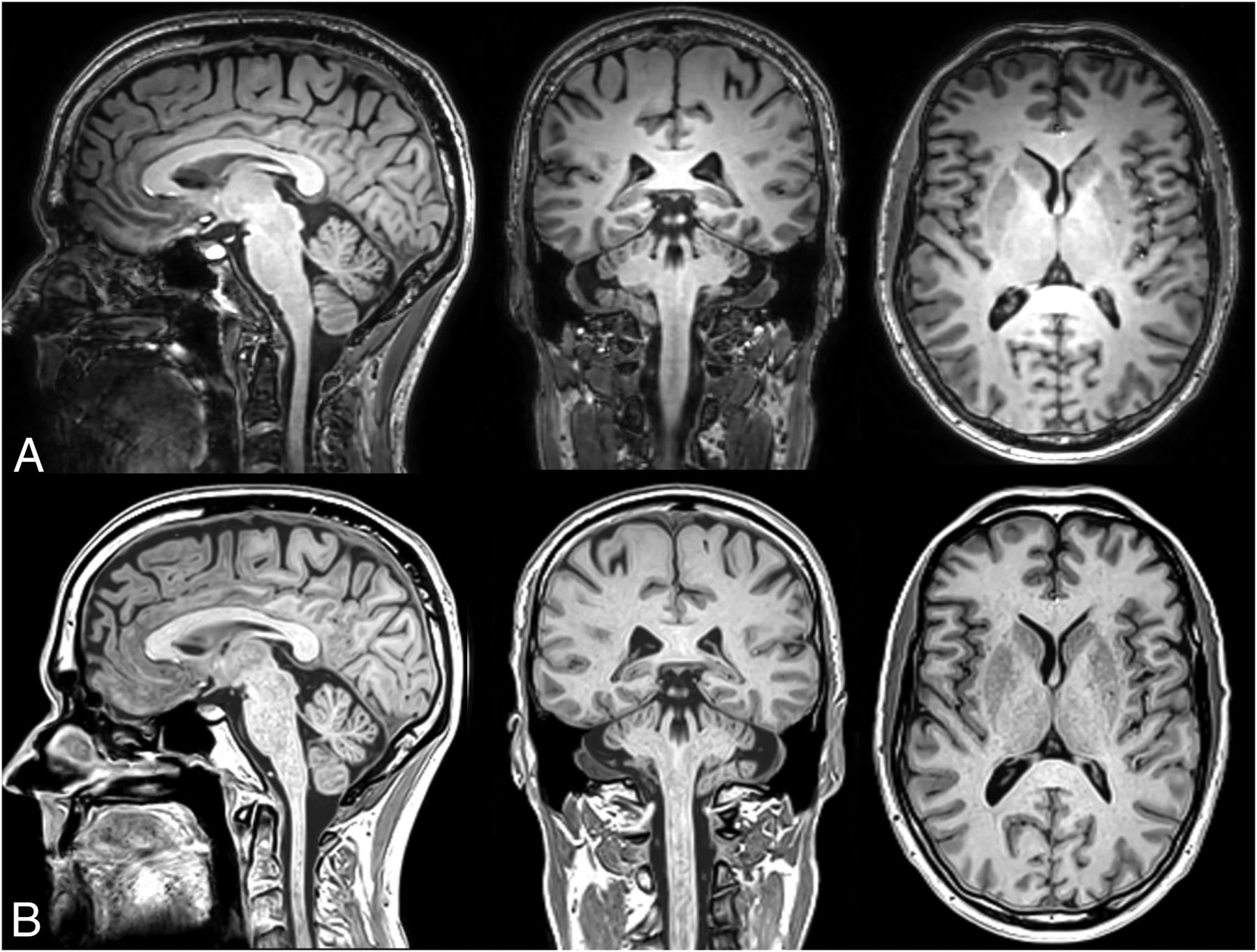
- FIG 1.
Representative example of a sagittal, coronal, and axial reformat of the 3D-T1-weighted image stack at 1.2-mm isotropic resolution at a scan time of 6 minutes 10 seconds. The subject is a man, 54 years of age. Upper row (A), conventional T1-weighted NeuroQuant sequence. Lower row (B), synthetic T1-weighted image, created from the R1, R2, and PD maps (SyMRI 22Q2).
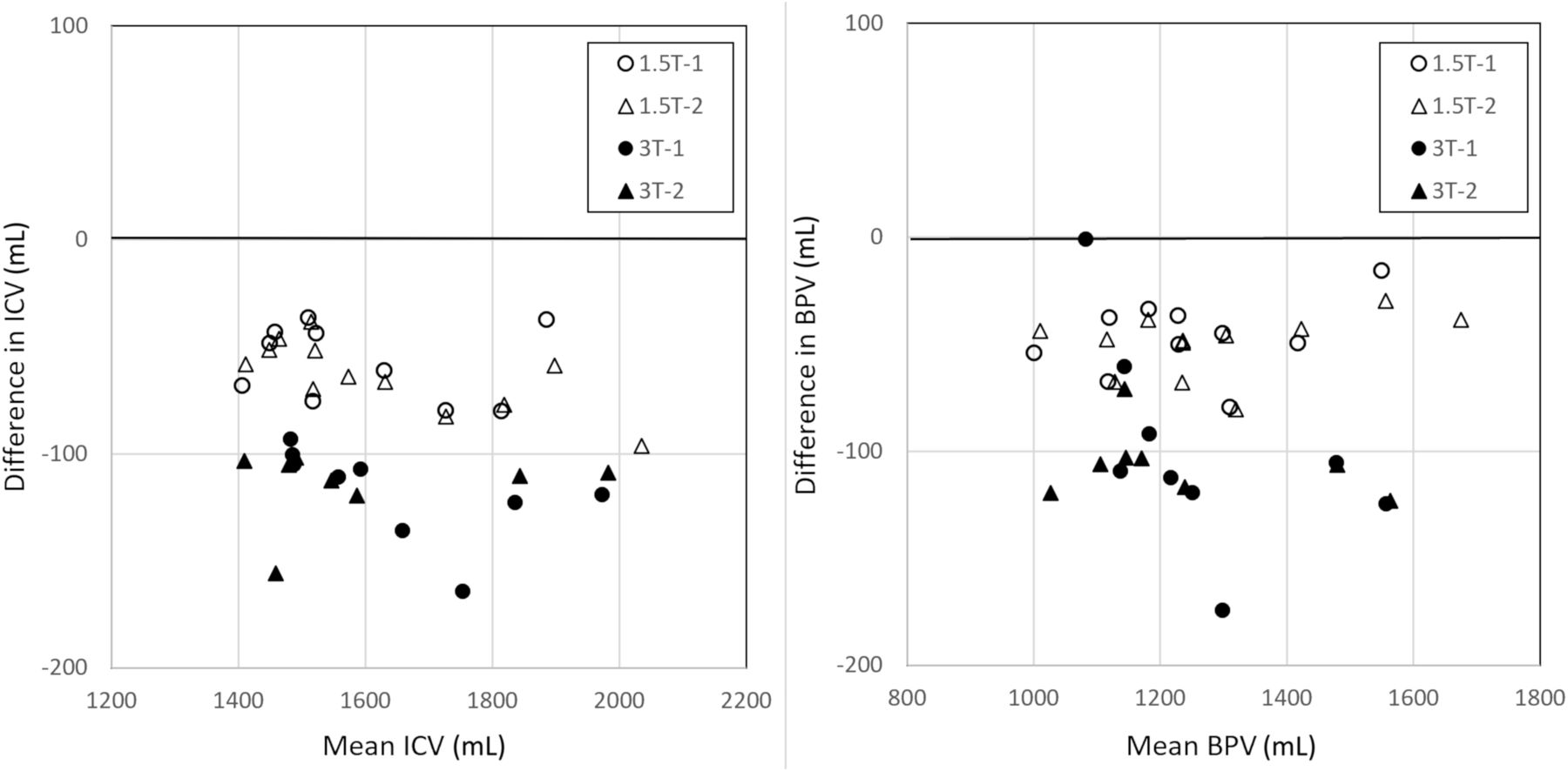
- FIG 2.
Bland-Atman plot with NQ segmentation results for ICV and BPV, in which T1-weighted and syT1WI were pooled as input data to focus on reproducibility at 2 different field strengths, irrespective of acquisition type. Measurements 1 (circles) and 2 (triangles) were plotted separately for both field strengths. There is a substantial bias, with lower ICV and BPV when using syT1WI. The bias is larger at 3T then at 1.5T.
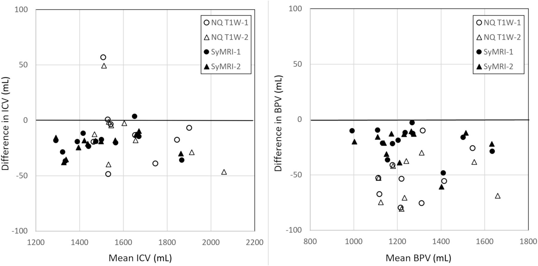
- FIG 3.
Bland-Atman plot with segmentation results for ICV and BPV in which data from 1.5T and 3T were pooled to focus on the reproducibility for the 2 different acquisition methods, irrespective of field strength NQ T1-weighted imaging and SyMRI. Measurements 1 (circles) and 2 (triangles) are plotted separately for both methods. There is a bias, with lower ICV and BPV when using 3T. The bias is largest for the BPV using NQ T1-weighted imaging.


Tables
- Table 1:
Reproducibility of volume estimation of various brain structures using NeuroQuant with either a conventional or synthetic T1-weighted image stack as inputa
syT1WI–T1-Weighted NQ 1.5T syT1WI–T1-Weighted NQ 3T Mean (mL) Bias (mL) SD (mL) r Mean (mL) Bias (mL) SD (mL) r Forebrain parenchyma 1084 −37 14 1.00 1041 −80 34 0.98 Cortical gray matter 506 8.3 14 0.98 492 −25 17 0.97 Superior lateral ventricle 29 −2.2 1.4 0.99 27 0.8 3.7 0.84 Inferior lateral ventricle 1.7 0.0 0.2 0.97 1.7 −0.1 0.3 0.78 Hippocampus 7.5 −0.8b 0.3 0.97 7.4 −0.7b 0.3 0.94 Amygdala 3.3 −0.2 0.2 0.95 3.5 −0.6b 0.2 0.84 Caudate 6.0 0.1 0.5 0.87 6.0 −0.3 0.6 0.83 Putamen 11 −2.1b 0.5 0.94 11 −2.2b 0.6 0.87 Pallidum 1.4 0.1 0.2 0.84 1.7 −0.1 0.1 0.90 Thalamus 15 −0.3 0.8 0.90 15 −2.1b 0.6 0.96 Cerebellum 145 −7.3 2.3 0.98 141 −16b 4.2 0.90 ICV 1619 −61b 17 1.00 1610 −116b 19 0.99 BPV 1273 −49b 16 1.00 1226 −103b 36 0.98 WMV 651 −50 22 0.97 622 −82 64 0.98 GMV 621 1 13 0.99 608 −62 72 0.97 CSFV 346 −13 20 0.93 384 −10 25 0.82 - Table 2:
Repeatability of volume estimation of various brain structures using NeuroQuant with either a conventional or synthetic T1-weighted image stack as inputa
T1-Weighted 1.5T syT1WI 1.5T T1-Weighted 3T syT1WI 3T Forebrain parenchyma 0.6 0.7 0.6 4.0 Cortical gray matter 1.2 1.0 1.5 4.4 Superior lateral ventricle 1.1 1.2 1.0 18.2 Inferior lateral ventricle 5.6 4.6 3.5 21.3 Hippocampus 1.2 1.2 2.4 3.8 Amygdala 3.6 2.3 4.0 5.2 Caudate 4.0 2.0 2.9 6.4 Putamen 2.6 1.3 1.6 6.2 Pallidum 12.8 5.3 8.8 6.3 Thalamus 3.4 2.9 3.3 6.1 Cerebellum 0.7 0.9 1.0 2.9 ICV 0.4 0.3 0.3 1.5 BPV 0.5 0.6 0.5 3.7 WMV 1.2 0.7 1.0 3.7 GMV 0.9 0.9 1.2 4.0 CSFV 2.1 2.4 1.6 6.0 Note:—WMV indicates white matter volume; GMV, gray matter volume; CSFV, CSF volume.
↵a Expressed as coefficient of variation (%), at 1.5T and 3T.
- Table 3:
Reproducibility of volume estimation of WM, GM, CSF, BPV, and ICV using NeuroQuant and SyMRI, between 1.5T and 3Ta
T1-Weighted NQ 1.5T-3T syT1WI NQ 1.5T-3T SyMRI 1.5T-3T Mean Bias SD r Mean Bias SD r Mean Bias SD r WM 661 27b 65 0.83 613 58b 23 0.97 547 0 39 0.89 GM 622 −6 58 0.83 609 55b 14 0.98 669 −7 33 0.95 CSF 368 –41b 34 0.76 359 –45b 22 0.88 236 1 16 0.97 BPV 1284 21 119 0.84 1223 113b 31 0.98 1258 21b 13 1.00 ICV 1652 −20 120 0.86 1581 68b 20 0.99 1494 21b 9 1.00 - Table 4:
Repeatability of the volume estimation of WM, GM, CSF, BPV, and ICV using NeuroQuant and SyMRIa
T1-Weighted NQ 1.5T syT1WI NQ 1.5T SyMRI 1.5T T1-Weighted NQ 3T syT1WI NQ 3T SyMRI 3T WM 1.2 0.7 4.8 1.0 3.7 4.4 GM 0.9 0.9 4.6 1.2 4.0 4.4 CSF 2.1 2.4 3.5 1.6 6.0 3.3 BPV 0.5 0.6 0.7 0.5 3.7 0.6 ICV 0.4 0.3 0.4 0.3 1.5 0.1 ↵a Expressed as CoV for 1.5T and 3T.


Jump to section
Related Articles
Cited By...
- No citing articles found.