
Article Figures & Data
Figures
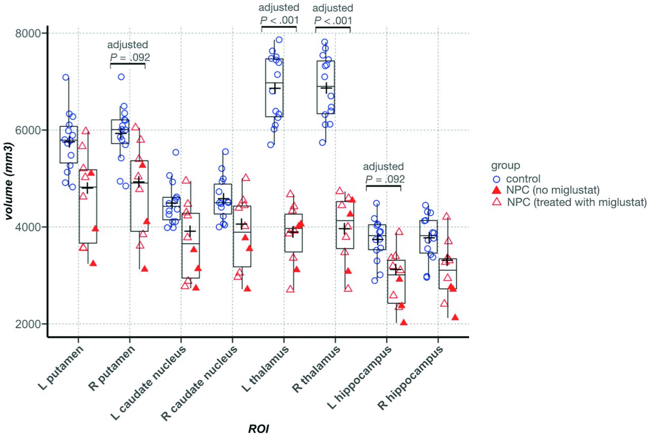
- FIG 1.
Between-group comparison of volume in the ROIs. In the Tukey boxplots, the middle line of each box indicates the median, upper and lower boundaries of the boxes show the upper and lower quartiles, respectively; the upper and lower whiskers show the maximum and minimum data points; and the cross signs indicate the mean in each group. Filled triangles represent data points from patients with NPC who were not taking miglustat. Error bars indicate SDs. L indicates left; R, right.
- FIG 2.
Voxelwise between-group comparison of QSM. A, Lightbox axial view with 4-mm section spacing. The yellow-red spectrum indicates clusters with higher QSM, and the blue-to-light blue spectrum shows clusters with lower QSM values in the NPC group compared with healthy controls. B, A visualization of the thalamus with clusters that show significant between-group QSM difference. Red and blue indicate higher and lower QSM in the NPC group compared with the control group, respectively. Clusters with increased QSM in NPC, depicted in red, are in the posterior part of the thalamus, consistent with the anatomic location of the pulvinar nucleus.
- FIG 3.
Representative QSM images from control and NPC groups. Colored lines mark the borders of segmentations labels: green, putamen; brown, caudate nucleus; red, thalamus; blue, globus pallidus.
- FIG 4.
ROI-based between-group comparison of mean QSM values. In the Tukey boxplots, the middle line of each box indicates the median, upper, and lower boundaries of the boxes and shows the upper and lower quartiles, respectively; the upper and lower whiskers show the maximum and minimum data points; and the cross signs indicate the mean in each group. Filled triangles represent data points from patients with NPC who were not taking miglustat. Error bars indicate SDs in the entire control and NPC groups. In the thalamus, patients with NPC who were not taking miglustat had higher mean QSM values than those who were prescribed miglustat. L indicates left; R, right.
- FIG 5.
Pearson correlation between volume and QSM in the brain regions that showed a difference in volume or QSM values between groups. Cells containing correlation coefficients are color-coded according to the value of the correlation coefficient. Blue indicates negative correlation, and red indicates positive correlation. Color intensities correspond to the value of the coefficient.


Tables
Control (n = 14) NPC (n = 10) P Value Age (mean) (SD) (yr) 32.6 (9.2) 33.1 (12.1) .92 Sex (male/female) 6:8 4:6 .89 Iturriaga score (mean) (SD) NA 9.1 (3) NA NUCOG (mean) (SD) NA 63.9 (18) NA SOFAS (mean) (SD) NA 42 (22.7) NA Miglustat dose NA 7 Patients taking 200 mg TID NA Note:—NA indicates not applicable; TID, 3 times per day.
- Table 2:
ROI-based between-group comparison of mean QSM values with age and ROI volume as covariates
ROI Control (Mean) (SD) (ppb) NPC (Mean) (SD) (ppb) FDR-Adjusted P Value Effect Size (η2p) Putamen Left 17 (5) 17.01 (9) .56 0.03 Right 15.6 (3.5) 15.8 (7.4) .79 0.003 Caudate Left 22.6 (3.4) 22 (7.7) .56 0.02 Right 22.2 (4.3) 20.4 (5.9) .63 0.01 Globus pallidus Left 81.3 (20.8) 95.1 (23) .56 0.06 Right 84.9 (16.6) 102.7 (19.5) .51 0.12 Thalamus Left 2.4 (4) 2.2 (6.1) .31 0.1 Right 1.5 (3.5) 1 (6.2) .56 0.04 Hippocampus Left –1.6 (2.2) –8.2 (3.6) .10 0.3 Right –1.9 (2.1) –5.7 (3.3) .04 0.2 Note:—ppb indicates parts per billion; FDR, false detection rate


Jump to section
Related Articles
Cited By...
- No citing articles found.