
Article Figures & Data
Figures
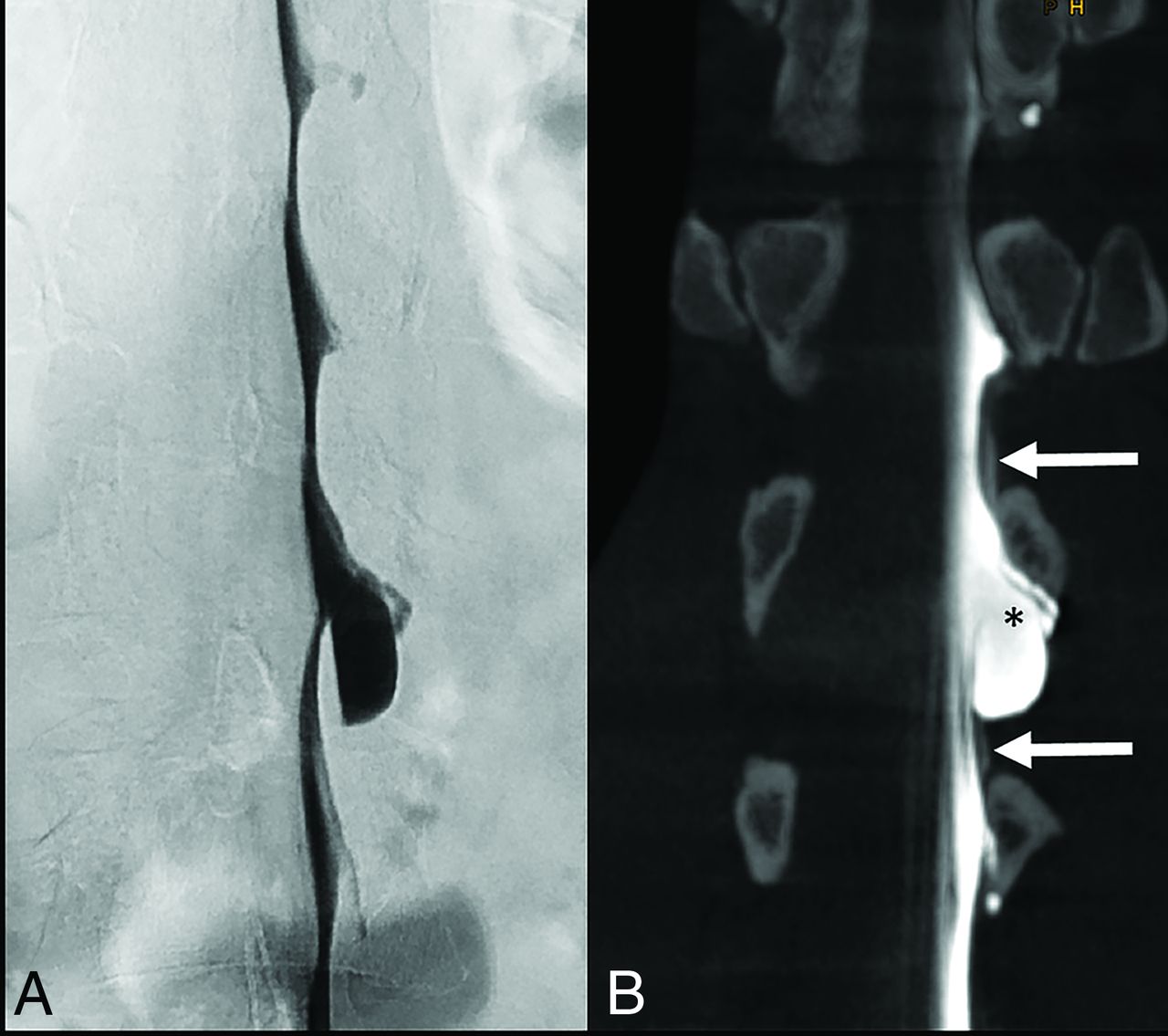
- FIG 1.
A 47-year-old man with SIH, resulting in bilateral subdural hematomas. DSM with the patient in left lateral decubitus position does not demonstrate a contrast extradural outflow at the level of a suspicious meningeal diverticulum at L1/2 (A). Subsequent CBCT in a coronal reconstruction reveals a subtle extradural contrast collection next to the diverticulum (arrows in B), confirming a slow-flow type 2 leak. The high spatial resolution of CBCT also indicates an accompanying arachnoid outpouching with an interrupted dura (medial-cranial to the asterisk in B), appearing like a meningeal diverticulum (later confirmed by surgery). CBCT settings are the following: 3D-(5sDCT Body), 49-cm zoom, 90 kV, 544 mA, 90 images/sec in a 4.83-second runtime (397 total images).
- FIG 2.
A 37-year-old woman with SIH and a persistent SLEC on MR imaging of the spine months after the epidural blood patch. Anterior-posterior DSM with the patient in the left lateral decubitus position shows a slightly contrast-filled prominent diverticulum at T7/8 (arrows in A) without extradural contrast egress. Coronal CBCT clearly demonstrates an epidural contrast collection (arrow in B) next to the diverticulum. Axial CBCT shows a type 2 leak with more posterior contrast leakage (arrow in C) (caused by encapsulation and neomembranes as reported from the operation), previously not visible on DSM due to superimpositions. CBCT settings are the following: 3D-(4sDCT Body Care), 49-cm zoom, 90 kV, 134 mA, 90 images/sec in 3.53-second runtime (248 total images).
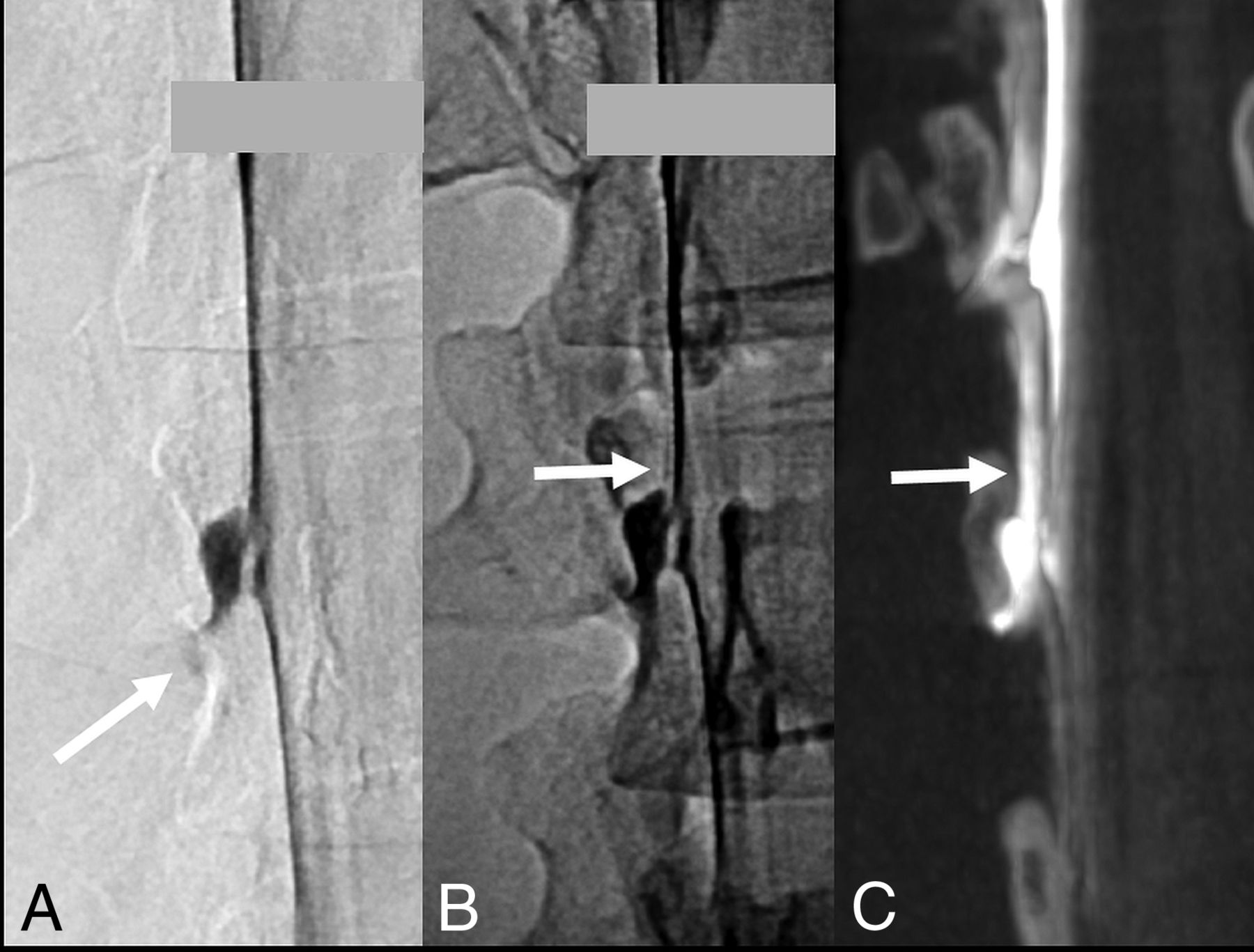
- FIG 3.
A 26-year-old woman severely impaired by SIH. DSM with the patient in a right lateral decubitus position shows a small but remarkable diverticulum at the L1/2 level. A subtle contrast flickering is visible at the bottom of the diverticulum (arrow in A). Delay single x-ray shows a faint hyperdense line as a questionable indication of extradural contrast outflow (arrow in B). Subsequent CBCT in a coronal reconstruction confirms the findings as a type 2 leak (arrow in C). CBCT settings are the following: 3D-(4sDCT Body Care) 49-cm zoom, 90 kV, 404 mA, 90 images/sec in 3.53-second runtime (248 total images).


