
Article Figures & Data
Figures
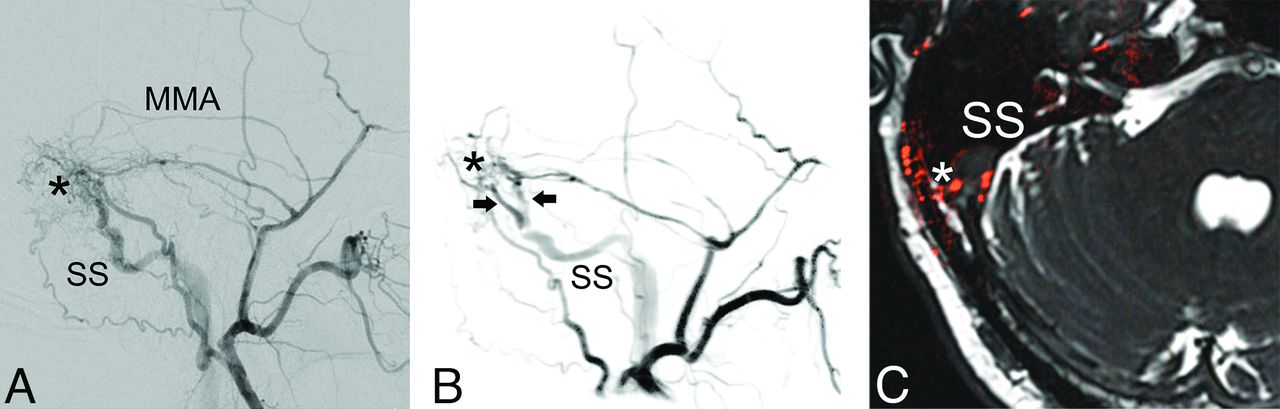
- FIG 1.
A DAVF between the middle meningeal artery (MMA), occipital artery and venous channels in the wall of the right lateral sinus with postthrombotic changes. Comparison of a standard lateral projection in 2D-DSA (A) and a slightly modified oblique projection of a virtual DSA-reconstruction (B) as well as an axial fusion image (C) derived from a 4D-DSA data set fused with a CISS MR imaging sequence. The possibility to choose an optimized projection on 4D-DSA improves the visualization of the arterial network at the fistulous point (asterisk) with connections to 2 venous channels (arrows) with Y-shaped convergence to a single venous channel in the wall of the sigmoid sinus (SS) with postthrombotic changes.
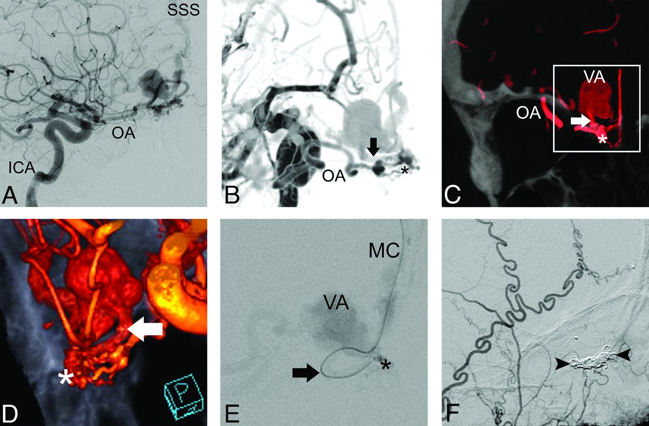
- FIG 2.
Comparison of a lateral standard projection in 2D-DSA (A) with an oblique projection in virtual DSA derived from 4D-DSA (B) and 4D-DSA coronal CT MIP (C) and volume rendering with an overlay of skull in an oblique zoomed-in projection (D). The 2D-DSA standard projection shows the frontoethmoidal DAVF supplied by the ophthalmic artery (OA) with drainage into the cortical veins and then the superior sagittal sinus (SSS). 4D-DSA images offer better visualization of the frontoethmoidal fistulous point (asterisk) and its anatomic relation to the draining vein (arrow), which is obscured in the standard projection of 2D-DSA, and the following venous aneurysm (VA). Note that the fistulous point is located within the olfactory groove. For endovascular therapy, a transvenous approach is selected with superselective positioning of the microcatheter (MC) in the draining vein past the VA (E), close to the fistulous point. After coiling (arrowheads) of the draining vein (F), we achieved complete embolization of the DAVF, as seen in the control angiogram of the ECA.


Tables
Age (yr) Sex Location Type Symptoms Therapy 62 Male Frontoethmoidal III None Transvenous coiling 64 Male Frontoethmoidal IV None Transvenous coiling 72 Male Frontoethmoidal IV None Transvenous coiling 51 Female Frontoethmoidal IV None Surgery 48 Male Frontal IV Headache, vertigo Transarterial Onyx 43 Male Frontal III None Transarterial Onyx 45 Male Frontal III Visual field loss Transarterial Onyx 66 Male Frontal III Acute cerebral hemorrhage Surgery 69 Male Infratentorial III None Transarterial Onyx 54 Male Infratentorial III Acute cerebral hemorrhage Surgery 57 Male Infratentorial IV Acute cerebral hemorrhage Surgery 75 Female Occipital IIa Pulsatile tinnitus Transarterial Onyx 47 Male Occipital I Pulsatile tinnitus Transarterial Onyx 77 Male Occipital IIa Pulsatile tinnitus Transarterial Onyx 79 Male Occipital IV Acute cerebral hemorrhage Transarterial Onyx 72 Male Occipital I Pulsatile tinnitus None 64 Male Occipital IIa Pulsatile tinnitus Transarterial Onyx 70 Male Occipital I Pulsatile tinnitus Transvenous coiling 84 Female Occipital IIb Pulsatile tinnitus None 59 Male Occipital I Pulsatile tinnitus None 31 Male Temporo-occipital III Acute cerebral hemorrhage Transarterial Onyx 31 Male Temporo-occipital III None Surgery 62 Male Tentorial IV Headache Transarterial Onyx 53 Male Tentorial IV Hemiparesis Radiosurgery 62 Male Carotid cavernous A Cranial nerve deficit (II) Transarterial coiling 68 Female Carotid cavernous A Cranial nerve deficit (III+IV) Transarterial coiling 67 Female Carotid cavernous D Cranial nerve deficit (IV) Transvenous coiling No. % 4D considered equal to 2D 13 48.1 4D considered advantageous to 2D 8 29.7 Improved projection 6 22.2 Reduction of vessel overlay 4 14.8 Display of adjacent anatomic structures 4 14.8 4D considered inferior to 2D 6 22.2 Incorrect grading 2 7.4 Endovascular access not displayed 2 7.4 Contrast injection too early 1 3.7 Incomplete display of arterial feeders 4 18.5 Pseudoretrograde display of small vessels 2 7.4 Motion artifacts 4 14.8


Jump to section
Related Articles
Cited By...
- No citing articles found.