
Article Figures & Data
Figures
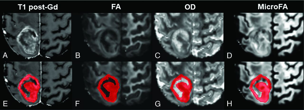
- FIG 1.
Presurgical (3T) MR imaging in a patient with a right parietal GBM. Representative axial images are shown in the upper row (A–D) with the corresponding ROI (of contrast-enhancing tumor components based on A) overlaid sections in the lower row (E–H). Gd indicates Gadoteridol.
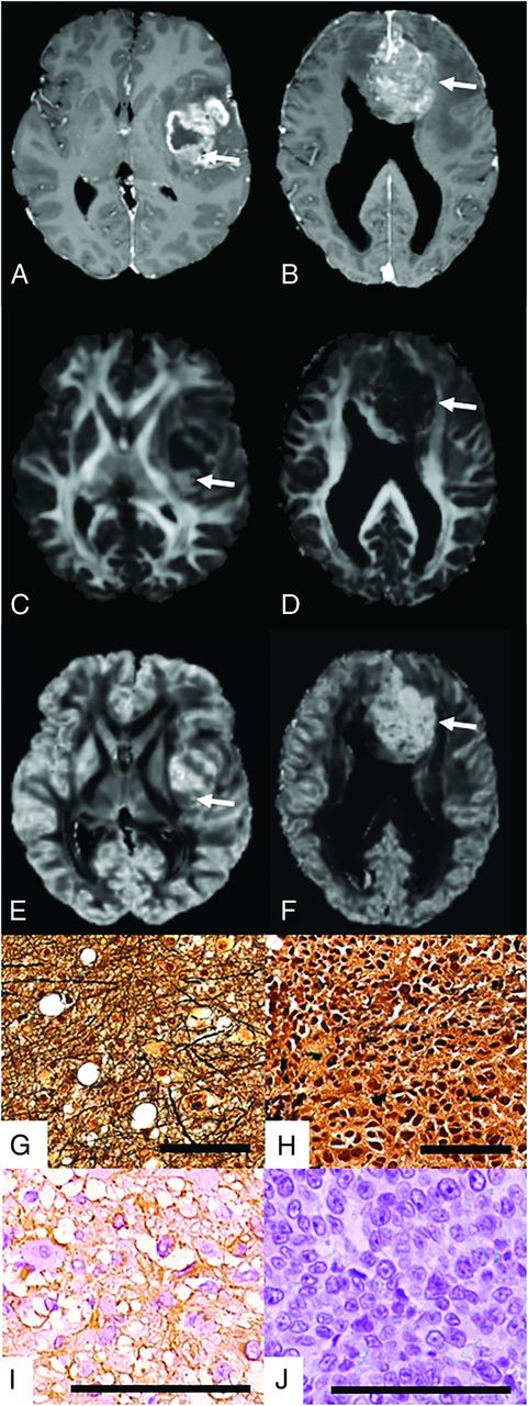
- FIG 2.
Representative MR images of patients with left insular GBM (A, T1WI post-Gd; C, FA; E, OD) and a large callosal melanoma metastasis (B, T1 post-Gd; D, FA; F, OD). Representative Bielschowsky silver staining to demonstrate nerve fibers and neurofibrillary tangles (G and H) and immunohistochemical labeling for GFAP (I and J). Scale bar = 100 μm. As illustrated by parametric maps for FA (C and D, arrows) and OD (E and F, arrows), values in contrast-enhancing solid tumor components (A and B, arrows) approximate normal-appearing white matter in GBM. This finding is accompanied by abundant axonal structures visualized by Bielschowsky silver staining (G) and high GFAP expression (I) in GBM compared with melanoma metastasis (H and J). Scale = 100 μm.
- FIG 3.
DTI, NODDI, and DMI metrics in contrast-enhancing tumor areas in patients with GBM (n = 22) and metastases (n = 21). Compared with metastases, GBM showed a significant shift toward increased FA and decreased OD, whereas no significant differences were found regarding microFA. Four asterisks indicate P ≤ .001. NAWM indicates normal-appearing white matter.
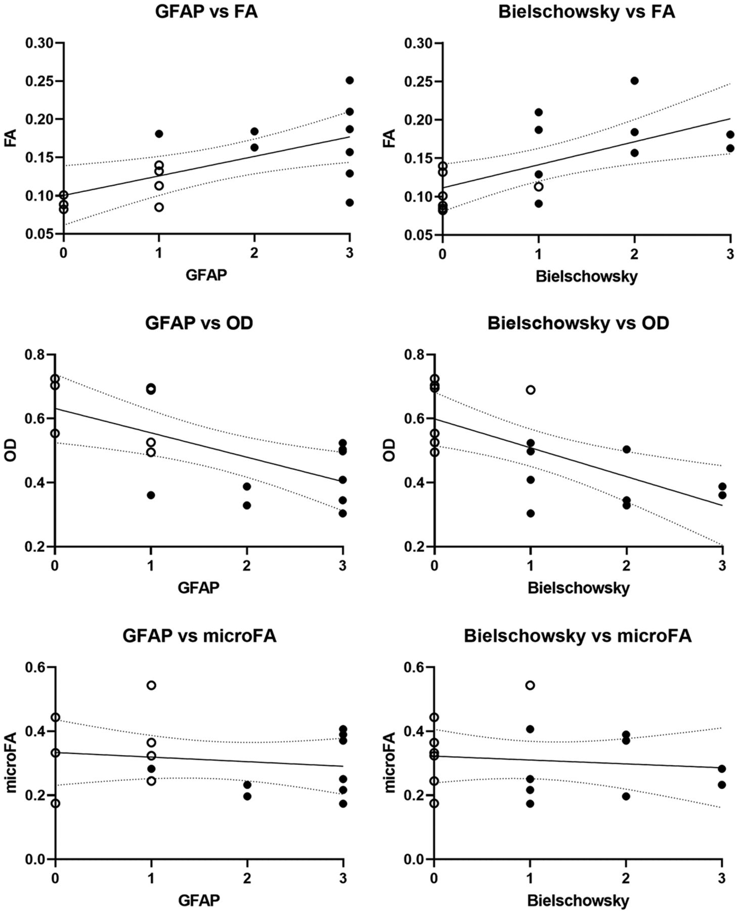
- FIG 4.
The Spearman rank correlation demonstrates a positive association of FA with GFAP and Bielschowsky scores (upper row) and a negative association of OD with GFAP and Bielschowsky scores (middle row). No association between microFA with GFAP and Bielschowsky scores can be detected (lower row). Open circles indicate metastasis cases; filled circles, GBM cases.
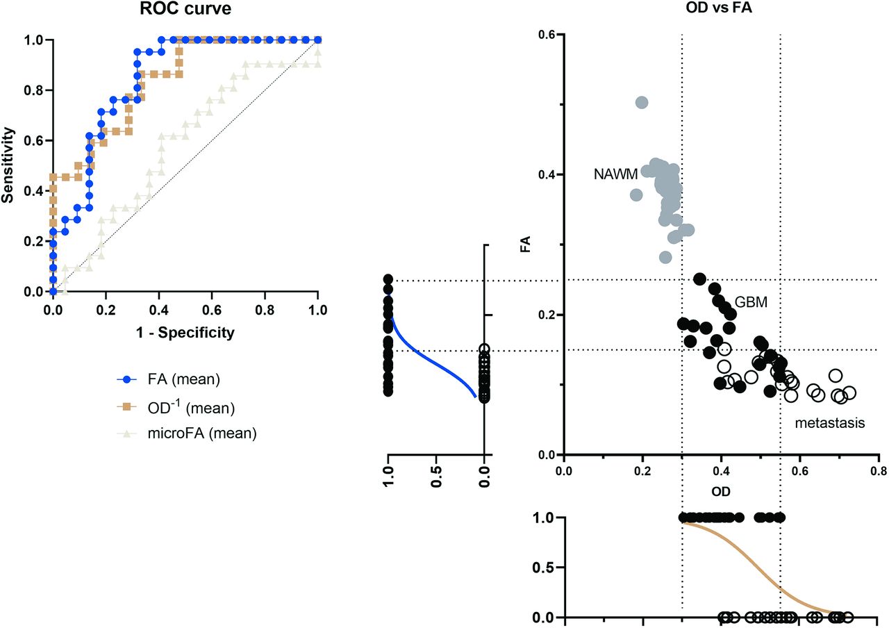
- FIG 5.
ROC curves of 22 patients with GBM and 21 with cerebral metastases showing a high predictive value of both FA (AUC = 0.8463) and OD (AUC = 0.8398) regarding the presence of GBM versus metastasis (left panel) and a scatterplot of OD and FA values for each sample (normal-appearing white matter controls = gray filled circles, GBM = black filled circles, metastasis = open circles) with arbitrary cutoff values of 0.3 and 0.55 for OD and 0.15 and 0.25 for FA. Logistic regression indicates the probability of GBM diagnosis compared with metastasis diagnosis at each individual OD or FA value (right panel).


Tables
Patient characteristics and ROI (contrast-enhancing tumor area) derived diffusion metricsa
GBM Metastasis P Value (GBM vs Met) No. 22 21 Sex (male/female) 11/11 12/9 P = .82 Age (yr) 65.6 (12.7) 66.6 (11.8) P = .99 FA 0.16 (0.04) 0.11 (0.02) P < .001 OD 0.43 (0.08) 0.56 (0.10) P < .001 microFA 0.29 (0.10) 0.27 (0.10) P = .56 Note:—Met indicates Metastasis.
↵a Data are given as mean and standard deviation (SD).


Jump to section
Related Articles
Cited By...
- No citing articles found.