Advertisement

Main menu

  • Home
  • Content
    • Current Issue
    • Accepted Manuscripts
    • Article Preview
    • Past Issue Archive
    • Video Articles
    • AJNR Case Collection
    • Case of the Week Archive
    • Case of the Month Archive
    • Classic Case Archive
  • Special Collections
    • AJNR Awards
    • Low-Field MRI
    • Alzheimer Disease
    • ASNR Foundation Special Collection
    • Photon-Counting CT
    • View All
  • Multimedia
    • AJNR Podcasts
    • AJNR SCANtastic
    • Trainee Corner
    • MRI Safety Corner
    • Imaging Protocols
  • For Authors
    • Submit a Manuscript
    • Submit a Video Article
    • Submit an eLetter to the Editor/Response
    • Manuscript Submission Guidelines
    • Statistical Tips
    • Fast Publishing of Accepted Manuscripts
    • Graphical Abstract Preparation
    • Imaging Protocol Submission
    • Author Policies
  • About Us
    • About AJNR
    • Editorial Board
    • Editorial Board Alumni
  • More
    • Become a Reviewer/Academy of Reviewers
    • Subscribers
    • Permissions
    • Alerts
    • Feedback
    • Advertisers
    • ASNR Home

User menu

  • Alerts
  • Log in

Search

  • Advanced search
American Journal of Neuroradiology

American Journal of Neuroradiology

ASHNR American Society of Functional Neuroradiology ASHNR American Society of Pediatric Neuroradiology ASSR
  • Alerts
  • Log in

Advanced Search

  • Home
  • Content
    • Current Issue
    • Accepted Manuscripts
    • Article Preview
    • Past Issue Archive
    • Video Articles
    • AJNR Case Collection
    • Case of the Week Archive
    • Case of the Month Archive
    • Classic Case Archive
  • Special Collections
    • AJNR Awards
    • Low-Field MRI
    • Alzheimer Disease
    • ASNR Foundation Special Collection
    • Photon-Counting CT
    • View All
  • Multimedia
    • AJNR Podcasts
    • AJNR SCANtastic
    • Trainee Corner
    • MRI Safety Corner
    • Imaging Protocols
  • For Authors
    • Submit a Manuscript
    • Submit a Video Article
    • Submit an eLetter to the Editor/Response
    • Manuscript Submission Guidelines
    • Statistical Tips
    • Fast Publishing of Accepted Manuscripts
    • Graphical Abstract Preparation
    • Imaging Protocol Submission
    • Author Policies
  • About Us
    • About AJNR
    • Editorial Board
    • Editorial Board Alumni
  • More
    • Become a Reviewer/Academy of Reviewers
    • Subscribers
    • Permissions
    • Alerts
    • Feedback
    • Advertisers
    • ASNR Home
  • Follow AJNR on Twitter
  • Visit AJNR on Facebook
  • Follow AJNR on Instagram
  • Join AJNR on LinkedIn
  • RSS Feeds

AJNR Awards, New Junior Editors, and more. Read the latest AJNR updates

Research ArticleNeurovascular/Stroke Imaging
Open Access

DWI-Detected Ischemic Lesions after Endovascular Treatment for Cerebral Aneurysms: An Updated Systematic Review and Meta-analysis

Abiel Berhe Habtezghi, Sherief Ghozy, Cem Bilgin, Hassan Kobeissi, Ramanathan Kadirvel and David F. Kallmes
American Journal of Neuroradiology November 2023, 44 (11) 1256-1261; DOI: https://doi.org/10.3174/ajnr.A8024
Abiel Berhe Habtezghi
aFrom the Department of Radiology (A.B.H., S.G., C.B., H.K., R.K., D.F.K.), Mayo Clinic, Rochester, Minnesota
  • Find this author on Google Scholar
  • Find this author on PubMed
  • Search for this author on this site
  • ORCID record for Abiel Berhe Habtezghi
Sherief Ghozy
aFrom the Department of Radiology (A.B.H., S.G., C.B., H.K., R.K., D.F.K.), Mayo Clinic, Rochester, Minnesota
  • Find this author on Google Scholar
  • Find this author on PubMed
  • Search for this author on this site
  • ORCID record for Sherief Ghozy
Cem Bilgin
aFrom the Department of Radiology (A.B.H., S.G., C.B., H.K., R.K., D.F.K.), Mayo Clinic, Rochester, Minnesota
  • Find this author on Google Scholar
  • Find this author on PubMed
  • Search for this author on this site
  • ORCID record for Cem Bilgin
Hassan Kobeissi
aFrom the Department of Radiology (A.B.H., S.G., C.B., H.K., R.K., D.F.K.), Mayo Clinic, Rochester, Minnesota
  • Find this author on Google Scholar
  • Find this author on PubMed
  • Search for this author on this site
  • ORCID record for Hassan Kobeissi
Ramanathan Kadirvel
aFrom the Department of Radiology (A.B.H., S.G., C.B., H.K., R.K., D.F.K.), Mayo Clinic, Rochester, Minnesota
bDepartment of Neurologic Surgery (R.K.), Mayo Clinic, Rochester, Minnesota.
  • Find this author on Google Scholar
  • Find this author on PubMed
  • Search for this author on this site
  • ORCID record for Ramanathan Kadirvel
David F. Kallmes
aFrom the Department of Radiology (A.B.H., S.G., C.B., H.K., R.K., D.F.K.), Mayo Clinic, Rochester, Minnesota
  • Find this author on Google Scholar
  • Find this author on PubMed
  • Search for this author on this site
  • ORCID record for David F. Kallmes
  • Article
  • Figures & Data
  • Info & Metrics
  • Responses
  • References
  • PDF
Loading

Abstract

BACKGROUND AND PURPOSE: DWI-detected ischemic lesions are potential complications of endovascular procedures that are performed to treat intracranial aneurysms. We completed a systematic review and meta-analysis to identify the occurrence of DWI-detected ischemic lesions after endovascular treatment for intracranial aneurysms.

MATERIALS AND METHODS: A systematic literature search of PubMed, the Web of Science, EMBASE, and Scopus between January 2000 and June 2022 of post-endovascular procedures for intracranial aneurysm studies was conducted using the Nested Knowledge AutoLit software. The main outcome was DWI-detected ischemic lesions within 5 days of the procedures. Information regarding associated risk factors such as the type of procedure, patient demographics, and aneurysm characteristics was also collected.

RESULTS: Twenty-nine studies with 2686 patients were included. The overall incidence of DWI ischemic lesions was 47.0% (95% CI, 39.6%–55.8%). The highest rate of lesions was seen with flow diversion at 62.4% (95% CI, 48.4%–80.5%), followed by complex procedures at 49.3% (95% CI, 29.5%–82.1%), stent-assisted coiling at 47.5% (95% CI, 34.6%–65.3%), simple coiling at 47.1% (95% CI, 35.7%–62.3%), and balloon-assisted coiling at 37.0% (95% CI, 28.3%–48.4%). The differences among different techniques were not statistically significant; however, there was significant heterogeneity and a significant risk of publication bias among included studies.

CONCLUSIONS: Many patients who undergo endovascular procedures for intracranial aneurysms present with new postprocedural DWI-detected ischemic lesions, regardless of the endovascular procedure used. Future studies and meta-analyses are needed to investigate early and long-term outcomes of such small infarcts.

ABBREVIATION:

APC
annual percentage change

Endovascular neurointerventional procedures have been used since the 1990s with the development of different techniques that obviate the need for a craniotomy. Endovascular techniques include simple coiling, stent-assisted coiling, balloon-assisted coiling, and flow diversion. Although less invasive than traditional surgery, these techniques have been associated with periprocedural complications ranging from aneurysmal rupture to different thromboembolism presentations. DWI-identified ischemic lesions after endovascular procedures have been reported to be a common occurrence with unknown clinical significance.1⇓⇓-4

The overall incidence of DWI-positive lesions previously reported after endovascular procedures was 1 in 2 patients; however, previous literature was based on outdated studies, older devices and techniques.1 We propose that the rates of DWI-detected lesions following endovascular procedures may vary with the introduction of new endovascular techniques and procedures. To determine the overall incidence of perioperative infarctions on DWI in patients undergoing endovascular treatment for intracranial aneurysms, we performed a systematic review and meta-analysis.

MATERIALS AND METHODS

Literature Search and Inclusion Criteria

In-depth article reviews were performed by A.B.H. and S.G. using keywords such “flow diverter,” “coiling,” “pipeline,” “endovascular,” “DWI-MR imaging,” “diffusion,” “restricted diffusion,” “MR imaging,” “diffusion-weighted imaging,” “cerebral aneurysm,” and “intracranial aneurysm.” During the review process, the last set of Boolean traits found in the Online Supplemental Data were applied. On June 10, 2022, this search algorithm was used to search PubMed, the Web of Science, EMBASE, and Scopus.

The inclusion criteria were observational studies or randomized controlled trials between January 2000 and June 2022 that consisted of at least 10 consecutive patients treated for intracranial aneurysms by endovascular means with endovascular coiling or flow diversion and DWI examinations performed within 5 days of endovascular treatment in all patients. In addition, the reference lists from included studies were retrieved for possible inclusion of missed publications. Exclusion criteria were studies that had <10 patients, reported outcomes of parent artery occlusion, included only symptomatic patients who underwent postoperative DWI, used intrasaccular devices (such as the Woven EndoBridge [WEB]; Sequent Medical), and conference proceedings. In addition, studies that did not provide the number of patients who had no lesions on DWI and studies for which an English translation was not available were excluded.

The current study used a novel semiautomated software platform (AutoLit, Nested Knowledge; https://wiki.nested-knowledge.com/doku.php?id=wiki:autolit) for screening of studies and extraction of data. Two independent reviewers performed the screening, and a third reviewer arbitrated disagreements. Baseline characteristics of patients and aneurysm types such as aneurismal size, location, and rupture status were extracted. During data extraction, whenever >2 means for subgroups were available, the formula found in the Online Supplemental Data from the Cochrane Guide for Systematic Reviews and Metanalysis was used.5 The guidelines provided by the Preferred Reporting Items for Systematic Reviews and Meta-Analyses (PRISMA) and Meta-analysis Of Observational Studies in Epidemiology (MOOSE) were followed, as well.

Outcomes and Patient Groups

DWI-detected ischemic lesions within 5 days of the endovascular procedure were the primary end point. Treatment groups were stratified by the type of procedure: simple coiling, stent-assisted coiling, balloon-assisted coiling, and flow diversion. When ≥2 procedures and multiple catheters were used, it was termed a complex procedure. Patient sex (male versus female), aneurysm size (considered small if <10 mm, giant if >10 mm),6 location (anterior or posterior circulation), and rupture status (ruptured or unruptured) were dichotomized.

Risk of Bias Assessment

We selected items from the Newcastle-Ottawa Quality Assessment Scale to fit the type of included studies. In 3 domains with 8 criteria and a maximum score of 8, we queried the following study characteristics using a previous study by one of the authors: 1) patient groups clearly defined (1 point given if patient groups were clearly defined by the type of endovascular procedure, zero points given if not); 2) outcomes reported (1 point for reports taken from hospital data, 2 points for reports by radiologists for the study using DWI); 3) outcomes reported for each patient group studied (1 point given if an outcome was reported for each procedure, zero points given if not); 4) imaging interpreted by an independent reader or interpreted by the operator (1 point if independent readers read it, zero points given if not); 5) readers blinded to the clinical status of the patient (1 point given if blinded, zero points given if not); 6) multiple readers used and interobserver agreement assessed (1 point given if interobserver agreement was assessed, zero points given if not); and 7) the study followed a specific study protocol in which all patients underwent MR imaging at the same time point (1 point given if the timing of the postprocedural MR imaging was provided, zero points given if not).1

Statistical Analysis

The cumulative incidence rates with corresponding 95% CIs were calculated using R statistical and computing software, Version 4.2.2 (http://www.r-project.org/) and the package ‘meta’ (https://cran.r-project.org/web//packages/meta/meta.pdf). A random-effects model was used to pool the data due to methodologic heterogeneity among the included studies, and subgroup analysis was performed to obtain the estimate per year. Heterogeneity was assessed using the Cochran Q and I2 tests. P values < .05 for the Q statistic indicated statistical significance. An I2 of >50% suggested moderate-to-high heterogeneity. Whenever there were ≥10 studies included, the Egger regression test was used to assess publication bias. If publication bias existed, we used the trim-and-fill method to adjust for funnel plot asymmetry and calculate the bias-adjusted estimates.7,8

By means of the Joinpoint Regression Trend Analysis Software, Version 4.9.1.0 (https://surveillance.cancer.gov/joinpoint/), the pooled cumulative incidence rates and their standard errors were used to conduct a joinpoint regression analysis to explore any trends across the years.9,10 This was conducted to calculate the average annual percentage change and annual percentage change (APC), selecting the best-fitting piecewise continuous log-linear model. The minimum number of “joinpoints” required to fit the data was determined using a permutation test.11,12 The weighted Bayesian Information Criterion was the model adopted, and the empirical quantile methods were used to estimate confidence intervals.13 The empirical method does not produce a test statistic or P value.13

RESULTS

Search Results and Patient Population

The literature search retrieved 491 studies, from which 209 duplicate studies were removed. From the remaining 282 studies, title and abstract screening resulted in 160 studies being excluded. Of the 122 studies remaining, full-text screening resulted in another 93 studies being excluded, as illustrated in Fig 1, which shows the process of identifying studies through databases and registers.

FIG 1.
  • Download figure
  • Open in new tab
  • Download powerpoint
FIG 1.

Identification of studies via databases and registers.

Risk of Bias

Of the 29 studies included in this meta-analysis, 2 had a very high risk of bias with scores between 0 and 3 points, 22 had a high risk of bias with scores of 4–6 points, and 5 had a low risk of bias scoring 7–8 points, as determined by the Modified Newcastle Ottawa Scale.14

Characteristics of the Included Studies

Twenty-nine studies with a total of 2686 patients presenting with 3687 intracranial aneurysms fulfilled our inclusion criteria and composed our analysis.15⇓⇓⇓⇓⇓⇓⇓⇓⇓⇓⇓⇓⇓⇓⇓⇓⇓⇓⇓⇓⇓⇓⇓⇓⇓⇓⇓-43 Endovascular procedures were simple coiling in 1184 patients, stent-assisted coiling in 662 patients, balloon-assisted coiling in 328 patients, flow diversion in 211 patients, and complex procedures in 301 patients. From studies that reported aneurysm rupture status, 275 were ruptured during treatment, while 2552 were unruptured. Among the studies that reported the location of the aneurysms, 1624 were in the anterior circulation and 510 were in the posterior circulation.

Study Outcomes

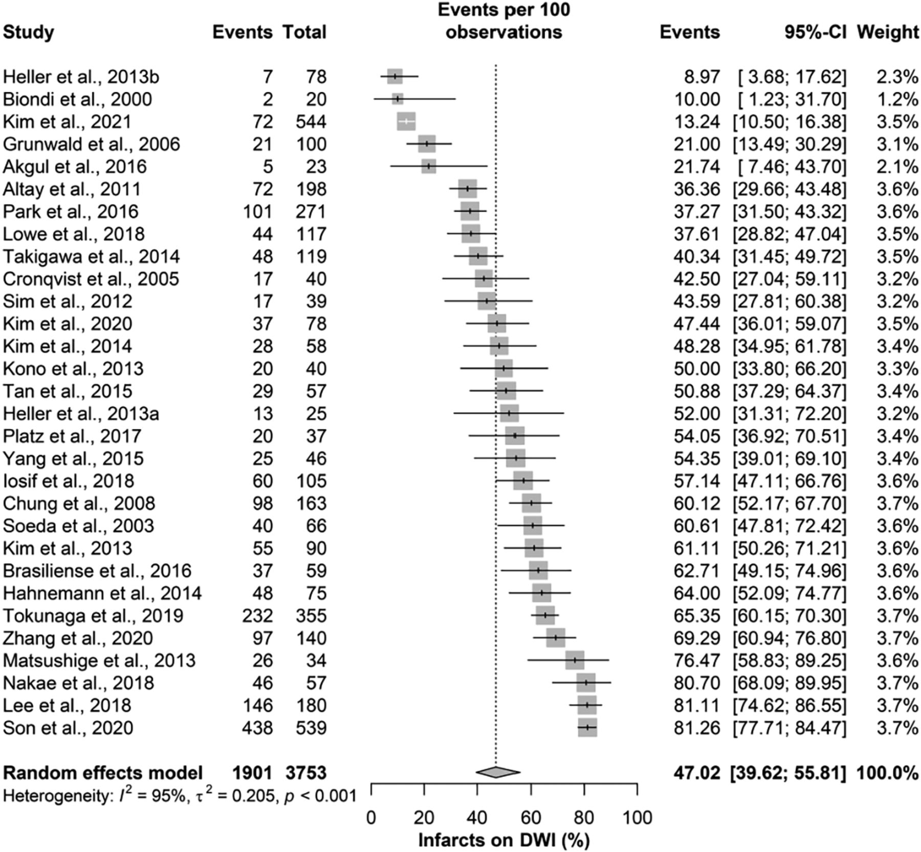
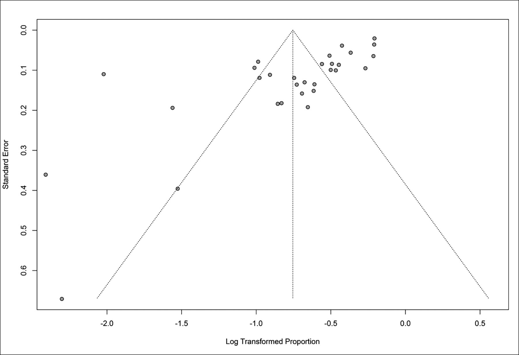
The overall incidence of DWI lesions was 47.0% (95% CI, 39.6%–55.8%), with significant heterogeneity among the included studies (I2 = 95%; P value < .001; Fig 2). Moreover, there was a significant risk of publication bias as assessed with the Egger regression test (P value < .001; Fig 3).

FIG 2.
  • Download figure
  • Open in new tab
  • Download powerpoint
FIG 2.

Infarcts on DWI.

FIG 3.
  • Download figure
  • Open in new tab
  • Download powerpoint
FIG 3.

Publication bias as assessed with the Egger regression test.

On further subgrouping based on the treatment technique used, the highest rate of lesions was seen with flow diversion at 62.4% (95% CI, 48.4%–80.5%), complex procedures at 49.3% (95% CI, 29.5%–82.1%), stent-assisted coiling at 47.5% (95% CI, 34.6%–65.3%), simple coiling at 47.3% (95% CI, 35.5%–63.7%), and balloon-assisted coiling at 35.9% (95% CI, 25.5%–50.6%). The differences among different techniques were not statistically significant (P value = .139), with significant heterogeneity among the included studies in all subgroups (Fig 4).

FIG 4.
  • Download figure
  • Open in new tab
  • Download powerpoint
FIG 4.

The subgroup of treatment technique used and DWI lesions.

For trend analysis, there was a significant reduction in DWI lesions through the full range of years from 2000 to 2022, with an average annual percent change of −5.0 (95% CI, −7.6 to −1.9). Moreover, there was 1 joinpoint identified in 2019, with a significant reduction in detected ischemic lesions (APC = −50.0; 95% CI, −63.1 to −35.0) after a relative stability in rates before that (APC = 1.6; 95% CI, −0.2−5.0; Online Supplemental Data).

Impact of Aneurysm Characteristics and Sex

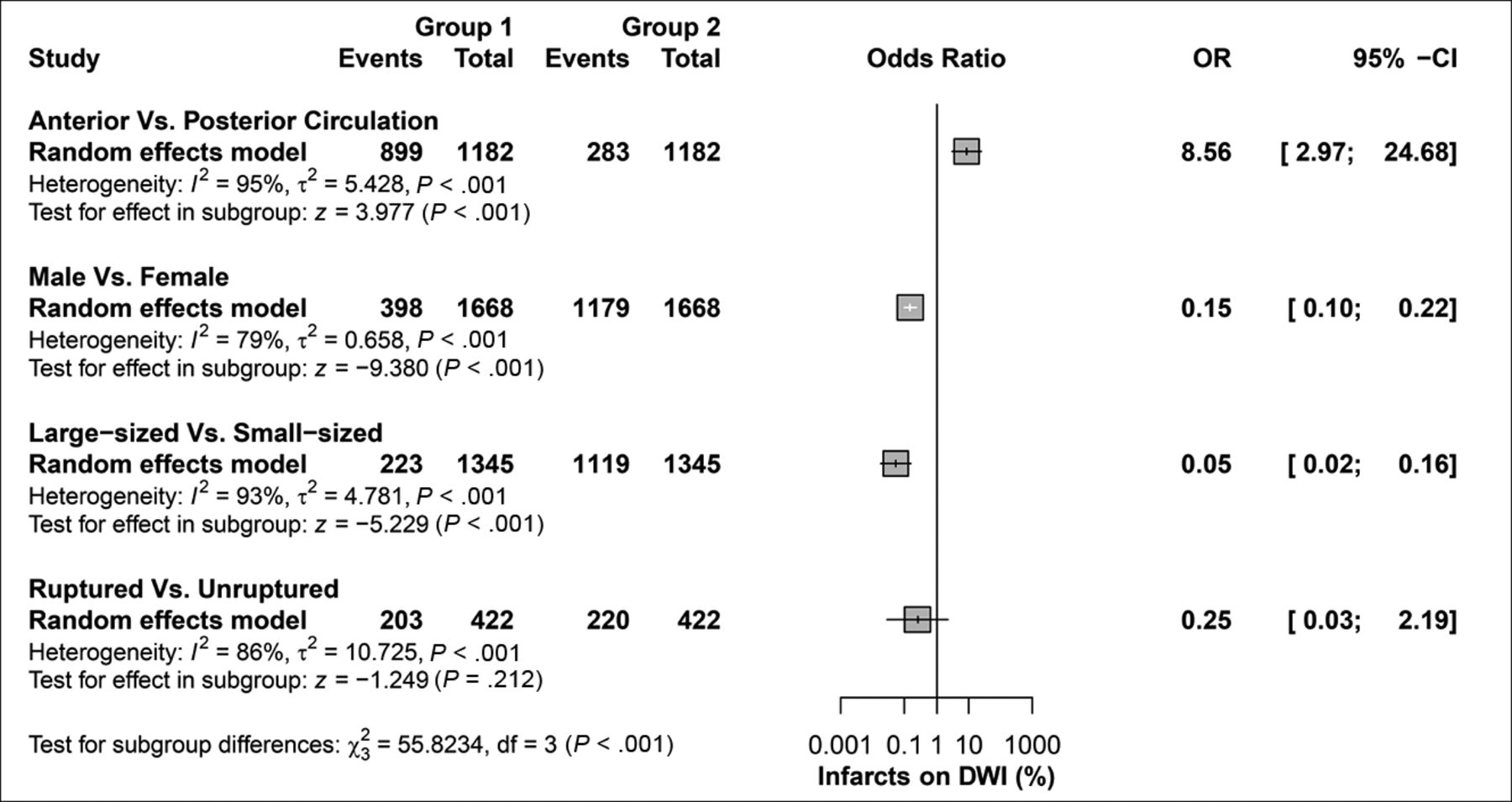
Aneurysms were more likely to be in the anterior circulation compared with the posterior circulation in DWI-detected lesions (OR = 8.56; 95% CI, 2.97–24.7, P value < .001). Men were less likely than women to have aneurysms with DWI-detected lesions (OR = 0.15; 95% CI, 0.1–0.22, P value < .001). Large-sized aneurysms were less common than small-sized aneurysms (OR = 0.05; 95% CI, 0.02–0.16, P value < .001), while rupture status was comparable (OR = 0.25; 95% CI, 0.02–2.2, P value = 0.21). For all aneurysm characteristics and sex of patients, there was significant heterogeneity among the included studies (Fig 5).

FIG 5.
  • Download figure
  • Open in new tab
  • Download powerpoint
FIG 5.

Risk factors and DWI lesions.

DISCUSSION

Our systematic review and meta-analysis found that the incidence of DWI-positive lesions after endovascular procedures for intracranial aneurysms was 47%. We found no statistically significant difference among techniques. These results highlight the high incidence of DWI-positive lesions, which are statistically similar among different endovascular treatments.

Previous studies have examined the clinical impact of DWI-detected lesions. Almost half of these lesions are asymptomatic, and ischemic lesions have been shown to regress if their size is small (<5 mm), resulting in favorable rates of good clinical outcomes.44 Studies are needed to further validate these findings using clinical outcome measures such as a standardized method of assessing the clinical relevance of lesions like the mRS. Other literature has evaluated the prevention of this postprocedural complication with remote ischemic preconditioning, which has been shown to be safe and effective.45

We found that the rate of silent infarcts in patients treated with flow diverters was higher than in those treated with coils alone, though this was not statistically significant. Flow diversion leads to more thromboembolic complications than coiling alone, despite the inherent thrombogenic properties of coils and flow diverters. Because flow diverters are high density and have large endoluminal surfaces, they have a high thrombosis risk in the parent arteries. An embolized thrombus can occur from the shearing stress caused by blood flowing through the device. Because lower density coils are placed in the aneurysm sac outside the cerebral circulation, thrombi forming on them are less likely to embolize.1

Studies have shown that DWI-positive lesions are not significantly different between ruptured and unruptured aneurysms (as shown in Fig 5), are associated with long procedural times, and decrease in incidence with postprocedural anticoagulation, all of which are consistent with the results of our present analysis.46 Additionally, previous literature has demonstrated that the presence or number of ischemic lesions on DWI is not related to cognitive changes following coil embolization.47 Periprocedural thromboembolic events identified on DWI have been shown to be reduced with a dual-antiplatelet agent (for unruptured aneurysms), and patients are given preoperative low-molecular-weight heparin to prevent this complication.48

We could not identify a discernible trend in the incidence of DWI lesions with time. The absence of a notable temporal trend suggests that the occurrence of DWI lesions did not change before and after 2017 (Online Supplemental Data).

Our study has limitations. We included retrospective, nonrandomized studies, and studies were inconsistent in their reporting of results. We did not consider the use of anticoagulants and individual antiplatelet therapy. We could not account for the differences among institutions performing endovascular procedures, the experience of the interventionalists who performed the procedures, and the radiologists who read the DWIs. There were associations between baseline characteristics such as female sex, anterior aneurysm, small aneurysm size, and the incidence of postprocedural DWI lesions. The studies included did not consistently report atherosclerotic risk factors, use of anticoagulants, and other aneurysmal characteristics.32 Finally, we did not have access to individual patient data, limiting the data that could be analyzed. In our study, the presence or absence of DWI lesions was used as the primary criterion for determining their occurrence. However, we acknowledge that a more nuanced approach, considering the size, number, and distribution of DWI lesions, could provide a more comprehensive understanding of the ischemic risk and its clinical implications. Because these details are currently lacking in the literature, further studies are needed to address these concerns.

CONCLUSIONS

In this systematic review and meta-analysis of 2686 patients, the rate of DWI-detected ischemic lesions was 47.0% (95% CI, 39.6%–55.8%). Rates of lesions were statistically similar among all included endovascular treatments. Further prospective and randomized studies are needed to elucidate the DWI-detected ischemic lesion rate after endovascular procedures and their lasting complications.

ACKNOWLEDGMENTS

The authors acknowledge Karl Holub, Stephen Mead, Jeffrey Johnson, and Darian Lehmann-Plantenberg for their design, development, and support of the Nested Knowledge meta-analytical software. The authors thank Desiree Lanzino, PhD, and Sonia Watson for their assistance in editing the manuscript.

Footnotes

  • Research reported in this publication was supported, in part, by the National Institute of Neurological Disorders and Stroke of the National Institutes of Health under award No. R01NS076491.

  • The content of this work is solely the responsibility of the authors and does not necessarily represent the official views of the National Institutes of Health.

  • Disclosure forms provided by the authors are available with the full text and PDF of this article at www.ajnr.org.

Indicates open access to non-subscribers at www.ajnr.org

References

  1. 1.↵
    1. Bond KM,
    2. Brinjikji W,
    3. Murad MH, et al
    . Diffusion-weighted imaging-detected ischemic lesions following endovascular treatment of cerebral aneurysms: a systematic review and meta-analysis. AJNR Am J Neuroradiol 2017;38:304–09 doi:10.3174/ajnr.A4989 pmid:27856436
    Abstract/FREE Full Text
  2. 2.↵
    1. Choi SH,
    2. Na DL,
    3. Chung CS, et al
    . Diffusion-weighted MRI in vascular dementia. Neurology 2000;54:83–89 doi:10.1212/wnl.54.1.83 pmid:10636130
    Abstract/FREE Full Text
  3. 3.↵
    1. Ihn YK,
    2. Shin SH,
    3. Baik SK, et al
    . Complications of endovascular treatment for intracranial aneurysms: management and prevention. Interv Neuroradiol 2018;24:237–45 doi:10.1177/1591019918758493 pmid:29466903
    CrossRefPubMed
  4. 4.↵
    1. Zhao J,
    2. Lin H,
    3. Summers R, et al
    . Current treatment strategies for intracranial aneurysms: an overview. Angiology 2018;69:17–30 doi:10.1177/0003319717700503 pmid:28355880
    CrossRefPubMed
  5. 5.↵
    1. Higgins JPT,
    2. Thomas J,
    3. Chandler J, et al.
    , eds. Cochrane Handbook for Systematic Reviews of Interventions. 2nd Edition. Chichester (UK): John Wiley & Sons; 2019
  6. 6.↵
    1. Merritt WC,
    2. Berns HF,
    3. Ducruet AF, et al
    . Definitions of intracranial aneurysm size and morphology: a call for standardization. Surg Neurol Int 2021;12:506 doi:10.25259/SNI_576_2021 pmid:34754556
    CrossRefPubMed
  7. 7.↵
    1. Duval S,
    2. Tweedie R
    . Trim and fill: A simple funnel-plot-based method of testing and adjusting for publication bias in meta-analysis. Biometrics 2000;56:455–63 doi:10.1111/j.0006-341x.2000.00455.x pmid:10877304
    CrossRefPubMed
  8. 8.↵
    1. Shi L,
    2. Lin L
    . The trim-and-fill method for publication bias: practical guidelines and recommendations based on a large database of meta-analyses. Medicine (Baltimore) 2019;98:e15987 doi:10.1097/MD.0000000000015987 pmid:31169736
    CrossRefPubMed
  9. 9.↵
    1. Dibas M,
    2. Doheim MF,
    3. Ghozy S, et al
    . Incidence and survival rates and trends of skull base chondrosarcoma: a population-based study. Clin Neurol Neurosurg 2020;198:106153 doi:10.1016/j.clineuro.2020.106153 pmid:32818757
    CrossRefPubMed
  10. 10.↵
    1. Ghozy S,
    2. Dibas M,
    3. Afifi AM, et al
    . Primary cerebral lymphoma' characteristics, incidence, survival, and causes of death in the United States. J Neurol Sci 2020;415:116890 doi:10.1016/j.jns.2020.116890 pmid:32428760
    CrossRefPubMed
  11. 11.↵
    1. Kim HJ,
    2. Fay MP,
    3. Feuer EJ, et al
    . Permutation tests for joinpoint regression with applications to cancer rates. Stat Med 2000;19:335–51 doi:10.1002/(SICI)1097-0258(20000215)19:3<335::AID-SIM336>3.0.CO;2-Z pmid:10649300
    CrossRefPubMed
  12. 12.↵
    1. Politis M,
    2. Higuera G,
    3. Chang LR, et al
    . Trend analysis of cancer mortality and incidence in Panama, using joinpoint regression analysis. Medicine (Baltimore) 2015;94:e970 doi:10.1097/MD.0000000000000970 pmid:26091467
    CrossRefPubMed
  13. 13.↵
    1. Kim HJ,
    2. Chen HS,
    3. Midthune D, et al
    . Data-driven choice of a model selection method in joinpoint regression. J Appl Stat 2023;50:1992–2013 doi:10.1080/02664763.2022.2063265 pmid:37378270
    CrossRefPubMed
  14. 14.↵
    1. Wells GA,
    2. Shea B,
    3. O'Connell D, et al
    . The Newcastle-Ottawa Scale (NOS) for assessing the quality of nonrandomised studies in meta-analyses. Clin Epidemiol 2000 https://www.ohri.ca/programs/clinical_epidemiology/oxford.asp. Accessed July 21, 2022
  15. 15.↵
    1. Akgul E,
    2. Onan HB,
    3. Akpinar S, et al
    . The DERIVO Embolization Device in the treatment of intracranial aneurysms: short- and midterm results. World Neurosurg 2016;95:229–40 doi:10.1016/j.wneu.2016.07.101 pmid:27514698
    CrossRefPubMed
  16. 16.↵
    1. Biondi A,
    2. Oppenheim C,
    3. Vivas E, et al
    . Cerebral aneurysms treated by Guglielmi detachable coils: evaluation with diffusion-weighted MR imaging. AJNR Am J Neuroradiol 2000;21:957–63 pmid:10815677
    Abstract/FREE Full Text
  17. 17.↵
    1. Brasiliense LB,
    2. Stanley MA,
    3. Grewal SS, et al
    . Silent ischemic events after Pipeline Embolization Device: a prospective evaluation with MR diffusion-weighted imaging. J Neurointerv Surg 2016;8:1136–39 doi:10.1136/neurintsurg-2015-012091 pmid:26747877
    Abstract/FREE Full Text
  18. 18.↵
    1. Cronqvist M,
    2. Wirestam R,
    3. Ramgren B, et al
    . Diffusion and perfusion MRI in patients with ruptured and unruptured intracranial aneurysms treated by endovascular coiling: complications, procedural results, MR findings and clinical outcome. Neuroradiology 2005;47:855–73 doi:10.1007/s00234-005-1408-2 pmid:16235046
    CrossRefPubMed
  19. 19.↵
    1. Grunwald IQ,
    2. Papanagiotou P,
    3. Politi M, et al
    . Endovascular treatment of unruptured intracranial aneurysms: occurrence of thromboembolic events. Neurosurgery 2006;58:612–18 doi:10.1227/01.NEU.0000204101.00996.D9 pmid:16575324
    CrossRefPubMed
  20. 20.↵
    1. Heller RS,
    2. Dandamudi V,
    3. Calnan D, et al
    . Neuroform intracranial stenting for aneurysms using simple and multi-stent technique is associated with low risk of magnetic resonance diffusion-weighted imaging lesions. Neurosurgery 2013;73:582–90; discussion 590–91 doi:10.1227/NEU.0000000000000053 pmid:23787881
    CrossRefPubMed
  21. 21.↵
    1. Heller RS,
    2. Dandamudi V,
    3. Lanfranchi M, et al
    . Effect of antiplatelet therapy on thromboembolism after flow diversion with the Pipeline Embolization Device. J Neurosurg 2013;119:1603–10 doi:10.3171/2013.7.JNS122178 pmid:23971953
    CrossRefPubMed
  22. 22.↵
    1. Iosif C,
    2. Lecomte JC,
    3. Pedrolo-Silveira E, et al
    . Evaluation of ischemic lesion prevalence after endovascular treatment of intracranial aneurysms, as documented by 3-T diffusion-weighted imaging: a 2-year, single-center cohort study. J Neurosurg 2018;128:982–91 doi:10.3171/2016.11.JNS161020 pmid:28598274
    CrossRefPubMed
  23. 23.↵
    1. Kim JK,
    2. Choi JH,
    3. Kim BS, et al
    . Association of anterior cerebral artery variants and cerebral infarction in patients with balloon-assisted coil embolization for unruptured internal carotid artery aneurysms. World Neurosurg 2021;147:e69–77 doi:10.1016/j.wneu.2020.11.118 pmid:33253946
    CrossRefPubMed
  24. 24.↵
    1. Kim MJ,
    2. Lim YC,
    3. Oh SY, et al
    . Thromboembolic events associated with electrolytic detachment of Guglielmi detachable coils and target coils: comparison with use of diffusion-weighted MR imaging. J Korean Neurosurg Soc 2013;54:19–24 doi:10.3340/jkns.2013.54.1.19 pmid:24044075
    CrossRefPubMed
  25. 25.↵
    1. Kim MS,
    2. Jo KI,
    3. Yeon JY, et al
    . Association between postprocedural infarction and antiplatelet drug resistance after coiling for unruptured intracranial aneurysms. AJNR Am J Neuroradiol 2016;37:1099–105 doi:10.3174/ajnr.A4777 pmid:27056423
    Abstract/FREE Full Text
  26. 26.↵
    1. Kim SH,
    2. Lee H,
    3. Kim SB, et al
    . Differences in thromboembolism after stent-assisted coiling for unruptured aneurysms between aspirin plus clopidogrel and ticagrelor. J Clin Neurosci 2020;82:128–33 doi:10.1016/j.jocn.2020.10.042 pmid:33317720
    CrossRefPubMed
  27. 27.↵
    1. Kono K,
    2. Shintani A,
    3. Yoshimura R, et al
    . Triple antiplatelet therapy with addition of cilostazol to aspirin and clopidogrel for Y-stent-assisted coil embolization of cerebral aneurysms. Acta Neurochir (Wien) 2013;155:1549–57 doi:10.1007/s00701-013-1771-4 pmid:23715948
    CrossRefPubMed
  28. 28.↵
    1. Lee SH,
    2. Jang MU,
    3. Kang J, et al
    . Impact of reducing the procedure time on thromboembolism after coil embolization of cerebral aneurysms. Front Neurol 2018;9:1125 doi:10.3389/fneur.2018.01125 pmid:30619075
    CrossRefPubMed
  29. 29.↵
    1. Lowe SR,
    2. Bhalla T,
    3. Tillman H, et al
    . A comparison of diffusion-weighted imaging abnormalities following balloon remodeling for aneurysm coil embolization in the ruptured vs unruptured setting. Neurosurgery 2018;82:516–24 doi:10.1093/neuros/nyx240 pmid:28520916
    CrossRefPubMed
  30. 30.↵
    1. Matsushige T,
    2. Kiura Y,
    3. Sakamoto S, et al
    . Multiple antiplatelet therapy contributes to the reversible high signal spots on diffusion-weighted imaging in elective coiling of unruptured cerebral aneurysm. Neuroradiology 2013;55:449–57 doi:10.1007/s00234-013-1137-x pmid:23314799
    CrossRefPubMed
  31. 31.↵
    1. Nakae R,
    2. Nagaishi M,
    3. Kawamura Y, et al
    . Microhemorrhagic transformation of ischemic lesions on T2*-weighted magnetic resonance imaging after Pipeline Embolization Device treatment. J Neurosurg 2018 May 1 [Epub ahead of print] doi:10.3171/2017.12.JNS172480 pmid:29999443
    CrossRefPubMed
  32. 32.↵
    1. Park JC,
    2. Lee DH,
    3. Kim JK, et al
    . Microembolism after endovascular coiling of unruptured cerebral aneurysms: incidence and risk factors. J Neurosurg 2016;124:777–83 doi:10.3171/2015.3.JNS142835 pmid:26381257
    CrossRefPubMed
  33. 33.↵
    1. Petrov A,
    2. Rentsenkhuu G,
    3. Nota B, et al
    . Initial experience with the novel p64MW HPC flow diverter from a cohort study in unruptured anterior circulation aneurysms under dual antiplatelet medication. Interv Neuroradiol 2021;27:42–50 doi:10.1177/1591019920939845 pmid:32640858
    CrossRefPubMed
  34. 34.↵
    1. Pikis S,
    2. Mantziaris G,
    3. Mamalis V, et al
    . Diffusion weighted image documented cerebral ischemia in the postprocedural period following Pipeline Embolization Device with shield technology treatment of unruptured intracranial aneurysms: a prospective, single center study. J Neurointerv Surg 2020;12:407–11 doi:10.1136/neurintsurg-2019-015363 pmid:31558655
    Abstract/FREE Full Text
  35. 35.↵
    1. Platz J,
    2. Wagner M,
    3. Güresir E, et al
    . Early diffusion-weighted MRI lesions after treatment of unruptured intracranial aneurysms: a prospective study. J Neurosurg 2017;126:1070–78 doi:10.3171/2016.2.JNS152456 pmid:27203140
    CrossRefPubMed
  36. 36.↵
    1. Sim SY,
    2. Shin YS
    . Silent microembolism on diffusion-weighted MRI after coil embolization of cerebral aneurysms. Neurointervention 2012;7:77–84 doi:10.5469/neuroint.2012.7.2.77 pmid:22970416
    CrossRefPubMed
  37. 37.↵
    1. Soeda A,
    2. Sakai N,
    3. Sakai H, et al
    . Thromboembolic events associated with Guglielmi detachable coil embolization of asymptomatic cerebral aneurysms: evaluation of 66 consecutive cases with use of diffusion-weighted MR imaging. AJNR Am J Neuroradiol 2003;24:127–32 pmid:12533341
    PubMed
  38. 38.↵
    1. Son W,
    2. Kang DH
    . Risk factor analysis of delayed intracerebral hemorrhage after coil embolization of unruptured cerebral aneurysms. Front Neurol 2020;11:584596 doi:10.3389/fneur.2020.584596 pmid:33193045
    CrossRefPubMed
  39. 39.↵
    1. Takigawa T,
    2. Suzuki K,
    3. Sugiura Y, et al
    . Thromboembolic events associated with single balloon-, double balloon-, and stent-assisted coil embolization of asymptomatic unruptured cerebral aneurysms: evaluation with diffusion-weighted MR imaging. Neuroradiology 2014;56:1079–86 doi:10.1007/s00234-014-1421-4 pmid:25185529
    CrossRefPubMed
  40. 40.↵
    1. Tan LA,
    2. Keigher KM,
    3. Munich SA, et al
    . Thromboembolic complications with Pipeline Embolization Device placement: impact of procedure time, number of stents and pre-procedure P2Y12 reaction unit (PRU) value. J Neurointerv Surg 2015;7:217–21 doi:10.1136/neurintsurg-2014-011111 pmid:24553344
    Abstract/FREE Full Text
  41. 41.↵
    1. Tokunaga K,
    2. Hatano T,
    3. Nakahara I, et al
    . Factors associated with postprocedural diffusion-weighted imaging-positive lesions in endovascular treatment for unruptured cerebral aneurysms. World Neurosurg 2019;130:e457–62 doi:10.1016/j.wneu.2019.06.114 pmid:31247348
    CrossRefPubMed
  42. 42.↵
    1. Yang H,
    2. Li Y,
    3. Jiang Y, et al
    . Thromboelastography for monitoring platelet function in unruptured intracranial aneurysm patients undergoing stent placement. Interv Neuroradiol 2015;21:61–68 doi:10.15274/inr-2014-10094 pmid:25934777
    CrossRefPubMed
  43. 43.↵
    1. Altay T,
    2. Kang HI,
    3. Woo HH, et al
    . Thromboembolic events associated with endovascular treatment of cerebral aneurysms. J Neurointerv Surg 2011;3:147–50 doi:10.1136/jnis.2010.003616 pmid:21990807
    Abstract/FREE Full Text
  44. 44.↵
    1. Iosif C,
    2. Camilleri Y,
    3. Saleme S, et al
    . Diffusion-weighted imaging-detected ischemic lesions associated with flow-diverting stents in intracranial aneurysms: safety, potential mechanisms, clinical outcome, and concerns. J Neurosurg 2015;122:627–36 doi:10.3171/2014.10.JNS132566 pmid:25559933
    CrossRefPubMed
  45. 45.↵
    1. Mohammad Seyedsaadat S,
    2. Rangel Castilla L,
    3. Lanzino G, et al
    . Remote ischemic preconditioning for elective endovascular intracranial aneurysm repair: a feasibility study. Neuroradiol J 2019;32:166–72 doi:10.1177/1971400919842059 pmid:30942660
    CrossRefPubMed
  46. 46.↵
    1. Lee SH,
    2. Kim SH,
    3. Jang JH, et al
    . Diffusion-weighted imaging-positive lesions following endovascular treatment for ruptured and unruptured aneurysms: its incidence according to antithrombotic drugs. J Cerebrovasc Endovasc Neurosurg 2022;24:249–56 doi:10.7461/jcen.2022.E2022.01.002 pmid:36065468
    CrossRefPubMed
  47. 47.↵
    1. Kang DH,
    2. Kim BM,
    3. Kim DJ, et al
    . MR-DWI-positive lesions and symptomatic ischemic complications after coiling of unruptured intracranial aneurysms. Stroke 2013;44:789–91 doi:10.1161/STROKEAHA.112.669853 pmid:23204052
    Abstract/FREE Full Text
  48. 48.↵
    1. Edwards NJ,
    2. Jones WH,
    3. Sanzgiri A, et al
    . Antiplatelet therapy for the prevention of peri-coiling thromboembolism in high-risk patients with ruptured intracranial aneurysms. J Neurosurg 2017;127:1326–32 doi:10.3171/2016.9.JNS161340 pmid:28059659
    CrossRefPubMed
  • Received February 23, 2023.
  • Accepted after revision September 8, 2023.
  • © 2023 by American Journal of Neuroradiology
PreviousNext
Back to top

In this issue

American Journal of Neuroradiology: 44 (11)
American Journal of Neuroradiology
Vol. 44, Issue 11
1 Nov 2023
  • Table of Contents
  • Index by author
  • Complete Issue (PDF)
Advertisement
Print
Download PDF
Email Article

Thank you for your interest in spreading the word on American Journal of Neuroradiology.

NOTE: We only request your email address so that the person you are recommending the page to knows that you wanted them to see it, and that it is not junk mail. We do not capture any email address.

Enter multiple addresses on separate lines or separate them with commas.
DWI-Detected Ischemic Lesions after Endovascular Treatment for Cerebral Aneurysms: An Updated Systematic Review and Meta-analysis
(Your Name) has sent you a message from American Journal of Neuroradiology
(Your Name) thought you would like to see the American Journal of Neuroradiology web site.
CAPTCHA
This question is for testing whether or not you are a human visitor and to prevent automated spam submissions.
Cite this article
Abiel Berhe Habtezghi, Sherief Ghozy, Cem Bilgin, Hassan Kobeissi, Ramanathan Kadirvel, David F. Kallmes
DWI-Detected Ischemic Lesions after Endovascular Treatment for Cerebral Aneurysms: An Updated Systematic Review and Meta-analysis
American Journal of Neuroradiology Nov 2023, 44 (11) 1256-1261; DOI: 10.3174/ajnr.A8024

Citation Manager Formats

  • BibTeX
  • Bookends
  • EasyBib
  • EndNote (tagged)
  • EndNote 8 (xml)
  • Medlars
  • Mendeley
  • Papers
  • RefWorks Tagged
  • Ref Manager
  • RIS
  • Zotero
0 Responses
Respond to this article
Share
Bookmark this article
DWI Lesions After Aneurysm Endovascular Treatment
Abiel Berhe Habtezghi, Sherief Ghozy, Cem Bilgin, Hassan Kobeissi, Ramanathan Kadirvel, David F. Kallmes
American Journal of Neuroradiology Nov 2023, 44 (11) 1256-1261; DOI: 10.3174/ajnr.A8024
del.icio.us logo Twitter logo Facebook logo Mendeley logo
  • Tweet Widget
  • Facebook Like
  • Google Plus One
Purchase

Jump to section

  • Article
    • Abstract
    • ABBREVIATION:
    • MATERIALS AND METHODS
    • RESULTS
    • DISCUSSION
    • CONCLUSIONS
    • ACKNOWLEDGMENTS
    • Footnotes
    • References
  • Figures & Data
  • Info & Metrics
  • Responses
  • References
  • PDF

Related Articles

  • No related articles found.
  • PubMed
  • Google Scholar

Cited By...

  • Pipeline shield reduces diffusion-weighted imaging-detected ischemia after intracranial aneurysm treatment compared with pipeline flex: a propensity score-matched retrospective cohort study
  • Endovascular treatment of unruptured intracranial aneurysms: Rate of thromboembolic events depicted by diffusion-weighted imaging in relation to different techniques
  • Crossref (6)
  • Google Scholar

This article has been cited by the following articles in journals that are participating in Crossref Cited-by Linking.

  • Changes in blood biochemistry thrombosis parameters 24 hours after stent-assisted endovascular treatment of intracranial aneurysms
    Celal Cinar, Ismail Oran, Halil Ibrahim Ozdemir, Mahmut Kusbeci, Ramazan Kaan Kavakli, Mahmut Tobu, Zuhal Parildar
    Interventional Neuroradiology 2024
  • Clinically Silent Microinfarct Incidence and Risk Factors After Treatment of Unruptured Intracranial Aneurysms with Hydrophilic Polymer-Coated Flow Diverters
    Ali Khanafer, Pablo Albiña-Palmarola, Philipp von Gottberg, Kamran Hajiyev, Sebastian J. Müller, Jose E. Cohen, Hans Henkes
    Clinical Neuroradiology 2025
  • DWI lesions after intracranial aneurysm treatment with contour or WEB—does the device matter?
    Friederike Gärtner, Tristan Klintz, Sönke Peters, Justus Mahnke, Karim Mostafa, Fernando Bueno Neves, Johannes Hensler, Naomi Larsen, Olav Jansen, Fritz Wodarg
    Interventional Neuroradiology 2024
  • Endovascular treatment of unruptured intracranial aneurysms: Rate of thromboembolic events depicted by diffusion-weighted imaging in relation to different techniques
    Laurent Pierot, Aymeric Rouchaud, Emmanuel Chabert, Hubert Desal, Frédéric Ricolfi, Coralie Barbe, Sébastien Soize, Adberrahim Zerroug, Francois Eugène, Frédéric Clarençon, Jean-Christophe Ferré
    Journal of NeuroInterventional Surgery 2024
  • Pathway plaques and diffusion-weighted lesion analysis after endovascular treatment of unruptured intracranial aneurysms: a prospective study
    Qianmei Jiang, Jing Jing, Zhu Hao, Yan Gao, Tao Liu, Xinjian Yang, Ming Lv, Shuo Chen, Zhe Zhang, Xinyao Liu, Xiaomeng Yang, Sili Jiang, Zhaobin Wang, Lian Liu
    European Radiology 2024 35 7
  • Pipeline shield reduces diffusion-weighted imaging-detected ischemia after intracranial aneurysm treatment compared with pipeline flex: a propensity score-matched retrospective cohort study
    Ryo Akiyama, Akira Ishii, Hideo Chihara, Chiaki Sakai, Takayuki Kikuchi, Masakazu Okawa, Taichi Ikedo, Shigeki Takada, So Matsukawa, Yoshiki Arakawa
    Journal of NeuroInterventional Surgery 2025

More in this TOC Section

  • ICH Shape & Density Predict Rebleeding after MICE
  • Circle of Willis Variants and Stroke Outcomes
  • P2-PCA VFR and BOLD-CVR in ICA Occlusion
Show more NEUROVASCULAR/STROKE IMAGING

Similar Articles

Advertisement

Indexed Content

  • Current Issue
  • Accepted Manuscripts
  • Article Preview
  • Past Issues
  • Editorials
  • Editor's Choice
  • Fellows' Journal Club
  • Letters to the Editor
  • Video Articles

Cases

  • Case Collection
  • Archive - Case of the Week
  • Archive - Case of the Month
  • Archive - Classic Case

More from AJNR

  • Trainee Corner
  • Imaging Protocols
  • MRI Safety Corner
  • Book Reviews

Multimedia

  • AJNR Podcasts
  • AJNR Scantastics

Resources

  • Turnaround Time
  • Submit a Manuscript
  • Submit a Video Article
  • Submit an eLetter to the Editor/Response
  • Manuscript Submission Guidelines
  • Statistical Tips
  • Fast Publishing of Accepted Manuscripts
  • Graphical Abstract Preparation
  • Imaging Protocol Submission
  • Evidence-Based Medicine Level Guide
  • Publishing Checklists
  • Author Policies
  • Become a Reviewer/Academy of Reviewers
  • News and Updates

About Us

  • About AJNR
  • Editorial Board
  • Editorial Board Alumni
  • Alerts
  • Permissions
  • Not an AJNR Subscriber? Join Now
  • Advertise with Us
  • Librarian Resources
  • Feedback
  • Terms and Conditions
  • AJNR Editorial Board Alumni

American Society of Neuroradiology

  • Not an ASNR Member? Join Now

© 2025 by the American Society of Neuroradiology All rights, including for text and data mining, AI training, and similar technologies, are reserved.
Print ISSN: 0195-6108 Online ISSN: 1936-959X

Powered by HighWire