Advertisement

Main menu

  • Home
  • Content
    • Current Issue
    • Accepted Manuscripts
    • Article Preview
    • Past Issue Archive
    • Video Articles
    • AJNR Case Collection
    • Case of the Week Archive
    • Case of the Month Archive
    • Classic Case Archive
  • Special Collections
    • AJNR Awards
    • Low-Field MRI
    • Alzheimer Disease
    • ASNR Foundation Special Collection
    • Photon-Counting CT
    • View All
  • Multimedia
    • AJNR Podcasts
    • AJNR SCANtastic
    • Trainee Corner
    • MRI Safety Corner
    • Imaging Protocols
  • For Authors
    • Submit a Manuscript
    • Submit a Video Article
    • Submit an eLetter to the Editor/Response
    • Manuscript Submission Guidelines
    • Statistical Tips
    • Fast Publishing of Accepted Manuscripts
    • Graphical Abstract Preparation
    • Imaging Protocol Submission
    • Author Policies
  • About Us
    • About AJNR
    • Editorial Board
    • Editorial Board Alumni
  • More
    • Become a Reviewer/Academy of Reviewers
    • Subscribers
    • Permissions
    • Alerts
    • Feedback
    • Advertisers
    • ASNR Home

User menu

  • Alerts
  • Log in

Search

  • Advanced search
American Journal of Neuroradiology

American Journal of Neuroradiology

ASHNR American Society of Functional Neuroradiology ASHNR American Society of Pediatric Neuroradiology ASSR
  • Alerts
  • Log in

Advanced Search

  • Home
  • Content
    • Current Issue
    • Accepted Manuscripts
    • Article Preview
    • Past Issue Archive
    • Video Articles
    • AJNR Case Collection
    • Case of the Week Archive
    • Case of the Month Archive
    • Classic Case Archive
  • Special Collections
    • AJNR Awards
    • Low-Field MRI
    • Alzheimer Disease
    • ASNR Foundation Special Collection
    • Photon-Counting CT
    • View All
  • Multimedia
    • AJNR Podcasts
    • AJNR SCANtastic
    • Trainee Corner
    • MRI Safety Corner
    • Imaging Protocols
  • For Authors
    • Submit a Manuscript
    • Submit a Video Article
    • Submit an eLetter to the Editor/Response
    • Manuscript Submission Guidelines
    • Statistical Tips
    • Fast Publishing of Accepted Manuscripts
    • Graphical Abstract Preparation
    • Imaging Protocol Submission
    • Author Policies
  • About Us
    • About AJNR
    • Editorial Board
    • Editorial Board Alumni
  • More
    • Become a Reviewer/Academy of Reviewers
    • Subscribers
    • Permissions
    • Alerts
    • Feedback
    • Advertisers
    • ASNR Home
  • Follow AJNR on Twitter
  • Visit AJNR on Facebook
  • Follow AJNR on Instagram
  • Join AJNR on LinkedIn
  • RSS Feeds

AJNR Awards, New Junior Editors, and more. Read the latest AJNR updates

Research ArticleNeurointervention

Association between Infarct Location and Hemorrhagic Transformation of Acute Ischemic Stroke following Successful Recanalization after Mechanical Thrombectomy

H. Ni, G.-D. Lu, Y. Hang, Z.-Y. Jia, Y.-Z. Cao, H.-B. Shi, S. Liu and L.-B. Zhao
American Journal of Neuroradiology January 2023, 44 (1) 54-59; DOI: https://doi.org/10.3174/ajnr.A7742
H. Ni
aFrom the Department of Interventional Radiology, The First Affiliated Hospital of Nanjing Medical University, Nanjing, China
  • Find this author on Google Scholar
  • Find this author on PubMed
  • Search for this author on this site
  • ORCID record for H. Ni
G.-D. Lu
aFrom the Department of Interventional Radiology, The First Affiliated Hospital of Nanjing Medical University, Nanjing, China
  • Find this author on Google Scholar
  • Find this author on PubMed
  • Search for this author on this site
  • ORCID record for G.-D. Lu
Y. Hang
aFrom the Department of Interventional Radiology, The First Affiliated Hospital of Nanjing Medical University, Nanjing, China
  • Find this author on Google Scholar
  • Find this author on PubMed
  • Search for this author on this site
  • ORCID record for Y. Hang
Z.-Y. Jia
aFrom the Department of Interventional Radiology, The First Affiliated Hospital of Nanjing Medical University, Nanjing, China
  • Find this author on Google Scholar
  • Find this author on PubMed
  • Search for this author on this site
  • ORCID record for Z.-Y. Jia
Y.-Z. Cao
aFrom the Department of Interventional Radiology, The First Affiliated Hospital of Nanjing Medical University, Nanjing, China
  • Find this author on Google Scholar
  • Find this author on PubMed
  • Search for this author on this site
  • ORCID record for Y.-Z. Cao
H.-B. Shi
aFrom the Department of Interventional Radiology, The First Affiliated Hospital of Nanjing Medical University, Nanjing, China
  • Find this author on Google Scholar
  • Find this author on PubMed
  • Search for this author on this site
  • ORCID record for H.-B. Shi
S. Liu
aFrom the Department of Interventional Radiology, The First Affiliated Hospital of Nanjing Medical University, Nanjing, China
  • Find this author on Google Scholar
  • Find this author on PubMed
  • Search for this author on this site
  • ORCID record for S. Liu
L.-B. Zhao
aFrom the Department of Interventional Radiology, The First Affiliated Hospital of Nanjing Medical University, Nanjing, China
  • Find this author on Google Scholar
  • Find this author on PubMed
  • Search for this author on this site
  • ORCID record for L.-B. Zhao
  • Article
  • Figures & Data
  • Supplemental
  • Info & Metrics
  • Responses
  • References
  • PDF
Loading

Abstract

BACKGROUND AND PURPOSE: The association between infarct location and hemorrhagic transformation of acute ischemic stroke after mechanical thrombectomy is not understood. We aimed to evaluate the association between CTP-based ischemic core variables at admission and hemorrhagic transformation after a successful thrombectomy.

MATERIALS AND METHODS: We retrospectively analyzed patients who underwent endovascular thrombectomy for acute anterior circulation large-vessel occlusion between October 2019 and June 2021. We enrolled 146 patients with visible ischemic core on pretreatment CTP who had successful reperfusion. The ischemic core infarct territories were classified into the cortical and subcortical areas and then qualitatively and quantitatively analyzed by CTP. Logistic regression and receiver operating characteristic curve analyses were performed to determine the association between ischemic core variables and hemorrhagic transformation.

RESULTS: Of the 146 patients analyzed, 72 (49.3%) had hemorrhagic transformation and 23 (15.8%) had symptomatic intracerebral hemorrhage. Multivariate analysis showed that subcortical infarcts were independently associated with hemorrhagic transformation (OR, 8.06; 95% CI, 2.31–28.10; P = .001) and subcortical infarct volume was independently linked to symptomatic intracerebral hemorrhage (OR, 1.05; 95% CI, 1.01–1.09; P = .039). The receiver operating characteristic curve indicated that subcortical infarcts can predict hemorrhagic transformation accurately (area under the curve = 0.755; 95% CI, 0.68–0.82; P < .001) and subcortical infarct volume can predict symptomatic intracerebral hemorrhage (area under the curve = 0.694; 95% CI, 0.61–0.77; P = .002).

CONCLUSIONS: Subcortical infarcts seen on CTP at admission are associated with hemorrhagic transformation in patients after successful thrombectomy, and subcortical infarct volume may influence the risk of symptomatic intracerebral hemorrhage.

ABBREVIATIONS:

HI
hemorrhagic infarction
HT
hemorrhagic transformation
ICC
intraclass correlation coefficient
IQR
interquartile range
PH
parenchymal hematoma
ROC
receiver operating characteristic
sICH
symptomatic intracerebral hemorrhage

Mechanical thrombectomy is a standard approach used in patients with acute ischemic stroke caused by anterior circulation large-vessel occlusion.1 Hemorrhagic transformation (HT) and symptomatic intracerebral hemorrhage (sICH) after thrombectomy may decrease or eliminate the benefits of reperfusion, resulting in poor functional outcomes for patients with acute ischemic stroke.2⇓⇓-5 The main mechanism of HT after thrombectomy is thought to be acute reperfusion injuries to the disrupted BBB.6,7 Data from the Contact Aspiration versus Stent Retriever for Successful Revascularization (ASTER) randomized trial showed that there was no significant lesion growth following successful recanalization with mechanical thrombectomy.8 Additionally, previous research reported that pretreatment BBB leakage within infarcted brain tissue before reperfusion was associated with HT.7 However, the evidence for the association between ischemic infarct location and size at admission and hemorrhagic events after thrombectomy is inconclusive.9⇓⇓⇓-13 A common limitation of previous studies was that they used the ASPECTS for grading the ischemic core, which does not provide detailed information on the location and volume of the ischemic core.

Our study sought to investigate the association between the pretreatment infarct core location and HT in patients with acute ischemic stroke after a successful thrombectomy. We classified infarcts into those in the cortical and subcortical areas, and we used CTP to qualitatively and quantitatively analyze core infarcts at admission. We hypothesized that the location and size of an infarct at admission are associated with HT after successful thrombectomy.

MATERIALS AND METHODS

Population Selection

Between October 2019 and June 2021, we retrospectively reviewed consecutive patients who underwent mechanical thrombectomy for acute anterior circulation large-vessel occlusions using our stroke database. The selection criteria were as follows: 1) 18 years or age or older; 2) an initial NIHSS score of ≥6; 3) a prestroke mRS score of 0 or 1; 4) last-known-well time of <24 hours; 5) occlusion of the ICA and/or MCA M1 or M2; 6) successful recanalization after thrombectomy; and 7) ischemic core volume evaluated by CTP at admission (CBF < 30%) > 0 mL.14 The exclusion criteria were as follows: 1) occlusion of posterior circulation; 2) mechanical thrombectomy not performed or failed recanalization; 3) CTP not performed due to renal failure, contrast medium allergy, or other reasons; 4) nondiagnostic quality of CTP or ischemic core volume of 0 mL; and 5) stroke recurrence during hospitalization. Because our study is a retrospective study, the requirement for informed consent from patients was waived. The study was approved by our institutional review board. Figure 1 shows the flow chart for patient selection.

FIG 1.
  • Download figure
  • Open in new tab
  • Download powerpoint
FIG 1.

Patient-selection flow chart. MT indicates mechanical thrombectomy; LVO, large-vessel occlusion.

Clinical Evaluation

We collected and reviewed demographic and clinical information including age, sex, medical history (hypertension, diabetes mellitus, myocardial infarction, hyperlipidemia, atrial fibrillation, history of ischemic stroke, smoking, coagulation index, baseline NIHSS score, baseline ASPECTS, treatment with IV alteplase before thrombectomy, and blood pressure at admission), imaging data, procedural details, workflow, and clinical outcomes at 90 days. The NIHSS score was used to evaluate stroke severity. A CT scan was obtained within the first 24 hours after the procedure and repeated after 5–7 days to discriminate HT from contrast staining, which disappears on a follow-up CT.15 HT subtypes (HI 1, HI 2, PH 1, and PH 2) were classified according to the European Cooperative Acute Stroke Study II (ECASS II) criteria.16 sICH was defined as hemorrhage seen on CT that was accompanied by deterioration in a patient’s neurologic status and an increase in the NIHSS score (≥ 4 points).16 Clinical outcomes were assessed with the mRS score; a good outcome was defined as an mRS score of 0–2 at 90 days after treatment. The mRS scores at 3 months were obtained from the clinic records or through telephone interviews.

Image Analysis

Multimodal CT-based images, including noncontrast CT, CTP, and postprocessed series, are routinely obtained in patients with suspected acute ischemic stroke at our institution. The automated ASPECTS with a standardized 10-point scale was obtained from noncontrast CT. We processed perfusion images with the commercial software RAPID (iSchemaView) and automatically obtained the colored parametric CTP maps. The volumes of the ischemic core infarcts (CBF < 30%) were obtained from the CTP maps. All CTP images were visually inspected, and qualitative assessments of the core infarcts were performed. Ischemic core infarct patterns were classified as follows: 1) involving the subcortical area (regardless of whether the cortical area was involved); and 2) involving the cortical area only. The subcortical infarct areas include the caudate, lentiform nucleus, internal capsule, and insular ribbon. The quantitative assessment of the cortical and subcortical core infarcts was performed by manually segmenting parametric CTP maps using the open-source software ITK-SNAP (http://www.itksnap.org). The collateral status from DSA was determined using the American Society of Interventional and Therapeutic Neuroradiology/Society of Interventional Radiology guidelines. A simplified dichotomized judgment was used to improve interobserver agreement: good collaterals (grade 3–4) and poor collaterals (grades 0–2).17 The imaging data were assessed by 2 experienced neuroradiologists who were blinded to the clinical treatment and outcomes. A third neuroradiologist helped them reach a consensus if there were any discrepancies between the 2 neuroradiologists. The results of 3 representative cases are shown in the Online Supplemental Data.

Intra-Arterial Therapy

According to current guidelines, within 4.5 hours of stroke onset, patients are eligible for IV recombinant tPA before endovascular therapy. Endovascular thrombectomy was performed with the patient under local anesthesia or conscious sedation. A stent retriever was recommended as a first-line thrombectomy device, but other devices were also permitted. The modified TICI score was used to assess the recanalization grade. Successful reperfusion was defined as a modified TICI score of 2b or 3. Rescue therapies performed after failed mechanical thrombectomy included permanent stent placement, balloon angioplasty, and use of a glycoprotein IIb/IIIa antagonist. Patients who had successful recanalization received standard care in the stroke unit, and the systolic blood pressure was controlled (<180 mm Hg) for the first 24 hours after mechanical thrombectomy.

Statistical Analysis

Continuous variables were described as means (SD) or medians (interquartile range [IQR]), and categoric variables, as frequencies (percentage). The Shapiro-Wilk test and histograms were used to assess the normality of distribution. The Student t test and the Mann-Whitney U test were performed to analyze continuous data. The Fisher exact test was used for the analyses of categoric data. Significant clinical factors (P < .1) identified using univariate analyses were included in the multivariate logistic regression model to determine ORs and CIs. The receiver operating characteristic (ROC) curve analyses were applied to identify the effectiveness of significant variables for predicting HT and sICH after successful endovascular therapy. The Cohen κ was used to assess the interrater reproducibility for ischemic core location and collateral status evaluation. The intraclass correlation coefficient (ICC) was performed to evaluate the interreader agreement for the ischemic core volume segment. Results of the Cohen κ coefficient and the ICC were interpreted as follows: κ/ICC < 0.4 was considered poor reproducibility; 0.4 < κ/ICC ≤ 0.75 was considered fair-to-good; and 0.75 < κ/ICC ≤1.00 was considered excellent. The SPSS 26.0 (IBM) and MedCalc for Windows (Version 11.0; MedCalc Software) software packages were used for analysis. A P value < .05 was considered statistically significant.

RESULTS

Of the 321 patients with acute ischemic stroke reviewed during the study period, 146 patients met the inclusion criteria and were enrolled in the study. The mean age of patients was 71 (SD, 11.7) years, and 49.3% were women. The median baseline NIHSS score was 17 (IQR, 12–21), the median baseline ASPECTS was 7 (IQR, 5–8), and the median American Society of Interventional and Therapeutic Neuroradiology/Society of Interventional Radiology grade was 2 (IQR, 1–3). The number of patients who had occlusions in the ICA, MCA, and ICA plus MCA was 47 (32.2%), 87 (59.6%), and 12 (8.2%), respectively. The causes of the stroke were cardioembolism (83, 56.8% of patients), large-artery atherosclerosis (38, 26.0% of patients), and undetermined (25, 17.1% of patients). Of the 146 enrolled patients who had a CTP at admission, 97 of them (66.4%) had symptom onset that was <6 hours. IV tPA was administered in 45 patients (30.8%) before mechanical thrombectomy.

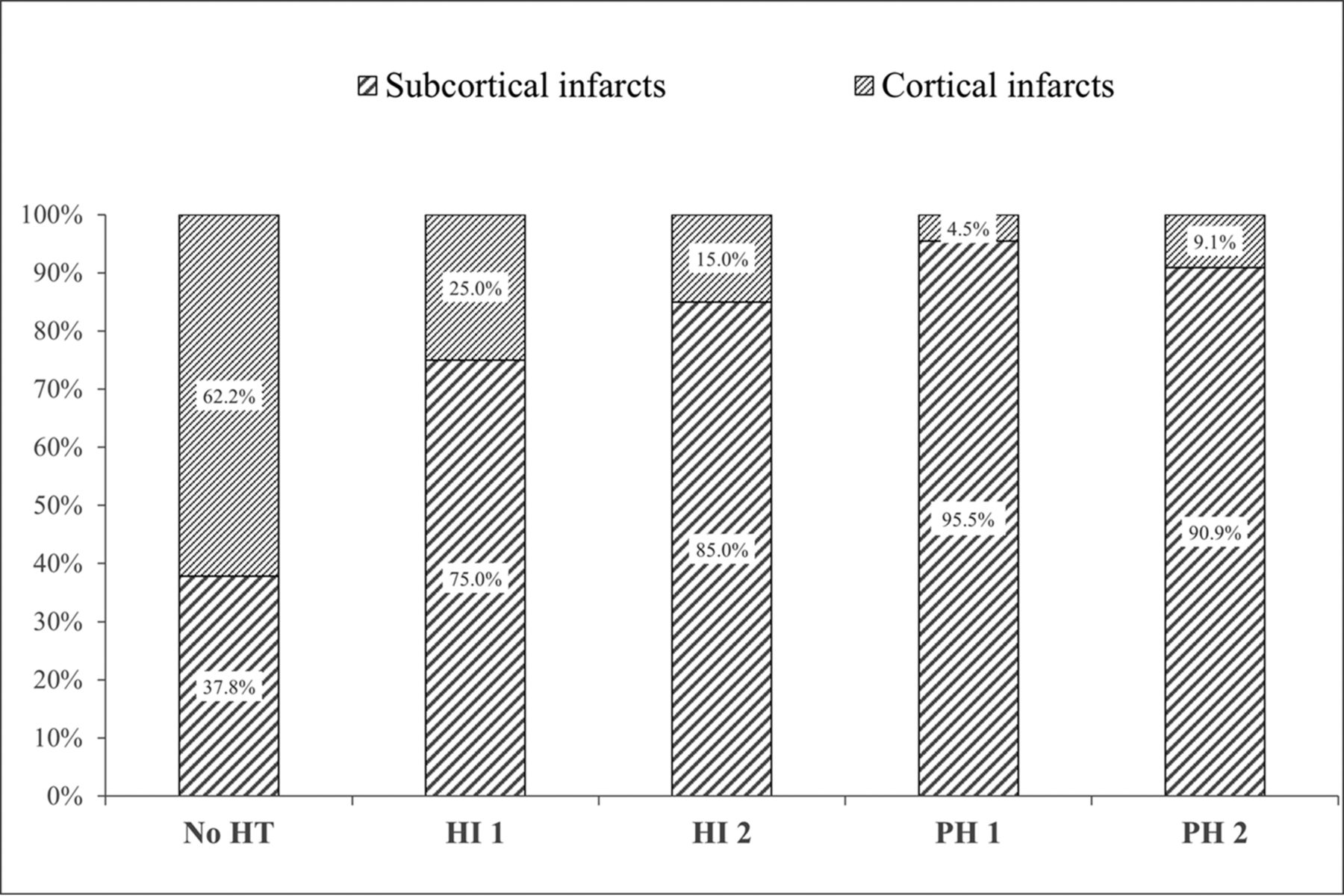
The assessment of infarct location and volume and collateral status showed excellent interrater agreement (κ = 0.908 and 0.826, respectively). The interreader reproducibility for the ischemic core volume segment was also excellent (ICC = 0.819). The CTP images of 92 patients (63.0%) showed a subcortical infarct. The median ischemic core volume determined by CTP maps was 19 mL (IQR, 9–44 mL), and the median infarct core volumes of the cortical and subcortical areas were 12 mL (IQR, 0–32 mL) and 7 mL (IQR, 0–14 mL), respectively. HT was identified in 72/146 patients (49.3%) after successful thrombectomy, and sICH was diagnosed in 23/146 patients (15.8%). In patients with HT, 5.5% (8/146) had HI 1, 13.7% (20/146) had HI 2, 15.1% (22/146) had PH 1, and 15.1% (22/146) had PH 2. The distribution of ischemic core infarct territories in patients with dichotomized HT and nonhemorrhagic transformation subtypes is shown in Fig 2. Patients with any HT subtype had a relatively high proportion of subcortical infarcts (75.0% in HI 1, 85.0% in HI 2, 95.5% in PH 1, and 90.9% in PH 2). This proportion was relatively low in patients with the nonhemorrhagic transformation subtype (37.8%). Functional independence at 3 months was achieved in 72 (49.3%) patients.

FIG 2.
  • Download figure
  • Open in new tab
  • Download powerpoint
FIG 2.

Distribution of ischemic core infarct patterns in patients with dichotomized HT and Non-HT subtypes.

The Online Supplemental Data show a comparison of characteristics between patients with and without hemorrhagic complications. Diabetes mellitus was more common in patients with HT than in those without HT (29.2% versus 10.8%, P < .01). Compared with patients without HT, patients with HT had a higher median NIHSS score (19 versus 16, P = .03), a lower median ASPECTS (6 versus 7, P < .01), worse collateral circulation (76.4% versus 36.5%, P < .01), and more passes of the retriever of ≥3 (34.7% versus 17.6%, P = .02). HT was more frequently observed in patients with subcortical infarcts (88.9% versus 37.8%, P < .01). Patients with HT had a larger median subcortical infarct volume compared with patients without HT (10 mL versus 0 mL, P < .01). In the multivariable regression analysis presented in the Online Supplemental Data, independent risk factors for HT were diabetes mellitus (OR, 4.54; 95% CI, 1.26–16.28; P = .020), core infarcts with subcortical involvement (OR, 8.06; 95% CI, 2.31–28.10; P = .001), poor collateral circulation (OR, 5.49; 95% CI, 2.15–14.01; P < .001), and >3 passes of a retriever (OR, 3.46; 95% CI 1.24–9.64; P = .018).

Patients with the sICH were older (76 versus 71 years of age, P = .04) and had a high prevalence of diabetes mellitus (39.1% versus 16.3%, P = .01), a higher baseline NIHSS score (21 versus 16, P < .01), a lower baseline ASPECTS (5 versus 7, P < .01), and worse collateral circulation (91.3% versus 49.6%, P < .01) than patients without sICH. Additionally, patients with sICH had more subcortical infarcts compared with those without sICH (87.0% versus 58.5%, P = .01). Subcortical infarct volume was higher in patients with sICH than in those without it (11 versus 6 mL, P < .01), whereas total core and cortical infarct volumes were not significantly associated with the occurrence of sICH (Online Supplemental Data). In the multivariable regression analysis, baseline ASPECTS (OR, 0.73; 95% CI, 0.57–0.93; P = .010), subcortical infarct volume (OR, 1.05; 95% CI, 1.01–1.09; P = .039), and collateral circulation (OR, 6.92; 95% CI, 1.47–32.73; P = .015) were significantly associated with sICH after successful thrombectomy (Online Supplemental Data).

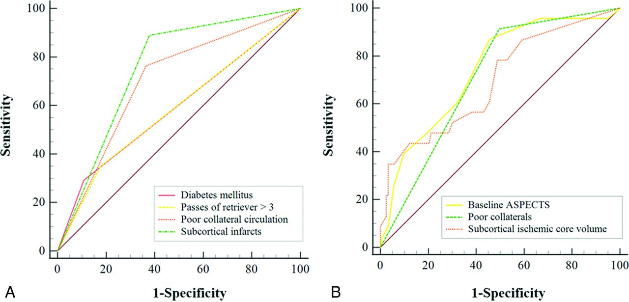
The ROC curves that were used for predicting HT and sICH are shown in Fig 3. The sensitivity and specificity for predicting HT and subcortical infarct involvement were 88.9% and 62.6% (area under the curve = 0.755; 95% CI, 0.68–0.82; P < .001), respectively. The cutoff subcortical infarct volume used for the prediction of sICH was ≥22 mL, and sensitivity and specificity were 34.8% and 96.8% (area under the curve = 0.694; 95% CI, 0.61–0.77; P = .002), respectively.

FIG 3.
  • Download figure
  • Open in new tab
  • Download powerpoint
FIG 3.

ROC analysis predicting HT (A) and sICH (B).

DISCUSSION

In this postthrombectomy patient cohort, we found that subcortical infarcts on admission CTP were associated with the occurrence of HT, and increased subcortical infarct core volume was associated with sICH. To the best of our knowledge, this study is the first to analyze the associations between CTP-based qualitative and quantitative core infarct variables and HT after successful thrombectomy.

The pathophysiologic mechanism of HT involves necrosis of cerebral infarct tissue, collateral circulation, BBB disruption, and infarct size.6,7,18⇓⇓⇓-22 Previous reports have shown an association between pretreatment BBB disruption and HT in patients with acute ischemic stroke after mechanical thrombectomy.7,22 However, the association of BBB disruption, infarction extent, and ischemic core at admission with HT after thrombectomy is unclear. Most studies reported that the ischemic core graded by ASPECTS is linked to HT or sICH after endovascular thrombectomy.11⇓-13 However, the multivariable regression analysis data from the Multicenter Randomized Clinical Trial of Endovascular Therapy for Acute Ischemic Stroke in the Netherlands (MR CLEAN) trial showed that the ASPECTS was not associated with HT or sICH.9 In this study, we quantified the pretreatment of ischemic core infarcts with CTP. We found that the total ischemic core and cortical infarct volumes did not show a positive association with HT or sICH after a successful thrombectomy. In contrast, subcortical infarct occurrence and volume were associated with HT and sICH risk, respectively. These findings may represent an imaging marker predictive of HT and sICH after mechanical thrombectomy that could help manage patients with acute ischemic stroke.

Reperfusion injury, including activation of the endothelium, excess production of oxygen-free radicals, inflammatory responses, leukocyte recruitment, increase in cytokine production, and edema formation could aggravate BBB disruption and potentially cause HT.23 Additionally, successful reperfusion after mechanical thrombectomy can cause acute hyperperfusion and hemodynamic changes to the infarct. The incidence of hyperperfusion after mechanical thrombectomy was relatively high in patients who achieved successful reperfusion.24 Postischemic hyperperfusion on arterial spin-labeled perfusion MR imaging is associated with HT in patients who underwent reperfusion therapy.25,26 These hemodynamic changes, especially in the lenticulostriate artery territories, may influence the rate of HT after reperfusion.27 Our findings support the hypothesis that subcortical infarcts, fed by perforating arteries, are associated with an increased incidence of HT.

Collateral circulation strongly influences the development of postrecanalization HT after acute large-vessel occlusion.9 The presence of unfavorable collaterals on angiography is one of the main reasons for HT after mechanical thrombectomy. Due to poor collateral flow, the hypoperfusion area may enhance the risk of HT after upstream recanalization. Arba et al7 found that pretreatment BBB disruption before reperfusion therapy was correlated with the extent of critically hypoperfused tissue. Subcortical tissue has poor leptomeningeal collaterals and more severe hypoperfusion compared with cortical tissue.18 BBB disruption and hemorrhagic conversion are more common in subcortical infarcts compared with cortical infarcts because of worse collateral anastomosis. Hence, our results show that subcortical infarction is associated with the occurrence of HT and increased subcortical core infarct volume is associated with sICH.

Several potential limitations should be mentioned in this study. First, it has the inherent limitations of its retrospective nature. Second, the boundaries between cortical and subcortical areas are sometimes difficult to distinguish accurately by manual infarct measurement. Artificial intelligence has enormous potential to achieve homogeneity in image interpretation in the future. Third, there may be certain differences between the core infarct volume obtained from CTP and the final true infarct size, though all patients underwent successful reperfusion. Fourth, the qualitative assessment by visual inspection could be feasible, but the quantitative infarct size assessment is not available immediately in clinical work.

CONCLUSIONS

This study shows that core infarct with subcortical involvement on CTP at admission is associated with an increased incidence of HT, and subcortical core infarct volume may influence the risk of sICH after a successful thrombectomy. The infarct location on CTP may be an imaging marker predictive of HT and sICH after mechanical thrombectomy that can help manage patients with acute stroke.

Footnotes

  • H. Ni and G.-D. Lu contributed equally to this work.

  • Disclosure forms provided by the authors are available with the full text and PDF of this article at www.ajnr.org.

References

  1. 1.↵
    1. Powers WJ,
    2. Rabinstein AA,
    3. Ackerson T, et al
    ; American Heart Association Stroke Council. 2018 Guidelines for the Early Management of Patients With Acute Ischemic Stroke: A Guideline for Healthcare Professionals From the American Heart Association/American Stroke Association. Stroke 2018;49:e46–110 doi:10.1161/STR.0000000000000158 pmid:29367334
    Abstract/FREE Full Text
  2. 2.↵
    1. Li W,
    2. Xing X,
    3. Wen C, et al
    . Risk factors and functional outcome were associated with hemorrhagic transformation after mechanical thrombectomy for acute large vessel occlusion stroke. J Neurosurg Sci 2020 Dec 15. [Epub ahead of print] doi:10.23736/S0390-5616.20.05141-3 pmid:33320467
    CrossRefPubMed
  3. 3.↵
    1. Lee YB,
    2. Yoon W,
    3. Lee YY, et al
    . Predictors and impact of hemorrhagic transformations after endovascular thrombectomy in patients with acute large vessel occlusions. J Neurointerv Surg 2019;11:469–73 doi:10.1136/neurintsurg-2018-014080 pmid:30291207
    Abstract/FREE Full Text
  4. 4.↵
    1. van Kranendonk KR,
    2. Treurniet KM,
    3. Boers AM, et al
    ; MR CLEAN Investigators. Hemorrhagic transformation is associated with poor functional outcome in patients with acute ischemic stroke due to a large vessel occlusion. J Neurointerv Surg 2019;11:464–68 doi:10.1136/neurintsurg-2018-014141 pmid:30297537
    Abstract/FREE Full Text
  5. 5.↵
    1. Nogueira RG,
    2. Gupta R,
    3. Jovin TG, et al
    . Predictors and clinical relevance of hemorrhagic transformation after endovascular therapy for anterior circulation large vessel occlusion strokes: a multicenter retrospective analysis of 1122 patients. J Neurointerv Surg 2015;7:16–21 doi:10.1136/neurintsurg-2013-010743 pmid:24401478
    Abstract/FREE Full Text
  6. 6.↵
    1. Hong JM,
    2. Kim DS,
    3. Kim M
    . Hemorrhagic transformation after ischemic stroke: mechanisms and management. Front Neurol 2021;12:703258 doi:10.3389/fneur.2021.703258 pmid:34917010
    CrossRefPubMed
  7. 7.↵
    1. Arba F,
    2. Piccardi B,
    3. Palumbo V, et al
    . Blood-brain barrier leakage and hemorrhagic transformation: the Reperfusion Injury in Ischemic StroKe (RISK) study. Eur J Neurol 2021;28:3147–54 doi:10.1111/ene.14985 pmid:34143500
    CrossRefPubMed
  8. 8.↵
    1. Gauberti M,
    2. Lapergue B,
    3. Martinez de Lizarrondo S, et al
    . Ischemia-reperfusion injury after endovascular thrombectomy for ischemic stroke. Stroke 2018;49:3071–74 doi:10.1161/STROKEAHA.118.022015 pmid:30571423
    CrossRefPubMed
  9. 9.↵
    1. van Kranendonk KR,
    2. Treurniet KM,
    3. Boers AM
    ; et al. MR CLEAN Investigators. Clinical and imaging markers associated with hemorrhagic transformation in patients with acute ischemic stroke. Stroke 2019;50:2037–43 doi:10.1161/STROKEAHA.118.024255 pmid:33755497
    CrossRefPubMed
  10. 10.↵
    1. Broocks G,
    2. Hanning U,
    3. Flottmann F, et al
    . Clinical benefit of thrombectomy in stroke patients with low ASPECTS is mediated by oedema reduction. Brain 2019;142:1399–407 doi:10.1093/brain/awz057 pmid:30859191
    CrossRefPubMed
  11. 11.↵
    1. Hao Y,
    2. Yang D,
    3. Wang H, et al
    ; ACTUAL Investigators (Endovascular Treatment for Acute Anterior Circulation Ischemic Stroke Registry). Predictors for symptomatic intracranial hemorrhage after endovascular treatment of acute ischemic stroke. Stroke 2017;48:1203–09 doi:10.1161/STROKEAHA.116.016368 pmid:28373302
    Abstract/FREE Full Text
  12. 12.↵
    1. Nawabi J,
    2. Kniep H,
    3. Schon G, et al
    . Hemorrhage after endovascular recanalization in acute stroke: lesion extent, collaterals and degree of ischemic water uptake mediate tissue vulnerability. Front Neurol 2019;10:569 doi:10.3389/fneur.2019.00569 pmid:31214107
    CrossRefPubMed
  13. 13.↵
    1. Kaesmacher J,
    2. Kaesmacher M,
    3. Maegerlein C, et al
    . Hemorrhagic transformations after thrombectomy: risk factors and clinical relevance. Cerebrovasc Dis 2017;43:294–304 doi:10.1159/000460265 pmid:28343220
    CrossRefPubMed
  14. 14.↵
    1. Campbell BC,
    2. Christensen S,
    3. Levi CR, et al
    . Cerebral blood flow is the optimal CT perfusion parameter for assessing infarct core. Stroke 2011;42:3435–40 doi:10.1161/STROKEAHA.111.618355 pmid:21980202
    Abstract/FREE Full Text
  15. 15.↵
    1. Yoon W,
    2. Seo JJ,
    3. Kim JK, et al
    . Contrast enhancement and contrast extravasation on computed tomography after intra-arterial thrombolysis in patients with acute ischemic stroke. Stroke 2004;35:876–81 doi:10.1161/01.STR.0000120726.69501.74 pmid:14988575
    Abstract/FREE Full Text
  16. 16.↵
    1. Hacke W,
    2. Kaste M,
    3. Fieschi C, et al
    . Randomised double-blind placebo-controlled trial of thrombolytic therapy with intravenous alteplase in acute ischaemic stroke (ECASS II): Second European-Australasian Acute Stroke Study Investigators. Lancet 1998;352:1245–51 doi:10.1016/S0140-6736(98)08020-9 pmid:9788453
    CrossRefPubMed
  17. 17.↵
    1. Higashida RT,
    2. Furlan AJ,
    3. Roberts H, et al
    ; Technology Assessment Committee of the Society of Interventional Radiology. Trial design and reporting standards for intra-arterial cerebral thrombolysis for acute ischemic stroke. Stroke 2003;34:e109–37 doi:10.1161/01.STR.0000082721.62796.09 pmid:12869717
    Abstract/FREE Full Text
  18. 18.↵
    1. Charbonnier G,
    2. Bonnet L,
    3. Biondi A, et al
    . Intracranial bleeding after reperfusion therapy in acute ischemic stroke. Front Neurol 2020;11:629920 doi:10.3389/fneur.2020.629920 pmid:33633661
    CrossRefPubMed
  19. 19.↵
    1. Arba F,
    2. Rinaldi C,
    3. Caimano D, et al
    . Blood-brain barrier disruption and hemorrhagic transformation in acute ischemic stroke: systematic review and meta-analysis. Front Neurol 2020;11:594613 doi:10.3389/fneur.2020.594613 pmid:33551955
    CrossRefPubMed
  20. 20.↵
    1. Sussman ES,
    2. Connolly ES Jr.
    . Hemorrhagic transformation: a review of the rate of hemorrhage in the major clinical trials of acute ischemic stroke. Front Neurol 2013;4:69 doi:10.3389/fneur.2013.00069 pmid:23772220
    CrossRefPubMed
  21. 21.↵
    1. Bai J,
    2. Lyden PD
    . Revisiting cerebral postischemic reperfusion injury: new insights in understanding reperfusion failure, hemorrhage, and edema. Int J Stroke 2015;10:143–52 doi:10.1111/ijs.12434 pmid:25598025
    CrossRefPubMed
  22. 22.↵
    1. Leigh R,
    2. Christensen S,
    3. Campbell BC, et al
    ; DEFUSE 2 Investigators. Pretreatment blood-brain barrier disruption and post-endovascular intracranial hemorrhage. Neurology 2016;87:263–69 doi:10.1212/WNL.0000000000002862 pmid:27316247
    Abstract/FREE Full Text
  23. 23.↵
    1. Khatri R,
    2. McKinney AM,
    3. Swenson B, et al
    . Blood-brain barrier, reperfusion injury, and hemorrhagic transformation in acute ischemic stroke. Neurology 2012;79:S52–57 doi:10.1212/WNL.0b013e3182697e70 pmid:23008413
    FREE Full Text
  24. 24.↵
    1. Shimonaga K,
    2. Matsushige T,
    3. Hosogai M, et al
    . Hyperperfusion after endovascular reperfusion therapy for acute ischemic stroke. J Stroke Cerebrovasc Dis 2019;28:1212–18 doi:10.1016/j.jstrokecerebrovasdis.2019.01.007 pmid:30704865
    CrossRefPubMed
  25. 25.↵
    1. Okazaki S,
    2. Yamagami H,
    3. Yoshimoto T, et al
    . Cerebral hyperperfusion on arterial spin labeling MRI after reperfusion therapy is related to hemorrhagic transformation. J Cereb Blood Flow Metab 2017;37:3087–90 doi:10.1177/0271678X17718099 pmid:28665168
    CrossRefPubMed
  26. 26.↵
    1. Yu S,
    2. Liebeskind DS,
    3. Dua S
    ; et al; UCLA Stroke Investigators. Postischemic hyperperfusion on arterial spin labeled perfusion MRI is linked to hemorrhagic transformation in stroke. J Cereb Blood Flow Metab 2015;35:630–37 doi:10.1038/jcbfm.2014.238 pmid:25564233
    CrossRefPubMed
  27. 27.↵
    1. Haussen DC,
    2. Ferreira IM,
    3. Barreira C, et al
    . Active reperfusion hemorrhage during thrombectomy: angiographic findings and real-time correlation with the CT “Spot Sign.” Interv Neurol 2018;7:370–77 doi:10.1159/000488084 pmid:30410514
    CrossRefPubMed
  • Received September 15, 2022.
  • Accepted after revision November 16, 2022.
  • © 2023 by American Journal of Neuroradiology
PreviousNext
Back to top

In this issue

American Journal of Neuroradiology: 44 (1)
American Journal of Neuroradiology
Vol. 44, Issue 1
1 Jan 2023
  • Table of Contents
  • Index by author
  • Complete Issue (PDF)
Advertisement
Print
Download PDF
Email Article

Thank you for your interest in spreading the word on American Journal of Neuroradiology.

NOTE: We only request your email address so that the person you are recommending the page to knows that you wanted them to see it, and that it is not junk mail. We do not capture any email address.

Enter multiple addresses on separate lines or separate them with commas.
Association between Infarct Location and Hemorrhagic Transformation of Acute Ischemic Stroke following Successful Recanalization after Mechanical Thrombectomy
(Your Name) has sent you a message from American Journal of Neuroradiology
(Your Name) thought you would like to see the American Journal of Neuroradiology web site.
CAPTCHA
This question is for testing whether or not you are a human visitor and to prevent automated spam submissions.
Cite this article
H. Ni, G.-D. Lu, Y. Hang, Z.-Y. Jia, Y.-Z. Cao, H.-B. Shi, S. Liu, L.-B. Zhao
Association between Infarct Location and Hemorrhagic Transformation of Acute Ischemic Stroke following Successful Recanalization after Mechanical Thrombectomy
American Journal of Neuroradiology Jan 2023, 44 (1) 54-59; DOI: 10.3174/ajnr.A7742

Citation Manager Formats

  • BibTeX
  • Bookends
  • EasyBib
  • EndNote (tagged)
  • EndNote 8 (xml)
  • Medlars
  • Mendeley
  • Papers
  • RefWorks Tagged
  • Ref Manager
  • RIS
  • Zotero
0 Responses
Respond to this article
Share
Bookmark this article
Infarct Location and Hemorrhagic Transformation
H. Ni, G.-D. Lu, Y. Hang, Z.-Y. Jia, Y.-Z. Cao, H.-B. Shi, S. Liu, L.-B. Zhao
American Journal of Neuroradiology Jan 2023, 44 (1) 54-59; DOI: 10.3174/ajnr.A7742
del.icio.us logo Twitter logo Facebook logo Mendeley logo
  • Tweet Widget
  • Facebook Like
  • Google Plus One
Purchase

Jump to section

  • Article
    • Abstract
    • ABBREVIATIONS:
    • MATERIALS AND METHODS
    • RESULTS
    • DISCUSSION
    • CONCLUSIONS
    • Footnotes
    • References
  • Figures & Data
  • Supplemental
  • Info & Metrics
  • Responses
  • References
  • PDF

Related Articles

  • PubMed
  • Google Scholar

Cited By...

  • Association between basal ganglia infarction and hemorrhagic transformation after mechanical thrombectomy in acute ischemic stroke: insights from the DIRECT-MT trial
  • Hypoperfusion Intensity Ratio and Hemorrhagic Transformation in Patients with Successful Recanalization after Thrombectomy
  • Association between ASPECTS region of infarction and clinical outcome in non-acute large vessel occlusion ischaemic stroke after endovascular recanalisation
  • Crossref (16)
  • Google Scholar

This article has been cited by the following articles in journals that are participating in Crossref Cited-by Linking.

  • Role of Brain Imaging in the Prediction of Intracerebral Hemorrhage Following Endovascular Therapy for Acute Stroke
    Pierre Seners, Anke Wouters, Benjamin Maïer, William Boisseau, Benjamin Gory, Jeremy J. Heit, Christophe Cognard, Mikael Mazighi, Brice Gaudilliere, Robin Lemmens, Greg Zaharchuk, Gregory W. Albers, Richard Leigh, Jean-Marc Olivot
    Stroke 2023 54 8
  • Association between infarct location and haemorrhagic transformation of acute ischaemic stroke after intravenous thrombolysis
    Y. Gu, C. Xu, Z. Zhang, C. Fang, J. Yu, D. He, G. Xu
    Clinical Radiology 2024 79 3
  • Endovascular thrombectomy for DAWN- and DEFUSE-3 ineligible acute ischemic stroke patients: a systematic review and meta-analysis
    Samah Morsi, Sherief Ghozy, Mohamed Elfil, Hatem Tolba, Alejandro Rabenstein, Ramanathan Kadirvel, David F. Kallmes
    Journal of Neurology 2024 271 5
  • Hypoperfusion Intensity Ratio and Hemorrhagic Transformation in Patients with Successful Recanalization after Thrombectomy
    Jiaxiang You, Xiaoxi Li, Jun Xia, Haopeng Li, Jun Wang
    American Journal of Neuroradiology 2024 45 10
  • Transcranial Doppler ultrasound to evaluate the risk of hyperperfusion after endovascular stroke thrombectomy
    Aristeidis H. Katsanos, Abhilekh Srivastava, Demetrios J. Sahlas, Kanjana Perera, Kelvin K. H. Ng, Raed A. Joundi, Brian Van Adel, Ramiro Larrazabal, Kanchana Ratnayake, Georgios Tsivgoulis, Oscar Benavente, Robert Hart, Mukul Sharma, Ashkan Shoamanesh, Luciana Catanese
    Journal of Neuroimaging 2024 34 1
  • Low blood flow ratio is associated with hemorrhagic transformation secondary to mechanical thrombectomy in patients with acute ischemic stroke
    Lufei Feng, Mengying Yu, Mo Zheng, Wangle Huang, Fei Yao, Chaomin Qiu, Ru Lin, Ying Zhou, Haoyu Wu, Guoquan Cao, Dexing Kong, Yunjun Yang, Haoli Xu
    Journal of Neuroradiology 2024 51 4
  • Assessment of the Risk of Cerebral Parenchymal Hemorrhage After Thrombectomy in Acute Ischemic Stroke With Dynamic Cerebral Autoregulation
    Ran Liu, Songwei Chen, Liuping Cui, Hongxiu Chen, Xijuan Pan, Fubo Zhou, Wenbo Zhao, Yingqi Xing
    Operative Neurosurgery 2025
  • Association between ASPECTS region of infarction and clinical outcome in non-acute large vessel occlusion ischaemic stroke after endovascular recanalisation
    Jialiang Lu, Ziwei Lu, Ye Li, Fangcun Li, Yuxuan Feng, Meijuan Dang, Yang Yang, Fan Tang, Tao Li, Lili Zhao, Yating Jian, Xiaoya Wang, Lei Zhang, Hong Fan, Guilian Zhang
    Stroke and Vascular Neurology 2024
  • Association between basal ganglia infarction and hemorrhagic transformation after mechanical thrombectomy in acute ischemic stroke: insights from the DIRECT-MT trial
    Rundong Chen, Weilong Hua, Yilei Zhang, Yongxin Zhang, Hongjian Zhang, Yongwei Zhang, Jianmin Liu, Pengfei Yang, Lei Zhang
    Journal of NeuroInterventional Surgery 2024
  • Hemorrhagic Infarction Does Not Worsen Functional Outcomes in Noncardioembolic Ischemic Stroke—Secondary Analysis From PACIFIC-STROKE
    Chih-Hao Chen, Ashkan Shoamanesh, Pablo Colorado, Feryal Saad, Robin Lemmens, Gian Marco De Marchis, Valeria Caso, Lizhen Xu, Laura Heenan, Jaime Masjuan, Hanne Christensen, Pooja Khatri, Hardi Mundl, Robert G. Hart, Eric E. Smith
    Stroke 2025

More in this TOC Section

Neurointervention

  • Factors Associated with Major Re-Recanalization following Second Coiling for Recanalized Aneurysms: A Multicenter Experience over 20 Years during Long-Term Follow-up
  • A Key Factor Shapes LS-DAVFs EVT Outcome
  • Optimizing Voxel Size in 3D Rotational Angiography
Show more Neurointervention

Adult Brain

  • Diagnostic Neuroradiology of Monoclonal Antibodies
  • Cerebral ADC Changes in Fabry Disease
  • ML for Glioma Molecular Subtype Prediction
Show more Adult Brain

Similar Articles

Advertisement

Indexed Content

  • Current Issue
  • Accepted Manuscripts
  • Article Preview
  • Past Issues
  • Editorials
  • Editor's Choice
  • Fellows' Journal Club
  • Letters to the Editor
  • Video Articles

Cases

  • Case Collection
  • Archive - Case of the Week
  • Archive - Case of the Month
  • Archive - Classic Case

More from AJNR

  • Trainee Corner
  • Imaging Protocols
  • MRI Safety Corner

Multimedia

  • AJNR Podcasts
  • AJNR Scantastics

Resources

  • Turnaround Time
  • Submit a Manuscript
  • Submit a Video Article
  • Submit an eLetter to the Editor/Response
  • Manuscript Submission Guidelines
  • Statistical Tips
  • Fast Publishing of Accepted Manuscripts
  • Graphical Abstract Preparation
  • Imaging Protocol Submission
  • Evidence-Based Medicine Level Guide
  • Publishing Checklists
  • Author Policies
  • Become a Reviewer/Academy of Reviewers
  • News and Updates

About Us

  • About AJNR
  • Editorial Board
  • Editorial Board Alumni
  • Alerts
  • Permissions
  • Not an AJNR Subscriber? Join Now
  • Advertise with Us
  • Librarian Resources
  • Feedback
  • Terms and Conditions
  • AJNR Editorial Board Alumni

American Society of Neuroradiology

  • Not an ASNR Member? Join Now

© 2025 by the American Society of Neuroradiology All rights, including for text and data mining, AI training, and similar technologies, are reserved.
Print ISSN: 0195-6108 Online ISSN: 1936-959X

Powered by HighWire