
Article Figures & Data
Figures
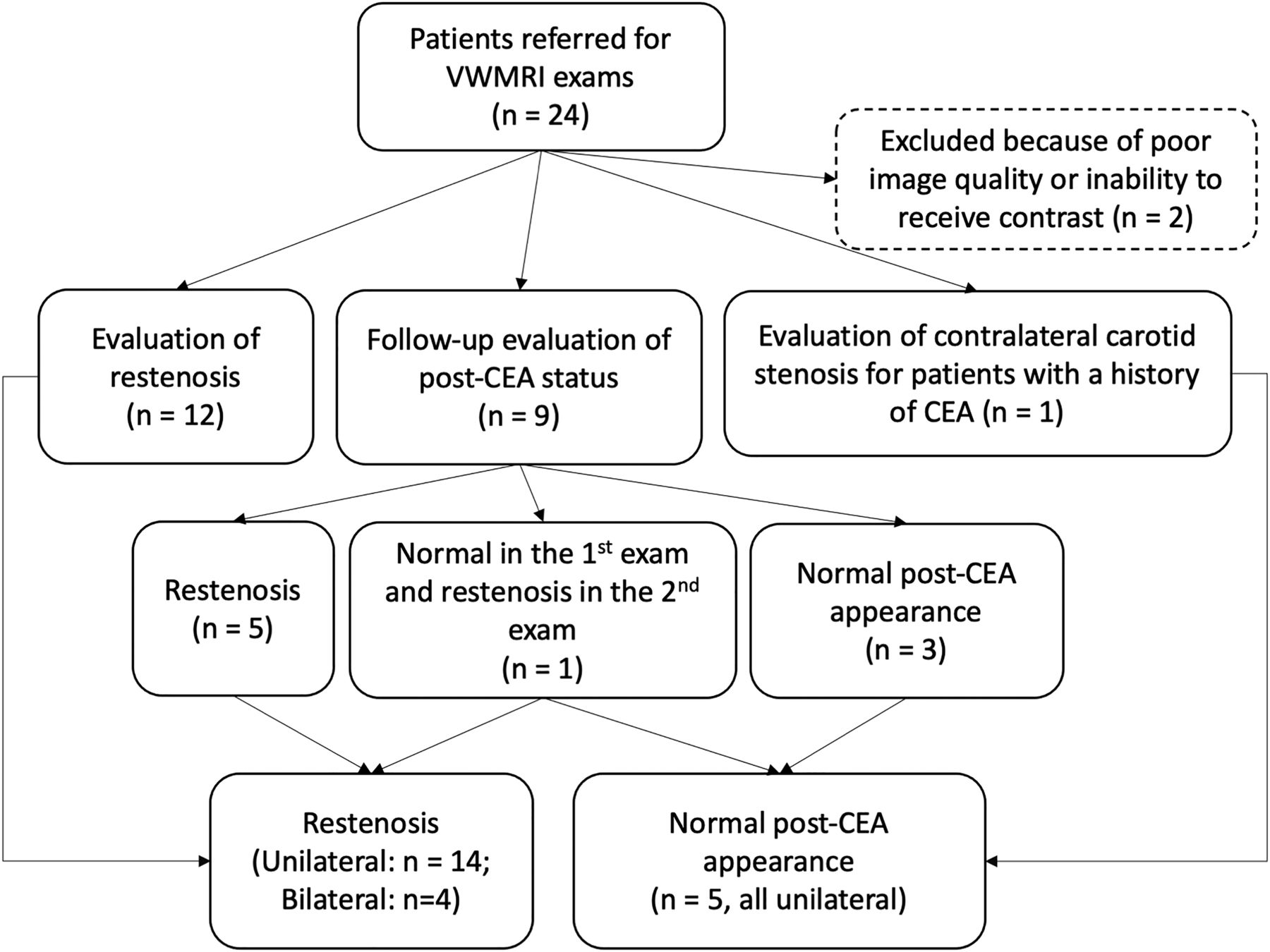
- FIG 1.
Flow chart of patient inclusion. The flow chart details referral reasons and exclusion criteria for patients with VWMRI examinations. N indicates number of patients.
- FIG 2.
Progression from normal post-CEA appearance to MH. TOF-MRA (A) of a post-CEA carotid artery shows an expected vessel geometry 2 months after the operation. Pre- (B) and postcontrast (C) VWMRI acquired at the proximal ICA (indicated by white line in A) shows vessel wall enhancement but no abnormal wall thickening of the proximal ICA (long arrows). TOF-MRA acquired 10 months after CEA (D) shows luminal stenosis. Pre- (E) and postcontrast (F) VWMRIs acquired at the same location (indicated by white line in D) show circumferential wall thickening with mild, homogeneous enhancement compatible with MH (long arrows). Short arrows in B, C, E, and F indicate the external carotid artery. VWMRIs were acquired using an electrocardiogram-gated double inversion recovery turbo spin-echo sequence (TR/TE/echo-train, 1 RR/9 ms/10; resolution, 0.35 × 0.35 × 2 mm3).
- FIG 3.
Representative images of MH. TOF-MRA (A) shows restenosis post-CEA extending from proximal to distal to the carotid bifurcation. Precontrast VWMRI (B) at the level of proximal ICA (indicated by white line in A) shows concentric homogeneous wall thickening (long arrow), indicative of MH. The lesion is enhanced on postcontrast VWMRI (C, long arrow). Short arrows in B and C indicate the external carotid artery. VWMRIs were acquired using an electrocardiogram-gated double inversion recovery turbo spin-echo sequence (TR/TE/echo-train, 1 RR/9 ms/10; resolution, 0.35 × 0.35 × 2 mm3).
- FIG 4.
Representative images of recurrent plaque. TOF-MRA (A) shows high-grade restenosis of the carotid bulb. The white line indicates the location of VWMRIs. Pre- (B, left image) and corresponding postcontrast (B, right image) VWMRIs show enhancing eccentric wall thickening with ulceration (asterisks), suggestive of a recurrent plaque. The corresponding specimen sections stained with MOVAT (C) confirm the diagnosis of recurrent plaque. The asterisk indicates ulceration. The arrowheads in B and C indicate the lumen. VWMRIs were acquired using an electrocardiogram-gated double inversion recovery turbo spin-echo sequence (TR/TE/echo-train, 1 RR/9 ms/10; resolution, 0.35 × 0.35 × 2 mm3).


Tables
- Table 1:
Comparison of clinical and imaging characteristics between recurrent and matched primary plaquesa
Recurrent Plaques Primary Plaques P Value Patient characteristicsb Age 75.8 (SD, 9.5) 74.3 (SD, 11.4) .736 Male 6 (60.0%) 25 (68.8%) .727 Hypertension 9 (90.0%) 29 (76.3%) .664 Hyperlipidemia 8 (80.0%) 23 (60.5%) .459 Diabetes 0 (0.0%) 7 (18.4%) .318 Plaque characteristicsc Plaque components Lipid core 12 (92.3%) 32 (82.1%) .662 Fibrous cap 12 (92.3%) 32 (82.1%) .662 Calcification 9 (69.2%) 31 (79.5%) .466 IPH 8 (61.5%) 12 (30.8%) .048d Ulceration 8 (61.5%) 20 (51.3%) .521 Maximum wall thickness (mm) 4.12 (SD, 1.74) 4.41 (SD, 1.50) .562 Remodeling ratio 1.57 (SD, 0.57) 1.49 (SD, 0.50) .662 Lesion length (cm) 2.26 (SD, 1.12) 1.47 (SD, 0.54) .001d Adventitial enhancement .358 Category 0 5 (38.5%) 7 (17.9%) Category 1 3 (23.1%) 14 (35.9%) Category 2 5 (38.5%) 18 (46.2%) Plaque position .002d Opposite flow divider 4 (30.8%) 31 (79.5%) Along flow divider/sidewalls 9 (69.2%) 8 (20.5%) - Table 2:
Multivariate logistic regression model for plaque-feature detection in recurrent plaques compared with primary plaques
Characteristics Multivariate OR 95% CI P Presence of IPH 1.63 0.36–7.47 .528 Plaque position Along flow divider/sidewalls vs opposite flow divider 6.96 1.37–35.28 .019a Plaque lengthb 4.27 1.32–13.85 .015a Maximum wall thicknessb 0.69 0.38–1.24 .210


Jump to section
Related Articles
Cited By...
- No citing articles found.